Ophthalmic imaging with k-mirror scanning, efficient interferometry, and pupil alignment through spatial frequency analysis

a technology of k-mirror scanning and ophthalmic imaging, applied in the field of ophthalmic imaging systems, can solve the problems of limiting the availability further complexity, and complexity of advanced optical imaging systems, and achieves simplified system-to-patient alignment, simplified rotational and/or, and efficient line-field

Pending Publication Date: 2022-08-18
CARL ZEISS MEDITEC INC +1
View PDF0 Cites 0 Cited by
  • Summary
  • Abstract
  • Description
  • Claims
  • Application Information

AI Technical Summary

Benefits of technology

[0009]The above objects are met in an ophthalmic imaging system with simplified rotational and / or linear scanning, relaxed design constraints, efficient line-field and / or full-field and / or partial-field interferometry, and simplified system-to-patient alignment. The complications and costs associated with galvanometer scanners may be avoided by using a K-mirror as the scanning component. Use of a K-mirror also avoids any aberration issues associated with prisms, such as from a dove prism, which permits the K-mirror to be located in a non-collimated optical path of the ophthalmic imaging system. For example, the K-mirror may be positioned such that an intermediate focal point of scattered light exiting an eye is located within the K-mirror (structure). Furthermore, the present implementation removes the task of pupil splitting from the scanning component, freeing it to be located at any convenient position and not being restricted to a pupil conjugate plane. Consequently, the present K-mirror may be positioned between a scan lens and an ocular lens, which is the lens closest to the eye. These reduced restrictions on the scanning component provide for reduced design constraints and ease of manufacture.
[0010]Using the K-mirror for rotation scanning allows it to be used for modulating the length of an available scan line. By moving one of the three mirrors of the K-mirror structure, the output scan line is offset along its radial direction from its center of rotation. This permits the creation of annular scan patterns, and / or the extension of the scan line length by as much as a factor of two.
[0012]Furthermore, one or more of the mirrors of the K-mirror structure may be made reflective on one side and transparent on an opposite side. The reflective side may be chosen to face the interior of the K-mirror structure. This may be achieved, for example, by applying to, or embedding within, a glass sheet a thin layer of a reflective and almost transparent material (e.g., a dielectric coating, such as aluminum). In this manner, a second input portal (from the transparent side) is created for the K-mirror structure. A fixation pattern may be inputted to the K-mirror through this second input portal. Additionally, by affixing the mechanism that generates the fixation pattern to the K-mirror such that both move in tandem, their positions relative to each other remain constant. In this manner, the position of a fixation pattern, as perceived by a patient during a scanning process, may remain constant.
[0015]Efficient interferometry may further be provided by positioning a physical collection aperture (e.g. system pupil stop) at a pupil conjugate plane of a patient's eye to define a mechanical collection pupil. Optionally, the physical collection aperture may be made to have a modular configuration. The collection aperture may selectively be changed from an on-axis configuration to an off-axis configuration, and vice versa. That is, the mechanical collection pupil may convert collected light (returning from the beam splitter toward a collector or camera) from having an on-axis configuration to having an off-axis configuration. The on-axis configuration permits the maximum amount of light available to reach the collector, but depth information provided by such light may be reduced by the appearance of a complex conjugate image. The off-axis configuration may reduce the amount of available light reaching the collector, but avoids the creation of a complex conjugate image and thereby achieve higher depth resolution (e.g., full-range A-scans or B-scans). That is, applying a 2D fast Fourier transfer (FFT) to the collected light from the off-axis configuration provides a spatial frequency distribution of scattered light at the collection aperture (e.g., corresponding to the patient's pupil conjugate). This spatial frequency distribution separates true signal components from their complex conjugate components, which facilitates the selection of true signal information (that does not include complex conjugate components) for processing.
[0016]As it would be understood by one versed in the art, a first type of spectral analysis (e.g., 1D FFT) is typically applied to the collected sample light (e.g. interference light) to recover depth information of a scan beam. However as stated above, by applying a second type of spectral analysis (e.g., 2D FFT) to the collected sample light, spatial characteristics of collected light (e.g., at the collection aperture on a pupil conjugate plane) returning from the sample arm may be recovered. As explained above, this information may be used to select spectral information that does not include complex conjugate components, but this information may further be used for alignment of the imaging system to a patient. That is, these spatial characteristics may be correlated to a position of a patient's eye (e.g., the patient's pupil) relative to the ophthalmic imaging device (e.g., the device's collection aperture). It has been found that this spectral analysis may be used to determine both translational and axial positions of an eye relative to the ophthalmic imaging device. In this manner, the scanning mechanism of the ophthalmic imaging device may, at least partially, determine proper alignment of the patient to the ophthalmic imaging device without the need for additional, customary alignment equipment, such as a pupil camera and its associated hardware / software. In this manner, the present system may provide automated pupil alignment based at least in part on the spectral analysis.

Problems solved by technology

An obstacle to this goal is the complexity and cost of advanced optical imaging systems for ophthalmic use (e.g., ophthalmic imaging systems), which limit their availability.
Further complexity may arise from the need to provide an alignment mechanism to align the ophthalmic imaging system to a patient's eye (e.g., pupil alignment), and / or to provide a fixation point to direct a patient's gaze.
These difficulties complicate their design and construction, leading to higher cost.

Method used


Image

  • Ophthalmic imaging with k-mirror scanning, efficient interferometry, and pupil alignment through spatial frequency analysis
  • Ophthalmic imaging with k-mirror scanning, efficient interferometry, and pupil alignment through spatial frequency analysis
  • Ophthalmic imaging with k-mirror scanning, efficient interferometry, and pupil alignment through spatial frequency analysis

Examples


Embodiment Construction

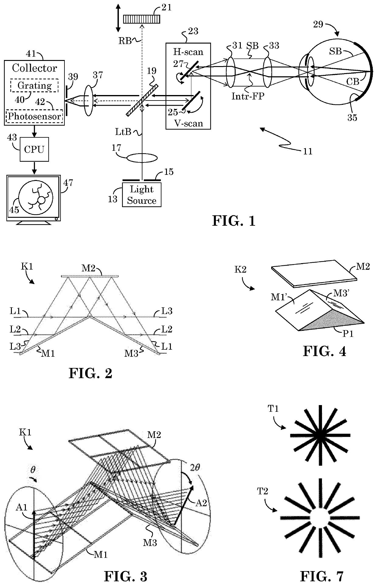
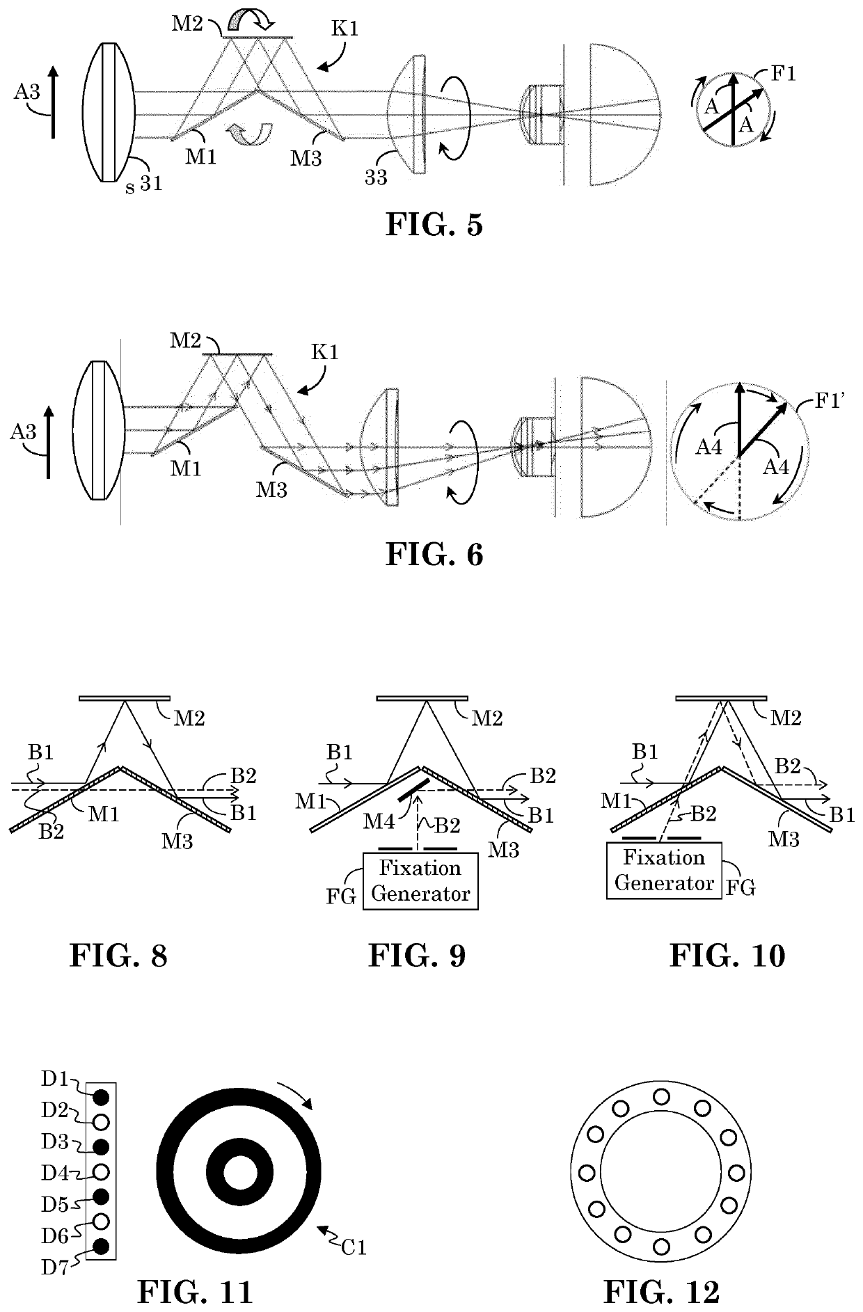
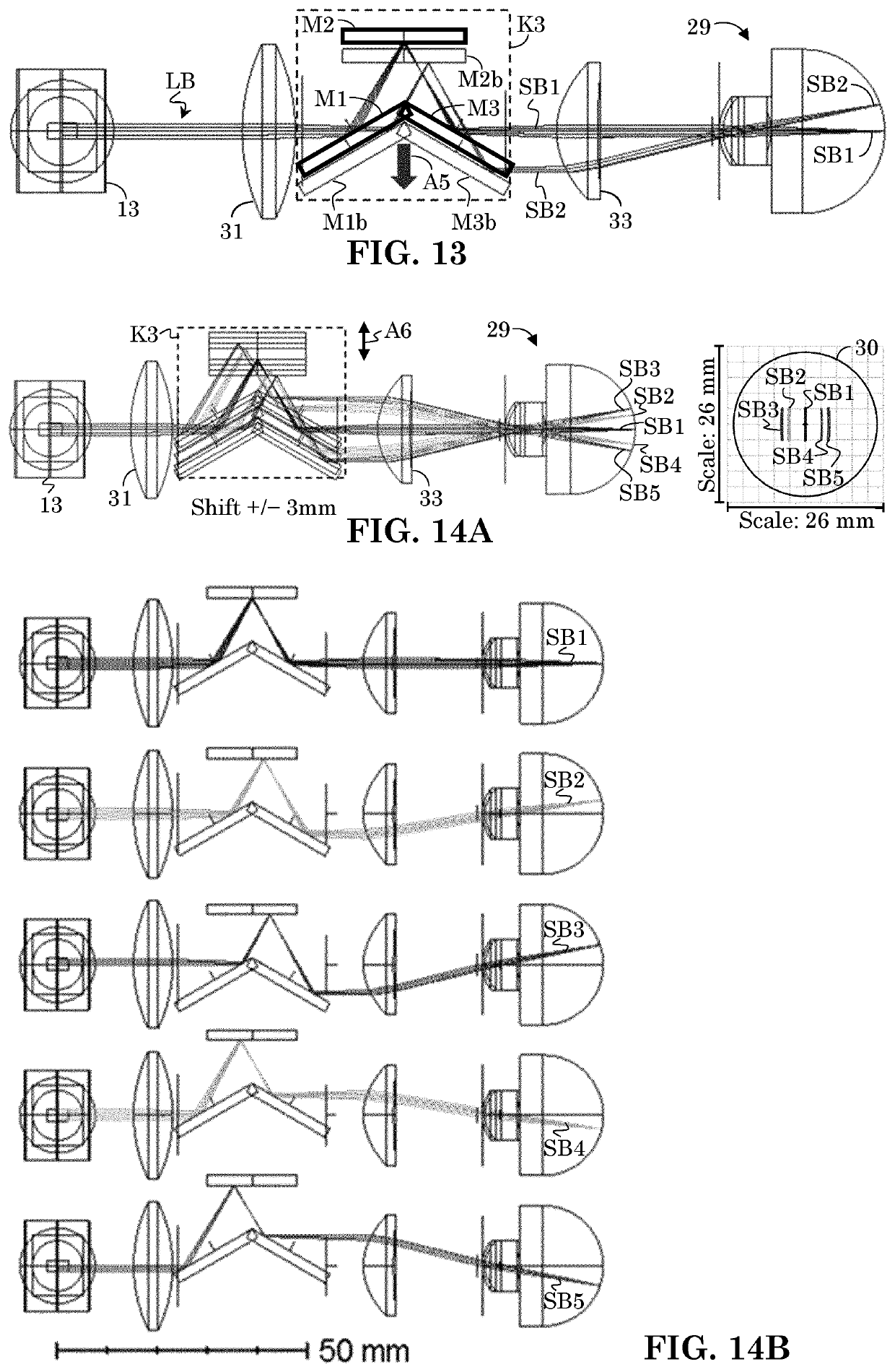
[0065]As is discussed more fully below, a fundus imager may provide high resolution grayscale or color images of the eye fundus, whereas optical coherence tomography (OCT) and optical coherence tomography angiography (OCTA) enable noninvasive, depth-resolved (e.g., A-scan), volumetric (e.g., C-scan), and two-dimensional (e.g., en face (frontal plane) or B-scan (transverse plane)) visualization of retinal vasculature. Generally, OCT provides structural images of tissue (e.g., vascular structure) whereas OCTA provides functional images of vasculature (e.g., blood flow). For example, OCTA may image vascular flow by using the motion of blood flow as an intrinsic contrast. For ease of discussion, a brief overview of such devices is presented, as it relates to the present improvements. A more detailed discussion of various types of ophthalmic imaging systems is provided below in section “Fundus Imaging System” and in section “Optical Coherence Tomography (OCT) Imaging System.” Aspects of ...


PUM


Abstract

Various techniques for providing a low-cost ophthalmic imaging system, such an OCT or fundus imagers are presented. Cost is reduced by using a K-minor as a scanning component. The K-mirror is positioned at a retina conjugate. A beam splitter is positioned at the pupil conjugate and may be used to provide pupil splitting functionality. The beam splitter's shape and area conform to the focal light footprint of an illumination source such that it spans only a fraction of a collection window. 2D FFT is applied to capture spectra for purposes of selectively removing complex conjugate components and for extracting patient pupil to system collection pupil alignment. Consequently, pupil alignment is achieved by use of captured OCT data without the need for additional pupil cameras.

Description

FIELD OF INVENTION[0001]The present invention is generally directed to the field of ophthalmic imaging systems. More specifically, it is directed to ophthalmic imaging systems with simplified scanning components, reduced designed constraints, and facilitated patient-to-system alignment.BACKGROUND[0002]Early diagnosis is critical for the successful treatment of various eye diseases. Optical imaging is a preferred method for non-invasive examination of the retina. Optical imaging may be used to identify major causes of vision loss, such as age-related macular degeneration, diabetic retinopathy, and glaucoma, but diagnosis is often not made until after damage has manifested itself. A goal of ophthalmic medical care is to identify possible pathologies at a pre-clinical stage of disease. An obstacle to this goal is the complexity and cost of advanced optical imaging systems for ophthalmic use (e.g., ophthalmic imaging systems), which limit their availability.[0003]There are various types...

Claims


Application Information

Patent Timeline
Patent Type & Authority Applications(United States)
IPC IPC(8): A61B3/10A61B3/00A61B3/12A61B5/00A61B3/15
CPCA61B3/102A61B3/0008A61B3/152A61B5/7257A61B3/0058A61B3/1233A61B3/112A61B3/12A61B3/14G02B26/105
Inventor WANG, YINGJIANLIU, YUANSTRAUB, JOCHENDARVISHZADEH VARCHEIE, MASHALEAHY, CONOR
Owner CARL ZEISS MEDITEC INC
Why Eureka
Try Eureka