Methods and systems for bicuspid valve detection with generative modeling

a bicuspid aortic valve and generative modeling technology, applied in image enhancement, instruments, ultrasonic/sonic/infrasonic image/data processing, etc., can solve the problem of difficult detection of the presence of the bicuspid aortic valv

Pending Publication Date: 2022-10-06
GE PRECISION HEALTHCARE LLC
View PDF15 Cites 0 Cited by
  • Summary
  • Abstract
  • Description
  • Claims
  • Application Information

AI Technical Summary

Benefits of technology

[0004]In one embodiment, a method comprises acquiring ultrasound video of a heart over at least one cardiac cycle, identifying frames in the ultrasound video corresponding to at least one cardiac phase, and classifying a cardiac structure in the identified frames as a bicuspid valve or a tricuspid valve. A

Problems solved by technology

However, the presence of a bicuspid aortic valve is difficult to detect during regular screening procedures due

Method used


Image

  • Methods and systems for bicuspid valve detection with generative modeling
  • Methods and systems for bicuspid valve detection with generative modeling
  • Methods and systems for bicuspid valve detection with generative modeling

Examples


Embodiment Construction

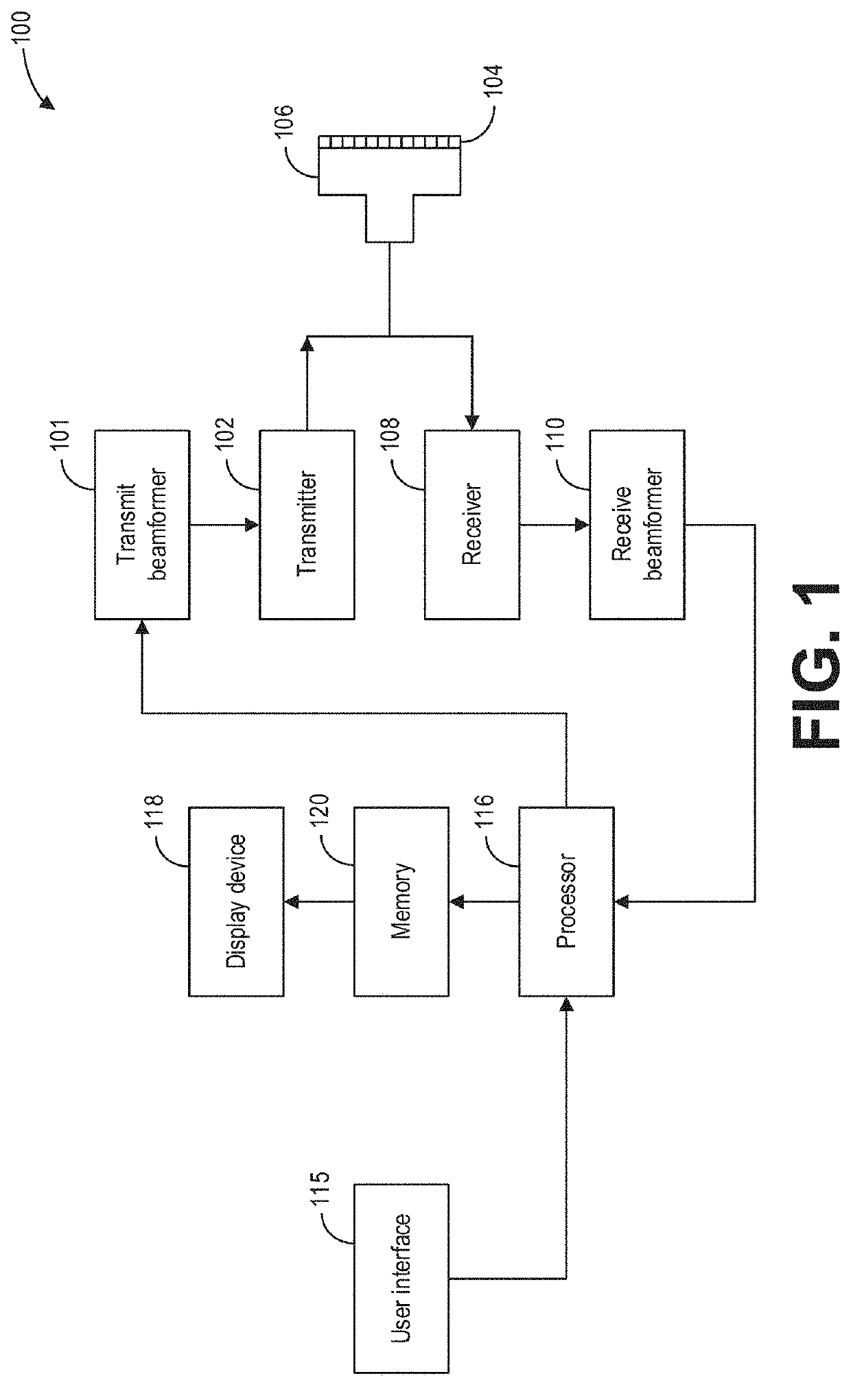
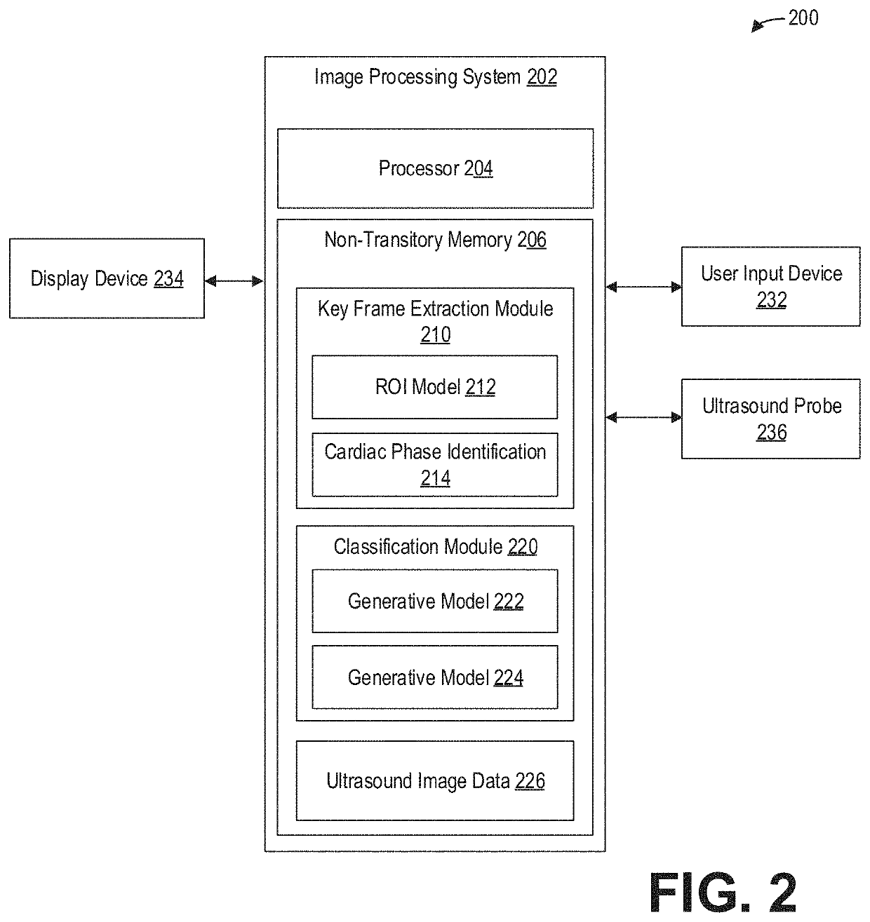
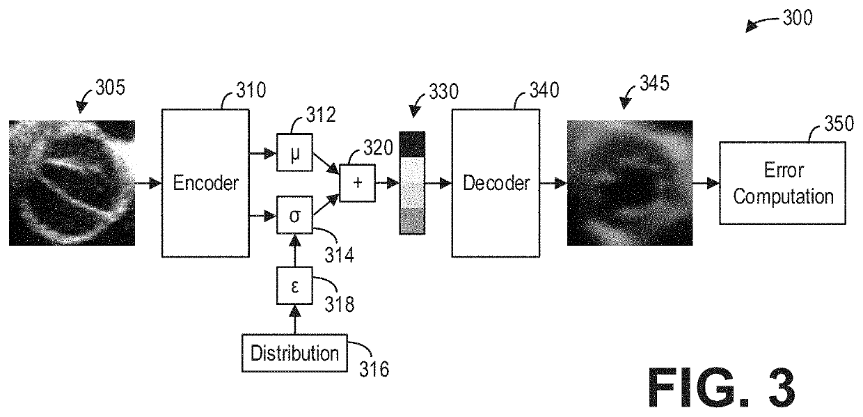
[0016]The following description relates to various embodiments of ultrasound imaging. In particular, methods and systems for automatic detection of bicuspid valves with an ultrasound imaging system are provided. An example of an ultrasound imaging system that may be used to acquire images in accordance with the present techniques is shown in FIG. 1. The ultrasound imaging system may be communicatively coupled to an image processing system, such as the image processing system of FIG. 2. The image processing system may include one or more deep learning models, such as one or more neural networks and one or more generative models, stored in non-transitory memory. A generative model configured as a variational autoencoder, as shown in FIG. 3, may be trained on ultrasound images of tricuspid valves at a select cardiac phase. Ultrasound video comprising a sequence of ultrasound image frames or ultrasound images may then be processed, for example as shown in FIG. 4, with such a trained gen...


PUM


Abstract

Various methods and systems are provided for bicuspid valve detection with ultrasound imaging. In one embodiment, a method comprises acquiring ultrasound video of a heart over at least one cardiac cycle, identifying frames in the ultrasound video corresponding to at least one cardiac phase, and classifying a cardiac structure in the identified frames as a bicuspid valve or a tricuspid valve. A generative model such as a variational autoencoder trained on ultrasound image frames at the at least one cardiac phase may be used to classify the cardiac structure. In this way, relatively rare occurrences of bicuspid aortic valves may be automatically detected during regular cardiac ultrasound screenings.

Description

FIELD[0001]Embodiments of the subject matter disclosed herein relate to ultrasound imaging, and more specifically to detecting bicuspid aortic valves with ultrasound imaging.BACKGROUND[0002]Aortic valve stenosis or aortic stenosis occurs when the aortic valve of a heart narrows, thereby resulting in an increased blood flow pressure gradient at the aortic valve opening. The aortic valve does not fully open and so blood flow from the heart to the aorta is reduced. The possibility of aortic stenosis is significantly elevated if the aortic valve is bicuspid rather than tricuspid, where the aortic valve includes only two leaflets rather than the typical three leaflets. Such bicuspid aortic valves are relatively rare and occur in less than two percent of the normal population.[0003]Ultrasound is the primary imaging modality used to screen for aortic stenosis. Early detection of a bicuspid aortic valve may enable proactive intervention and treatment to potentially reduce the likelihood of ...

Claims


Application Information

Patent Timeline
IPC IPC(8): G06T7/00A61B8/08G06K9/62G06K9/03G06K9/32G06N3/04G06N3/08
CPCG06T7/0014A61B8/0883A61B8/5223G06K9/6267G06K9/03G06K9/6257G06K9/3233G06N3/0454G06N3/088G06K2209/051G06T2207/30048G06T2207/20081G06T2207/10132G06T2207/10016A61B8/0891A61B8/5215G06T7/0012G06T2207/20084A61B8/486A61B8/488G06N3/047G06N3/045G06V2201/031G06V10/25G06V10/764G06V10/98G06V10/82G06F18/24G06F18/2148
Inventor ANNANGI, PAVANVENKATARAMANI, RAHULANAND, DEEPASAMSET, EIGIL
Owner GE PRECISION HEALTHCARE LLC
Why Eureka
Try Eureka