Intratumoral TFR Cells Curtail Anti-PD-1 Treatment Efficacy

a tfr cell and anti-pd-1 technology, applied in the field of cancer immunotherapy, can solve the problems that the anti-pd-1 therapy cannot only facilitate, but also dampen the anti-tumor immune attack, and limit its us

Pending Publication Date: 2022-11-17
LA JOLLA INST FOR IMMUNOLOGY +1
View PDF0 Cites 2 Cited by
  • Summary
  • Abstract
  • Description
  • Claims
  • Application Information

AI Technical Summary

Benefits of technology

[0006]The present invention pertains to T follicular regulatory cells (TFR cells) and their functional role in cancer. To date, TFR cells have been overlooked in cancer. This is a critical oversight, as these cells account for a substantial proportion of tumor-infiltrating CD4+ T cells, and importantly, are highly responsive to immune checkpoint blockade. It was found that TFR cells play a pivotal role in anti-tumor immunity and in determining anti-PD-1 treatment efficacy. The present inventors demonstrate that among tumor-infiltrating lymphocytes, TFR cells express the highest levels of the checkpoint receptor PD-1, making them highly susceptive to anti-PD-1 therapy and in turn lead to an accelerated accumulation of highly suppressive intratumoral TFR cells. Thus, by increasing the abundance and / or activity of intratumoral TFR cells, anti-PD-1 therapy cannot only facilitate, but also dampen anti-tumor immune attack. Moreover, it is shown herein that the efficacy of anti-PD-1 therapy can be improved by depleting TFR cells prior to initiating anti-PD1 treatment (e.g., with anti-IL1R2 antibodies).
[0007]Combination therapy utilizing anti-CTLA-4 and nivolumab (anti-PD-1) induces more frequent and severe immune related adverse events (irAEs), thus limiting its use. The inventors have found that selective depletion of TFR cells with novel immunotherapy drugs (e.g., anti-IL1R2) that do not significantly affect or deplete TREG cells, result in fewer and less severe irAEs while maintaining treatment efficacy.
[0009]In another embodiment, the present invention includes a method of diagnosing and treating a cancer in a patient, the method comprising the steps of: determining whether the patient has an increase in PD-1 expressing follicular regulatory T (TFR) cells in or about the cancer by: obtaining or having obtained a biological sample from the patient; performing or having performed an assay on the biological sample to determine if the patient has an increase in PD-1 expressing TFR cells, wherein the TFR cells are CD3+CD4+FOXP3+BCL6+ T cells, CD3+CD4+CXCR5+GITR+ T cells, CD3+CD4+CXCR5+FOXP3+BCL6+ T cells, CD3+CD4+CXCR5+FOXP3+ T cells, or CD3+CD4+CXCR5+BCL6+GITR+ T cells or any combination thereof, when compared to a reference level generated for specific tumor types or a healthy patient by: identifying that the patient has an increase in TFR cells that will limit the effectiveness of anti-PD-1 cancer therapy; and if the patient has TFR cells or shows an increase in TFR cells, then internally administering a selective TFR cell depleting therapy to the patient, and if the patient does not have TFR cells, an increase in the TFR cells, or if the TFR cells have been depleted by administering a TFR cell depleting therapy to the patient, then administering anti-PD-1 therapy to the patient in an amount sufficient to treat the cancer, wherein a failure to control cancer growth, or an immune related adverse effects (irAE), is lower following the depletion of FoxP3-expressing regulatory T (TREG) cells and the TFR cells in the patient. In one aspect, the presence of TFR cells is determined in a tumor biopsy. In another aspect, the step of detecting is measuring mRNA, protein, or both. In another aspect, the selective TFR cell depleting therapy is at least one of anti-CTLA-4, anti-IL1R2, anti-4-1BB, anti-ICOS, anti-GITR, anti-OX40, or anti-IL1R2, anti-CCR8 therapy, or other targets specifically expressed or enriched on TFR cells when compared to TREG cells and other T cell populations. In another aspect, the cancer is selected from a colorectal, a melanoma, a lung, a liver, a head and neck, and a breast cancer. In another aspect, the TFR cells express one or more of the following markers: FOXP3, GITR, CTLA-4, 4-1BB, ICOS, Tox, Ki67, and TCF1. In another aspect, the presence of TFR cells is further determined by measuring the expression of one or more genes selected from Tnfrsf1b, Lag3, Tigit, Batf, Illr2, Ccr8, Pdcd1, Tox, CCR8, TNFRSF1B, DUSP14, CLP1. In another aspect, the selective TFR cell depleting therapy does not reduce or eliminate TREGS.

Problems solved by technology

Thus, by increasing the abundance and / or activity of intratumoral TFR cells, anti-PD-1 therapy cannot only facilitate, but also dampen anti-tumor immune attack.
Combination therapy utilizing anti-CTLA-4 and nivolumab (anti-PD-1) induces more frequent and severe immune related adverse events (irAEs), thus limiting its use.

Method used


Image

  • Intratumoral TFR Cells Curtail Anti-PD-1 Treatment Efficacy
  • Intratumoral TFR Cells Curtail Anti-PD-1 Treatment Efficacy
  • Intratumoral TFR Cells Curtail Anti-PD-1 Treatment Efficacy

Examples


example 1

Cells Inhibit Anti-Tumor Immunity and are Responsive to Immune Checkpoint Blockade

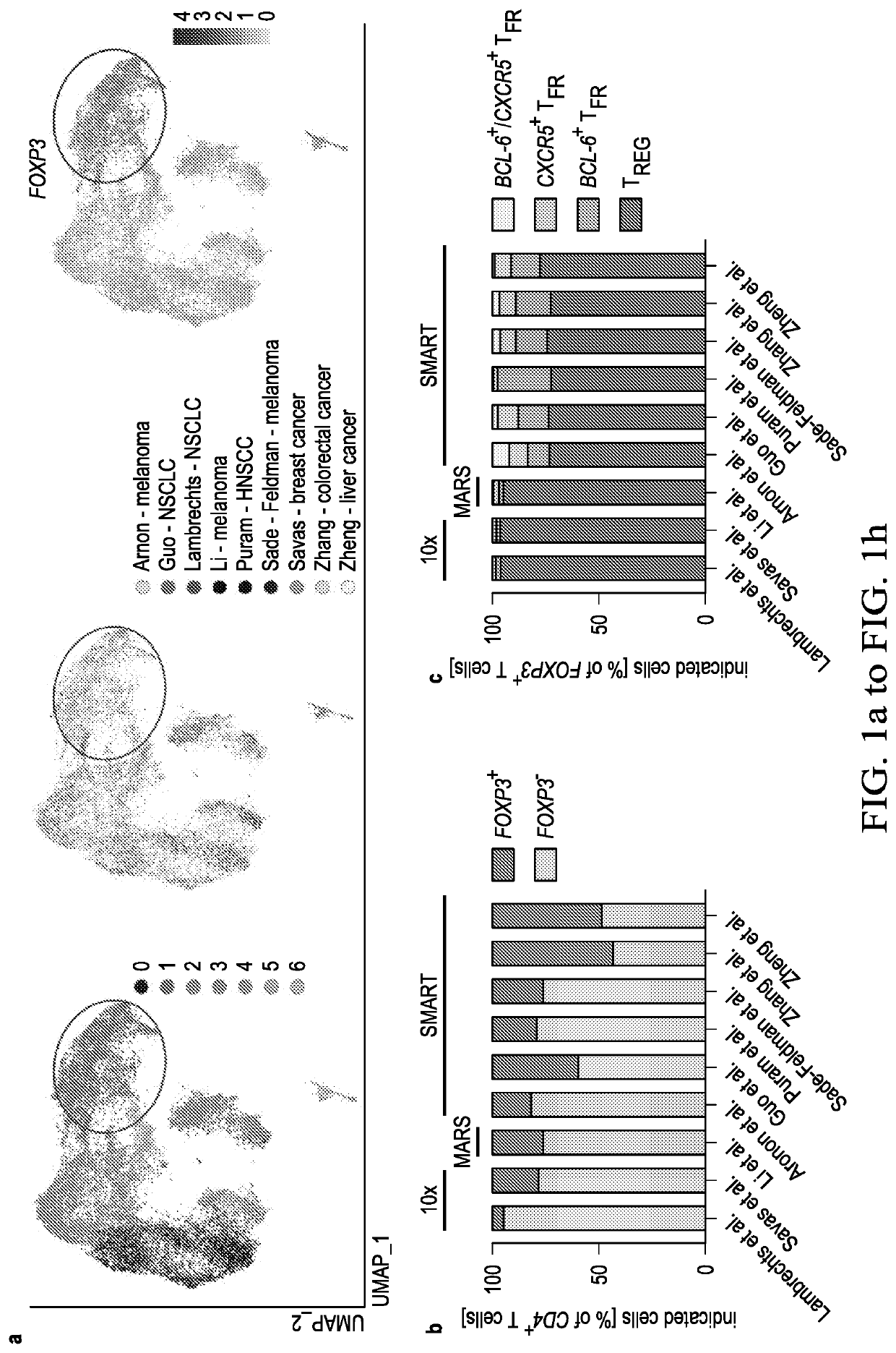
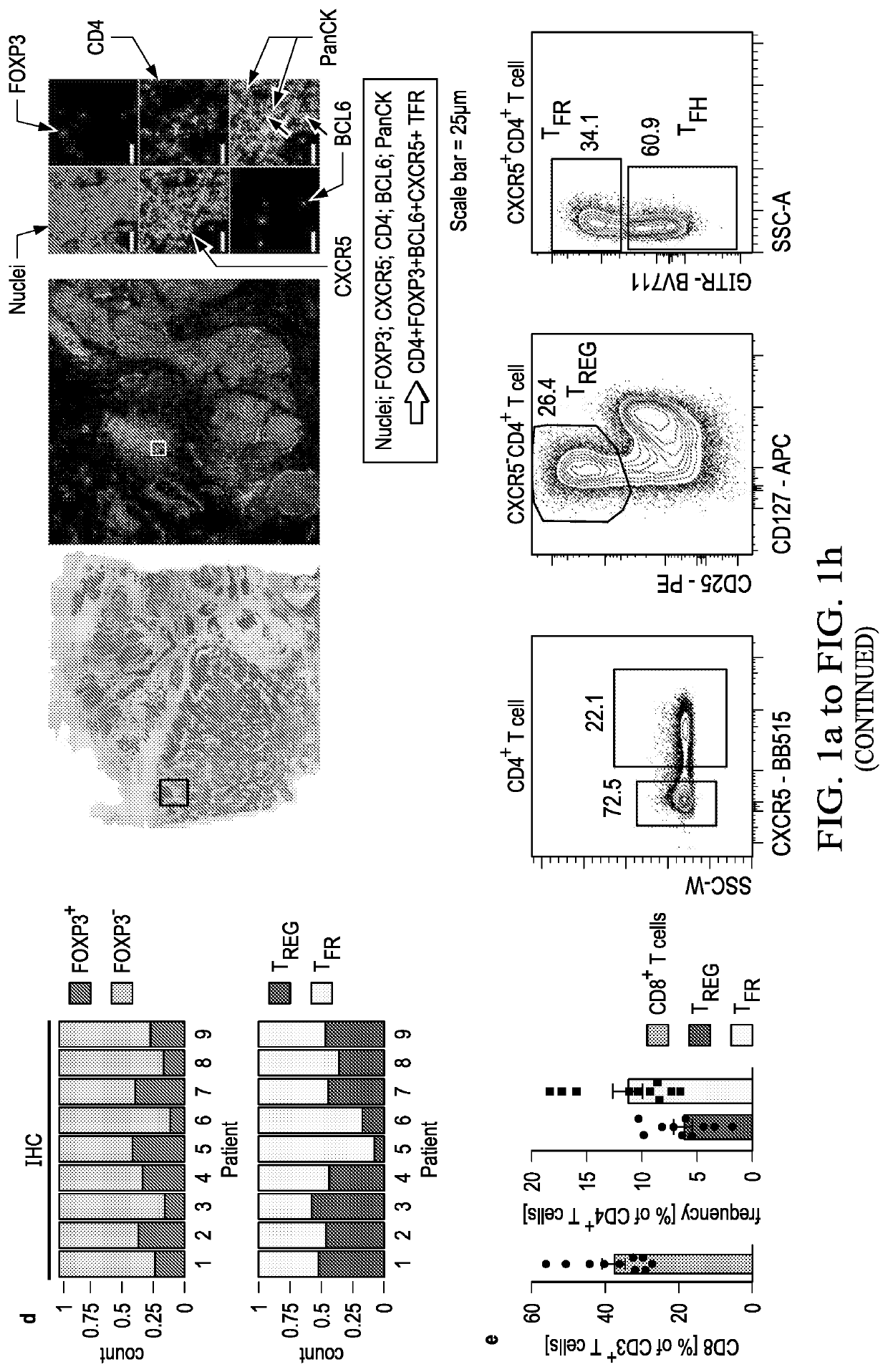
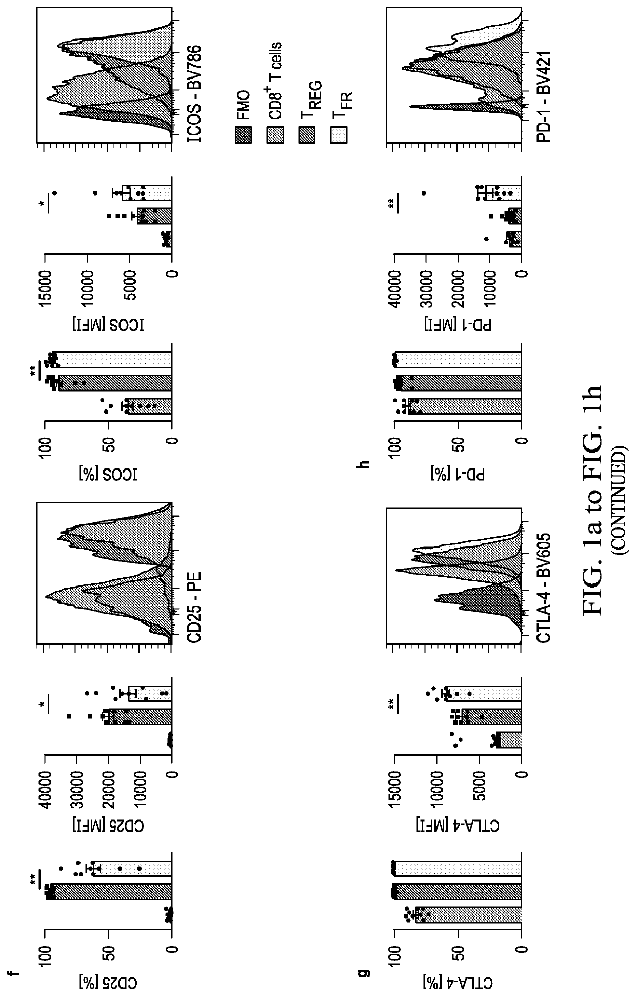
[0046]An increased density of regulatory T cells (TREG) in tumors has been linked to poor survival outcomes1. In non-cancer settings, TREG cells have been shown to differentiate into PD-1 expressing follicular regulatory T cells (TFR) that restrain germinal center responses2. It is not known whether such differentiation also occurs in the tumor microenvironment, and if so, whether such tumor-infiltrating TFR cells are molecularly distinct from TREG cells or are activated by anti-PD1 therapy. In this example, the inventors show that TFR cells are present in high numbers in human and murine tumor tissues, share T cell receptor (TCR) clonotypes with intratumoral TREG cells and express high levels of PD-1. Single-cell TCR data, trajectory analyses and adoptive transfer studies indicate intratumoral conversion of TREG to TFR cells. When compared to TREG cells, TFR cells exhibited enhanced suppressive capaci...

example 2

of TFR but not Tregs to Prevent Severe Immune-Related Adverse Events (irAEs)

[0085]Immune checkpoint blockade (ICB) targeting CTLA-4 or PD-1 can lead to dramatic, long-lasting responses; nonetheless, fewer than 30% of patients respond to monotherapy with either agent. While anti-CTLA-4 therapy is believed to deplete T regulatory (TREG) cells, anti-PD-1 blocking antibodies are thought to primarily activate CD8+ T cells. Combination therapy, though more effective, causes more frequent and severe immune-related adverse events (irAEs), potentially caused by undirected anti-CTLA-4-mediated TREG cell depletion and subsequent uninhibited anti-PD-1-mediated activation of effector T cells. As described hereinabove, a novel population of T cells, follicular regulatory T cells (TFR), are a district population of regulatory T cells that inhibit CD8 T cells. As shows in the example above, by increasing the abundance of TFR cells, anti-PD-1 therapy not only facilitates, but also dampens anti-tumor...


PUM

PropertyMeasurementUnit
densityaaaaaaaaaa
frequencyaaaaaaaaaa
mean fluorescence intensityaaaaaaaaaa

Abstract

The present invention includes methods of detecting follicular regulatory T cells (TFR) comprising: obtaining a biological sample from a subject and detecting whether TFR are increased in the tumor sample by contacting the biological sample with antibodies that detect CD3+CD4+ FOXP3+BCL6+ T cells CD3+CD4+CXCR5+GITR+ T cells, or both, when compared to a healthy subject, and detecting the increase of TFR in the tumor sample. The present invention also includes combination therapy that depletes follicular regulatory T cells (TFR) with minimal effect on regulatory T cells (TREGS) to prevent or reduce immune related adverse effects (irAEs).

Description

CROSS-REFERENCE TO RELATED APPLICATIONS[0001]This application claims priority to U.S. Provisional Patent Application Nos. 62 / 873,185, filed Jul. 11, 2019, and 62 / 971,603, filed Feb. 7, 2020, the entire contents of each of which are incorporated herein by reference.TECHNICAL FIELD OF THE INVENTION[0002]The present invention relates in general to the field of cancer immunotherapy, and more particularly, to the use of intratumoral TFR cells as a biomarker informing the choice of immune checkpoint blockade therapy. It moreover pertains to the occurrence of immunotherapy-mediated immune related adverse events (irAEs) and minimization thereof.BACKGROUND OF THE INVENTION[0003]Without limiting the scope of the invention, its background is described in connection with anti-PD-1 therapy.[0004]An increased density of T regulatory cells (TREG) in tumors has been linked to poor survival outcomes1. In non-cancer settings, TREG cells have been shown to differentiate into PD-1 expressing follicular...

Claims


Application Information

Patent Timeline
Patent Type & Authority Applications(United States)
IPC IPC(8): G01N33/574C12Q1/6886
CPCG01N33/57492C12Q1/6886C12Q2600/158C12Q2600/106C12Q1/6881G01N33/57484A61P35/00
Inventor VIJAYANAND, PANDURANGANOTTENSMEIER, CHRISTIANESCHWEILER, SIMON
Owner LA JOLLA INST FOR IMMUNOLOGY
Why Eureka
Try Eureka