Contrast agent imaging with destruction pulses in diagnostic medical ultrasound

a technology of diagnostic ultrasound and contrast agent, applied in the field of diagnostic medical ultrasound imaging, can solve the problems of increasing motion artifacts, reducing system frame rate, and many pulses needed to destroy contrast agents, and achieve the effect of enhancing the difference between contrast agents and tissue without sacrificing system frame rate or imaging resolution

Inactive Publication Date: 2002-01-22
SIEMENS MEDICAL SOLUTIONS USA INC
View PDF34 Cites 128 Cited by
  • Summary
  • Abstract
  • Description
  • Claims
  • Application Information

AI Technical Summary

Benefits of technology

is to enhance the difference between contrast agent and tissue without sacrificing the system frame rate or imaging resolution.
Another object of the invention is to improve the efficiency of contrast agent destruction.

Problems solved by technology

As a result, many pulses are needed to destroy the contrast agent.
Firing many pulses along the same line will reduce the system frame rate, and thereby increase motion artifacts due to tissue motion, especially in cardiac applications.
Secondly, the pulses which best destroy contrast agent are low frequency, long duration (low bandwidth) pulses.
Usually, a compromise is made between the requirements for imaging and destruction of contrast agent, further reducing the efficiency of contrast agent destruction by ultrasound pulses.
However, this does not overcome the system frame rate problem, but in fact may make it worse.
Furthermore, this approach to LOC imaging does not allow contrast agent to perfuse into a region of interest between firings of pulses for destroying contrast agent, so there is little contrast agent to image after destruction.
The problem with this approach is that the same high power pulses are simultaneously used to both image and destroy the contrast agent.

Method used


Image

  • Contrast agent imaging with destruction pulses in diagnostic medical ultrasound
  • Contrast agent imaging with destruction pulses in diagnostic medical ultrasound
  • Contrast agent imaging with destruction pulses in diagnostic medical ultrasound

Examples


Embodiment Construction

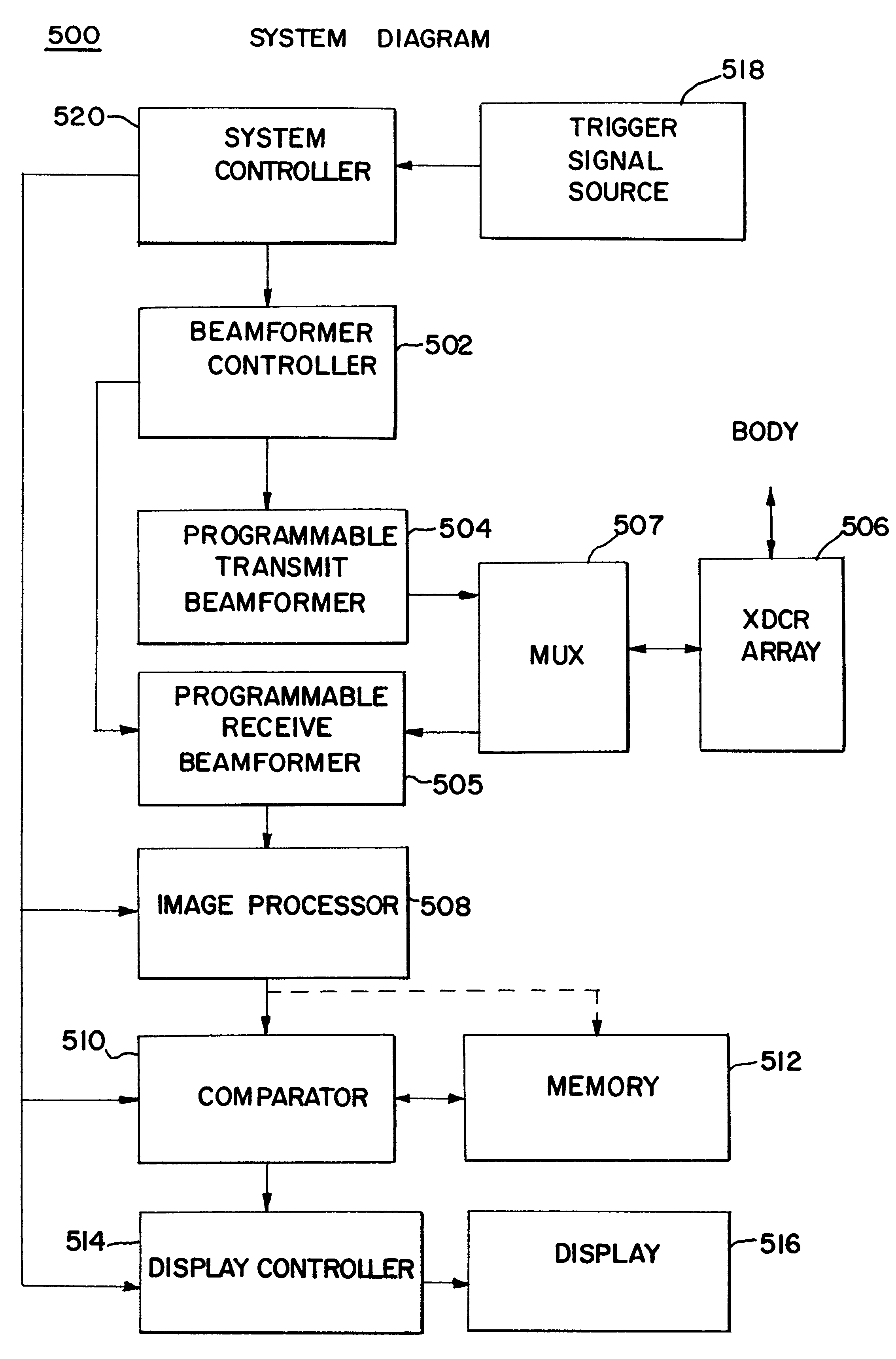
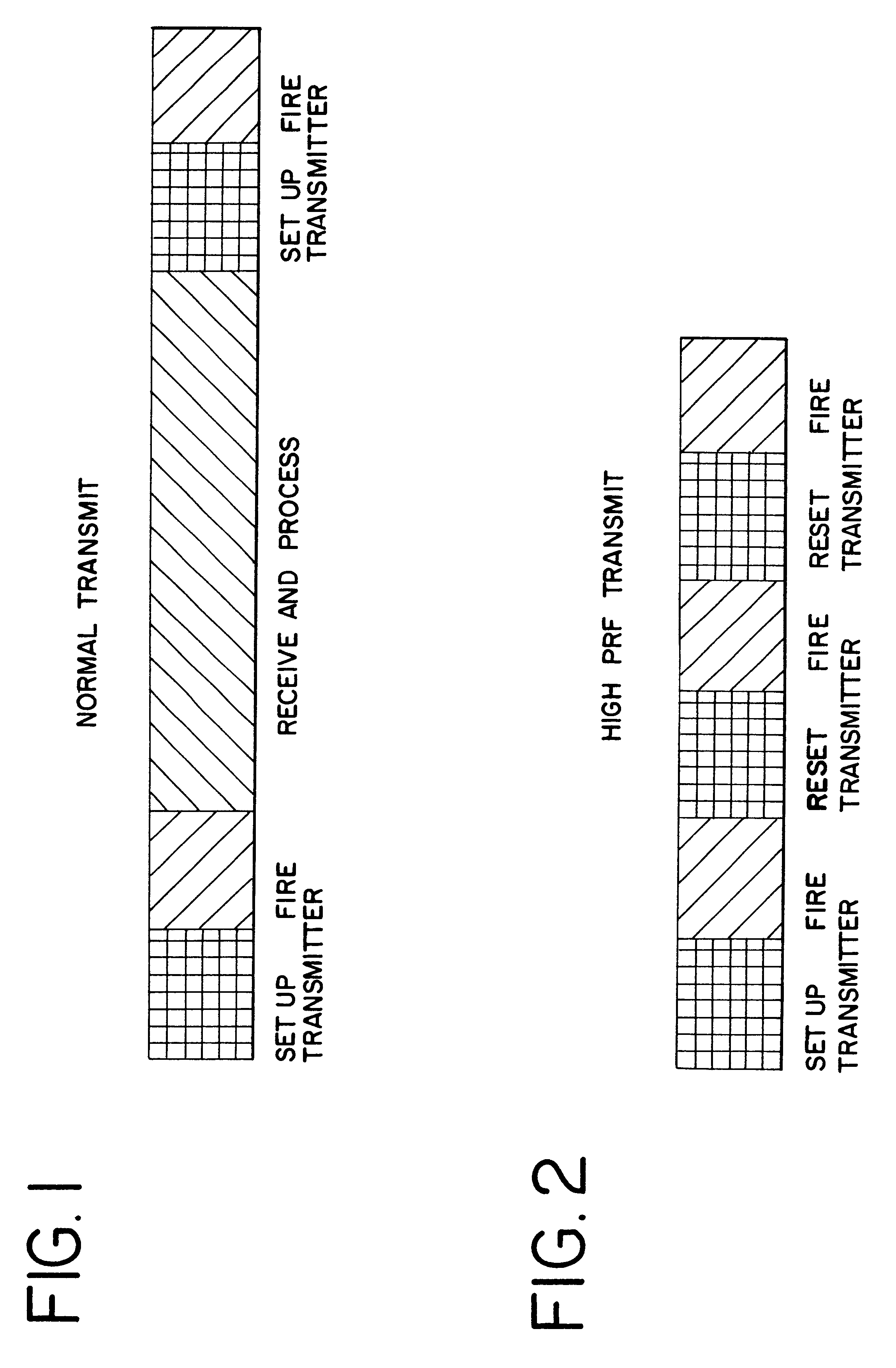
Imaging pulses are pulses which are transmitted, received, processed and displayed. Imaging pulses in some cases can also be destructive to contrast agent. On the other hand, destruction pulses are herein defined as pulses which are transmitted into a body, but not used for imaging. Since destruction pulses are not used for imaging, there is no need to optimize destruction pulse parameters for imaging. These destruction pulses can be optimized for destruction of contrast agent by using low frequency, low bandwidth and high power pulses.

The first aspect of the invention is directed to improving the efficiency of destruction of contrast agent by using high pulse repetition frequency (HPRF) destruction pulses. Here, HPRF destruction pulses are defined as pulses which are transmitted at a rate faster than required to allow the pulses to propagate to the farthest boundary of a region of interest and return to the transducer. Since destruction pulses are not processed or displayed, it is ...


PUM


Abstract

The invention is directed to improvements in diagnostic medical ultrasound contrast agent imaging. In a preferred embodiment, high pulse repetition frequency (HPRF) destruction pulses are fired at a rate higher than necessary for receiving returning echoes. Pulse parameters can also be changed between the plurality of contrast agent-destroying pulses. Other preferred embodiments of the invention are directed to simultaneous transmission of multiple beams of destruction pulses. Destruction frames that consist of a plurality of destruction pulses can be triggered and swept over the entire region of tissue being imaged and at a variety of focal depths from the transmitter. The destruction frames are fired at some time triggered from a timer or some fixed part of a physiological signal, such as an ECG signal. Other preferred embodiments of the invention are directed to continuous low power imaging pulses alternating with destruction pulses triggered at a fixed point of a physiological signal, and a comparison of the received signals from imaging pulses fired before and after the destruction pulses. Alternatively, destruction pulses are triggered at a fixed point on a physiological signal different from the fixed point of a physiological signal used to trigger imaging pulses. In another embodiment, triggered destruction frames are used to enable a comparison of imaging frames in order to determine physiological functions, such as perfusion of blood in cardiac tissue. Finally, in another embodiment, destruction pulses are combined with subharmonic imaging.

Description

1. Field of InventionThis invention relates generally to an improvement in diagnostic medical ultrasound imaging, and more specifically to more effective medical imaging of a human or animal due to improved utilization of an ultrasound contrast agent.2. Description of the Prior ArtA major goal of contrast agent imaging in diagnostic medical ultrasound is the measurement of physiological functions, such as perfusion of blood into tissue, by measuring the flow of contrast agent into tissue. In order to accomplish this, it is important to differentiate contrast agent from tissue.One method for achieving the differentiation of contrast agent and tissue is the loss-of-correlation (LOC) method in which multiple pulses are fired into a body containing contrast agent. The first pulse produces a return signal from both contrast agent and tissue, and simultaneously destroys contrast agent. Since contrast agent is destroyed, the second pulse produces relatively more signal from tissue. By subt...

Claims


Application Information

Patent Timeline
Patent Type & Authority Patents(United States)
IPC IPC(8): A61B8/00A61B17/22
CPCA61B8/481A61B8/543A61B2017/22008A61B17/22004
Inventor KRISHNAN, SRIRAMHOLLEY, GREGORY L.GARDNER, EDWARD A.MASLAK, SAMUEL H.
Owner SIEMENS MEDICAL SOLUTIONS USA INC
Why Eureka
Try Eureka