Surgical drape for colonoscopy

a surgical drape and colonoscopy technology, applied in the field of protective barriers, can solve the problems of contaminating the air of the examining room, presenting a serious health risk to medical personnel, and contaminating the health of the patient, so as to facilitate the attachment of the scope to the patient, increase the visibility of the doctor and the maneuverability of the scope

Inactive Publication Date: 2002-03-19
SHAW TIMOTHY A
View PDF13 Cites 50 Cited by
  • Summary
  • Abstract
  • Description
  • Claims
  • Application Information

AI Technical Summary

Benefits of technology

According to the present invention, a flexible surgical drape has an adhesive side and a non-adhesive side. The adhesive side is adhesive across substantially its entire face. The drape consists of a flexible sheet suitable for covering a substantial portion of a patient's body. A pocket for catching patient secretions and other fluids is attached to the adhesive side of the drape. The pocket is also adhesive so that it can be securely affixed to the patient's body in the location where it will best catch such secretions. The drape contains an opening capable of receiving a removable insert which, when in place, seals off the opening and prevents the passage of gases, secretions, and fluids that are typically present during a colonoscopy. The insert is made to lock into the opening so as to reduce the likelihood of its inadvertent removal. The insert may also be thought of as a valve, in that an examining tool, such as a colonoscope, fits through a hole extending through the valve and thus can be passed from one side of the drape to the other. The insert will be referred to hereinafter as a valve.
The continuously adhesive face of the drape facilitates its attachment to the patient in a way such that it contains patient secretions and keeps them away from the medical personnel performing the procedure. The fact that the valve is removable increases doctor visibility and the maneuverability of the scope, both of which aid in the scope insertion process. In rare cases, perhaps one out of a thousand, the physician will collect from the colon a specimen, possibly a polyp or part of a tumor, that is so large it must be removed before the examination can proceed. A removable valve in those instances provides the further advantage of allowing specimen removal after merely unlocking and displacing the valve. The entire drape need not be withdrawn and repositioned but can be left undisturbed.

Problems solved by technology

These expelled secretions obviously present a risk of contamination, a risk that becomes even greater if, as is often the case, the secretions are aerosolized, thereby contaminating the air of the examining room.
Because of the requirement of close doctor-patient proximity during a colonoscopy, such expelled and aerosolized excretions present a serious health risk to medical personnel.
Basic precautions against contamination such as gloves and face masks offer some protection but are imperfect in that they permit the passage of contaminated substances around their edges or through tears or other openings in them.
Surgical drapes generally provide a sanitary work area but do not provide protection from bodily fluids and excretions.
During a colonoscopy an examining tool is inserted into the colon through the anus, a delicate and potentially painful process.
This can lead to the need to stop the examination in order to reposition the drape if the seal between the patient and the drape is lost.

Method used


Image

  • Surgical drape for colonoscopy
  • Surgical drape for colonoscopy
  • Surgical drape for colonoscopy

Examples


Embodiment Construction

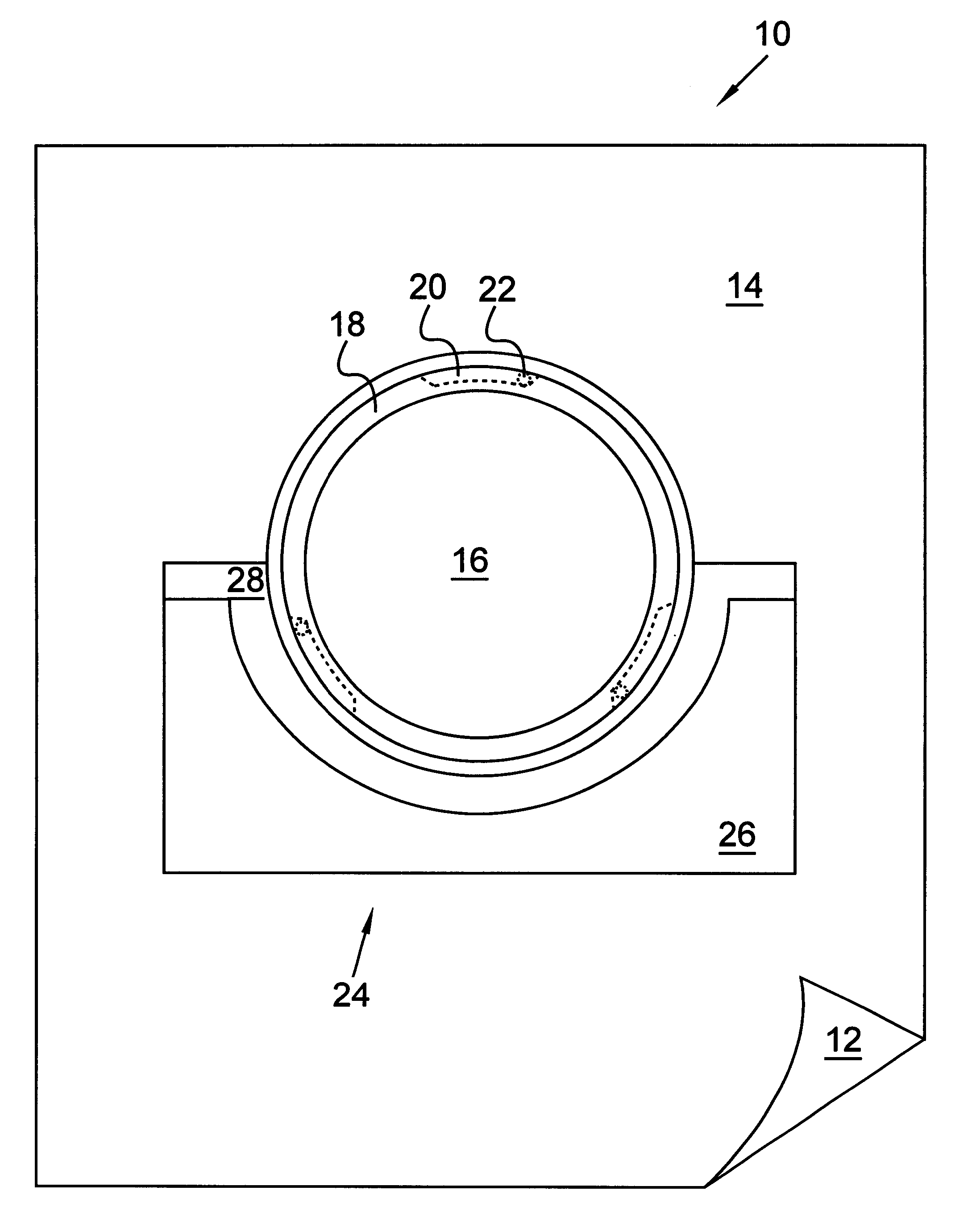
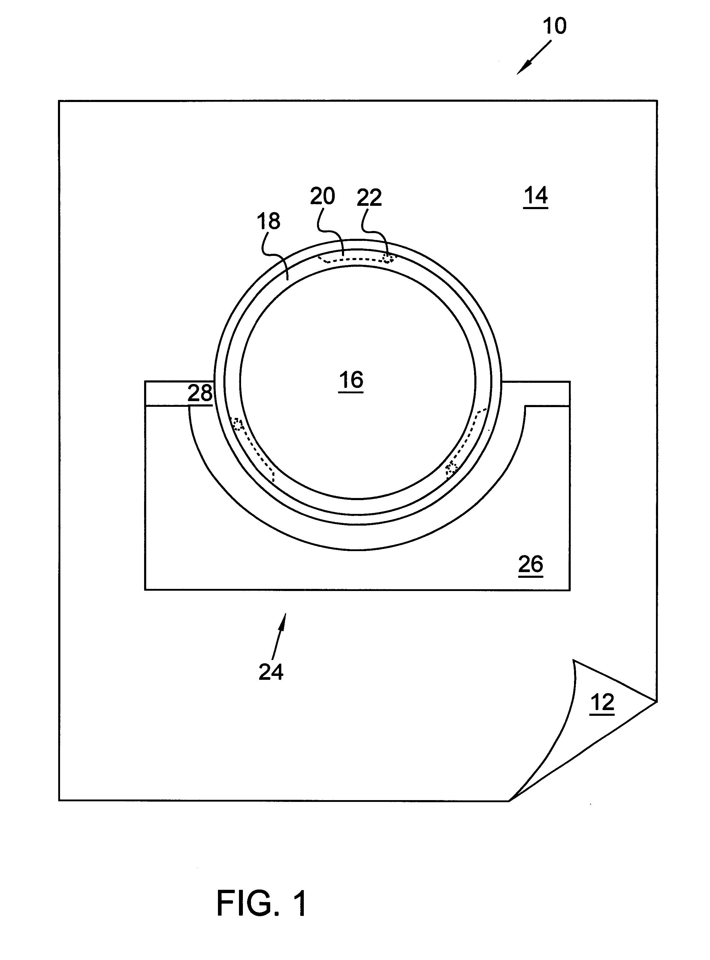
Referring now to FIG. 1, surgical drape 10 has non-adhesive side 12 and adhesive side 14. Drape 10 forms a substantially fluid-impermeable barrier between medical personnel and patient during a medical procedure. Adhesive side 14 is adhesive across substantially its entire face. Drape 10 is provided with opening 16 and ledge 18, opening 16 capable of receiving a removable valve and having flanges 20 which form part of the mechanism that locks the removable valve in place. Each flange 20 includes a flange finger 22. Flanges 20 and flange fingers 22 are drawn with dashed lines to indicate that in this view they are behind ledge 18 and would not be visible when looking at adhesive side 14 of drape 10. The locking mechanism will be further illustrated in connection with FIG. 4. Opening 16 is preferably located near the center of drape 10, but can be placed at other locations.

Ledge 18 provides a seat for a gasket attached to the removable valve. When the valve is inserted in opening 16, ...


PUM


Abstract

A flexible surgical drape has an adhesive side and a non-adhesive side. The adhesive side is adhesive across substantially its entire face. The drape consists of a flexible sheet suitable for covering a substantial portion of a patient's body. A pocket for catching patient secretions and other fluids is attached to the adhesive side of the drape. The drape contains an opening capable of receiving a removable valve, which, when in place, seals off the opening and prevents the passage of gases, secretions, and fluids that are typically present during a medical procedure. The valve can be made to lock into the opening so as to reduce the likelihood of its inadvertent removal. An examining tool such as a colonoscope fits through a hole extending through the valve and thus can be passed from one side of the drape to the other. A reservoir suitable for holding a lubricating substance is securely attached to and forms part of the valve, and the examining tool when inserted through the valve passes through this lubricating substance and is coated by it.

Description

1. Technical FieldThis invention generally relates to a protective barrier for use in a medical setting, and more specifically relates to a surgical drape designed to protect medical personnel from bodily fluids, excretions, and other contaminants likely to be present during a medical procedure. The invention is particularly suited for use during a colonoscopy but would be useful in conjunction with any procedure in which contamination from patient secretions is a concern and where a tubular examining tool is used.2. Background ArtA colonoscopy is a procedure in which the inside of the colon is examined using a long, flexible, fiber-optic viewing instrument called a colonoscope. The colon is located in the abdominal cavity and forms part of the large intestine. It is subject to various disorders, the diagnosis and treatment of which can sometimes be aided by the close-up view that a colonoscope provides. Besides providing a clearer visual picture of the colon, a colonoscope can also...

Claims


Application Information

Patent Timeline
Patent Type & Authority Patents(United States)
IPC IPC(8): A61B19/00A61B19/08
CPCA61B19/087A61B2019/086A61B2019/085A61B2046/201A61B2046/205A61B46/30
Inventor SHAW, TIMOTHY A.
Owner SHAW TIMOTHY A
Why Eureka
Try Eureka