Equine rhinovirus 1 proteins

a technology of equine rhinovirus and protein sequence, which is applied in the field of nucleotide and protein sequence, can solve the problems of difficult primary isolation of erhv1 from clinical specimens, limited analysis, and small number of reported isolations of erhv1

Inactive Publication Date: 2003-03-11
UNIVERSITY OF MELBOURNE
View PDF0 Cites 14 Cited by
  • Summary
  • Abstract
  • Description
  • Claims
  • Application Information

AI Technical Summary

Problems solved by technology

However, such analysis is restricted because there are only a few complete polyprotein sequences available.
There is serological evidence that the incidence of ERhV1 infection is as high as 50% in some horse populations however, the number of reported isolations of ERhV1 is very small.
We have clear evidence that primary isolation of the virus from clinical specimens is known to be difficult, suggesting that the true incidence of ERhV1 disease is much greater than reported.

Method used


Image

  • Equine rhinovirus 1 proteins
  • Equine rhinovirus 1 proteins
  • Equine rhinovirus 1 proteins

Examples


Embodiment Construction

The sequence of specific oligonucleotide primers used for the construction of expression plasmids are:

Virus growth and purification

ERhV1 strain 393 / 76 was isolated from a nasal swab taken from a thoroughbred horse in South Australia while it was being held in quarantine following importation from the United Kingdom. The mare had an acute, systemic febrile illness. The virus was passaged 14 times in quine fetal kidney (EFK) monolayer cell cultures and then once in Vero cells. ERhV1 virions were purified by a modification of the procedure described by Abraham and Colonno. Cells were harvested 48 hours after infection. The infected cells and supernatant fluid were frozen and thawed three times and clarified by centrifuging at 2,000.times. g for 20 min at 4 C. Polyethylene glycol 6000 and NaCl were added to the supernatant to final concentrations of 7% and 380 mM, respectively, and the mixture was stirred overnight at 4 C. The precipitated virions were recovered by centrifuging at 10,00...


PUM

PropertyMeasurementUnit
volumeaaaaaaaaaa
sizeaaaaaaaaaa
morphologyaaaaaaaaaa

Abstract

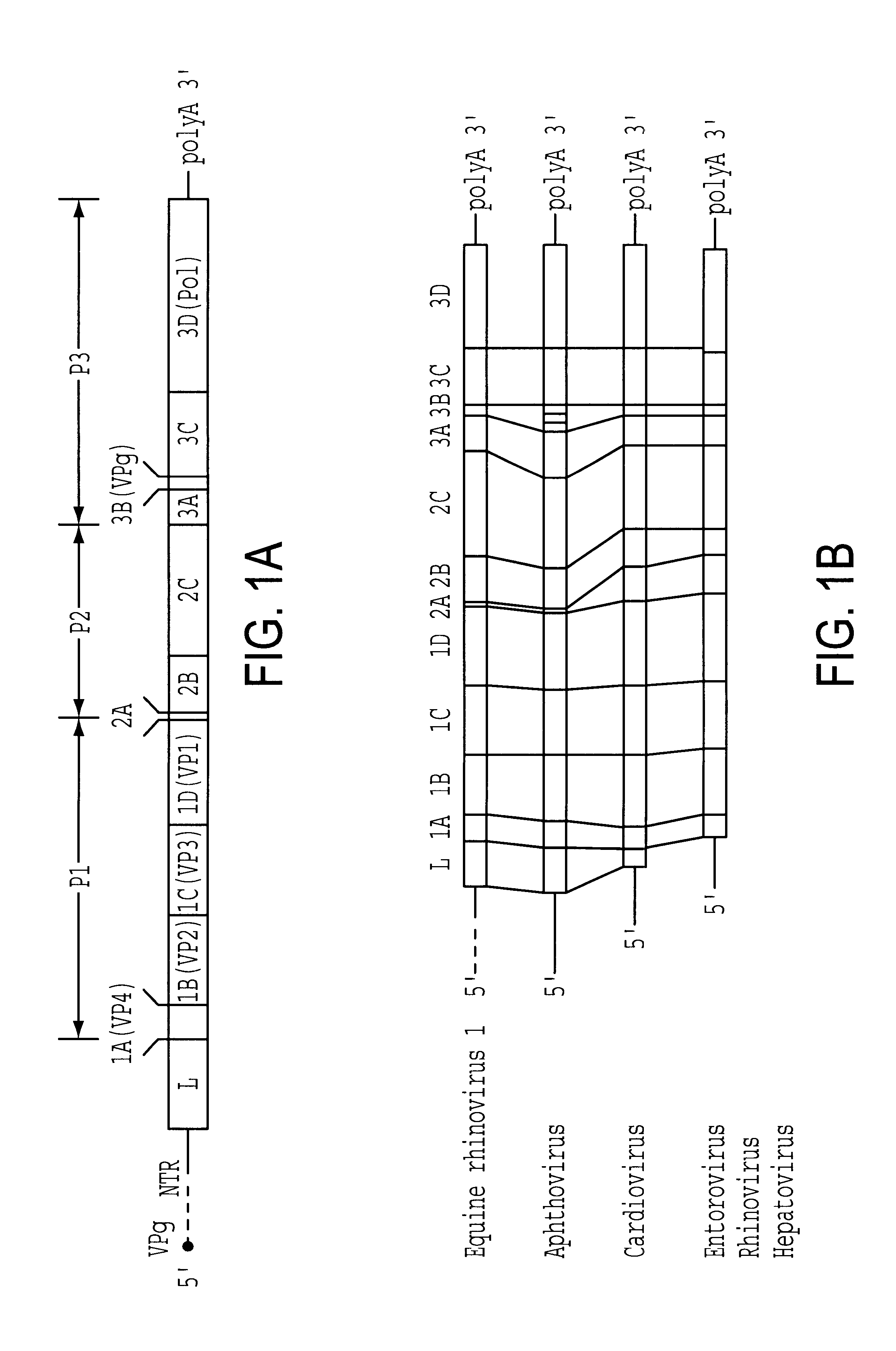
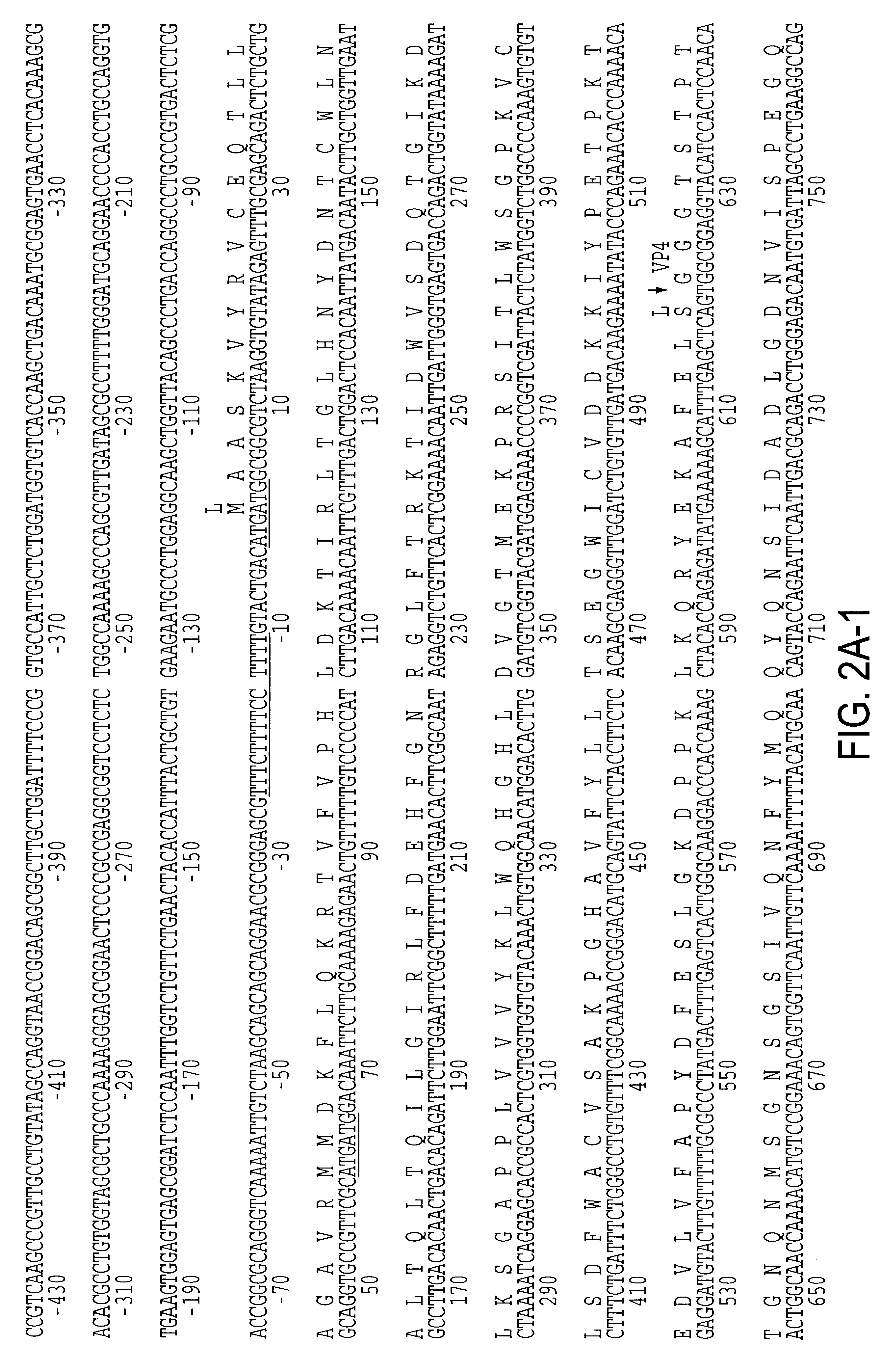
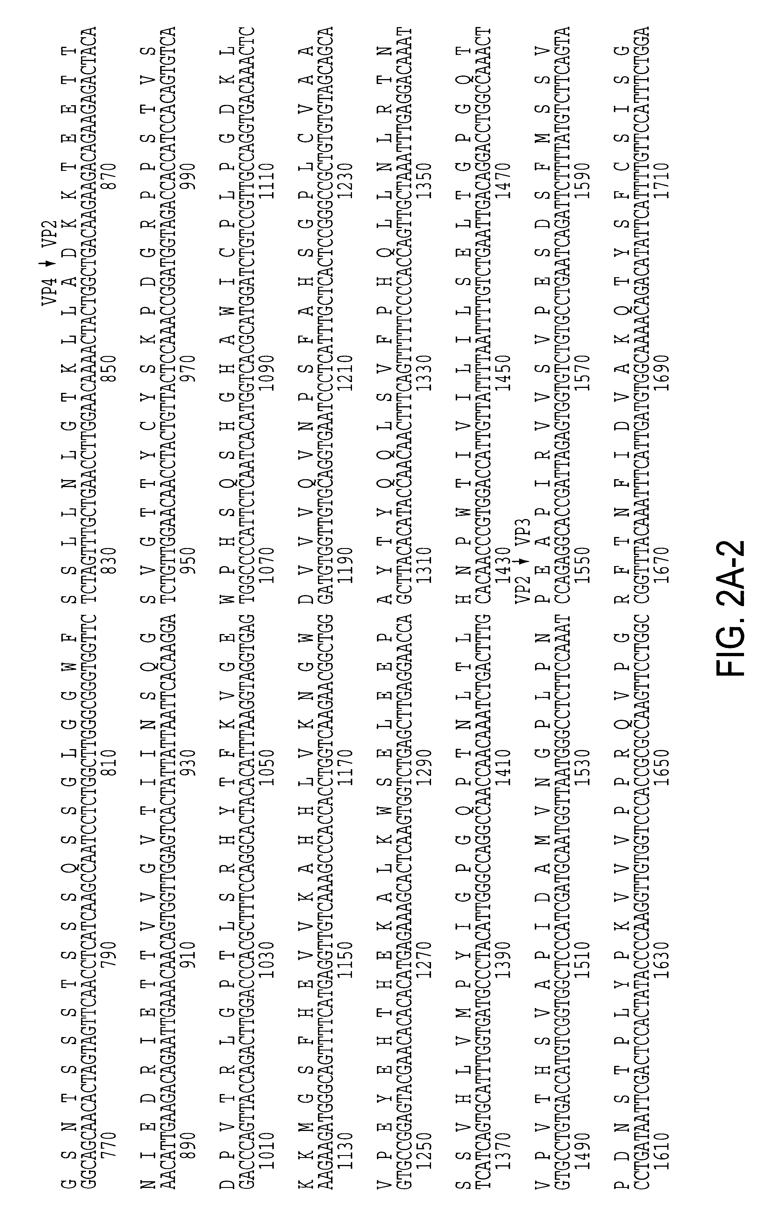
A determination of the nucleotide sequence for the genome of Equine rhinovirus 1 (ERhV1), a horse-respiratory pathogen of heretofor uncertain taxonomic status, reveals a structure and predicted amino acid sequence that are more similar to those of foot-and-mouth disease viruses, the only members of the aphthovirus genus, than of other picornaviruses. These insights inform an understanding of ERhV1 virion proteins and virus-like particles, as well as the design of probes, primers, antigens, vectors, diagnostics and tests of relevance to ERhV1.

Description

INTRODUCTION TO INVENTIONThis invention relates to the equine rhinovirus 1 (ERhV1) which has been sequenced and characterized. In particular, the invention relates to nucleotide and protein sequences of ERhV1 and a range of clinical and diagnostic products derived from ERhV1.BACKGROUND OF INVENTIONEquine rhinovuris 1 (ERhV1) was first isolated from horses in the United Kingdom and subsequently from horses in mainland Europe, the USA and Australia. Most isolates were from the nasopharynx of horses with an acute, febrile respiratory disease. Virions had the characteristic size and morphology of picornaviruses and were acid-labile. Two other serologically distinct, acid-labile picornaviruses, ERhV2 and ERhV3, have also been isolated from horses.Considerable uncertainity has surrounded the classification of ERhV1. Physicochemical studies have shown that the nucleic acid density and base composition of ERhV1 differ from those of rhinoviruses. In contrast to rhinoviruses, ERhV1 has a broa...

Claims


Application Information

Patent Timeline
Patent Type & Authority Patents(United States)
IPC IPC(8): C07K14/005C07K14/095G01N33/569A61K38/00A61P31/16C12N15/41
CPCC07K14/005G01N33/56983A61K38/00C12N2770/32734C12N2770/32722A61P31/16
Inventor STUDDERT, MICHAEL JCRABB, BRENDAN SFENG, LI
Owner UNIVERSITY OF MELBOURNE
Why Eureka
Try Eureka