Stereo image processing for radiography

a stereo image processing and radiography technology, applied in the field of three-dimensional imaging and analysis, can solve the problems of difficult implementation of binocular x-ray imaging, many missed information in conventional x-ray diagnostic and interventional procedures, and relatively fast methods, so as to reduce and eliminate illumination errors, remove keystone distortion, and reduce the effect of reducing and eliminating illumination errors

Inactive Publication Date: 2005-03-01
CANON KK
View PDF3 Cites 107 Cited by
  • Summary
  • Abstract
  • Description
  • Claims
  • Application Information

AI Technical Summary

Benefits of technology

The first step requires adjusting differently illuminated pairs of stereo images (602, 604) of an object (112, 206, 802) to reduce and eliminate illumination errors. In one embodiment, the pair of stereo images (602, 604) is obtained by an X-ray imaging device (110, 304) that is coupled to a processing computer graphics engine (130, 302). In this embodiment, the pair of stereo radiographs is generated from X-ray sources emitting X-rays from two distinct locations in space and is processed digitally into a pair of stereo images. In another embodiment, the pair of X-ray radiographs appears on film or paper and is converted into a computer-readable form (i.e., pair of stereo images in digital format) by an input device (310) which may be an image converter or a graphics engine. Pixels within each image are assigned to groups (606-1, . . . , 606-n, 608-1, . . . ,608-n) of a selected width. Each group forms a line through the image. The lines may be either straight or curved, although a selection of longitudinally curved lines allows for greater reduction in illumination errors. Each group (606-1, . . . ,606-n) in the first image (602) is associated with a corresponding group (608-1, . . . , 608-n) in the other image (604). Next, the gray-scale intensity levels are determined for each pixel in each group (e.g., 606-1 and 608-1). From this data, the mean intensity level and variance for each group (e.g., 606-1 and 608-1) are determined. Thereafter, the images are equalized based on the mean intensity and variance, and then the pixel illumination amongst the pair of images is adjusted accordingly.
The second step involves removing distortion from the radiographs that appear in the corresponding pair of ster

Problems solved by technology

This much-needed information is missed in many conventional X-ray diagnostic and interventional procedures, e.g., in orthopedic or chest X-ray imaging.
Binocular X-ray imaging has generally not been easy to implement because there are no visible surfaces on the radiograph that may be recreated in a three-dimensional image, as compared to reflective photography of images.
These methods are relatively fast, because only small subsets of the image pixels are used, but often fail because the chosen primitives cannot be reliably detected in the images.
Moreover, conventional methods of X-ray stereoscopic imaging have been unsatisfactory due to the insufficient manner of addressing the differences in illumination (i.e., brightness) amongst stereo image pairs.
Such differences in illumination of the stereo images is problematic because, for example, the right part of the right image may be darker than the right part of the left image.
More specifically, the illumination differences result in inconsistent brightness and contrast of the same area amongst the image pair.
These illumination errors also lead to difficulties in observing images in stereo, in visual comparison of two images taken at different times, and in mathematical analysis such as s

Method used


Image

  • Stereo image processing for radiography
  • Stereo image processing for radiography
  • Stereo image processing for radiography

Examples


Embodiment Construction

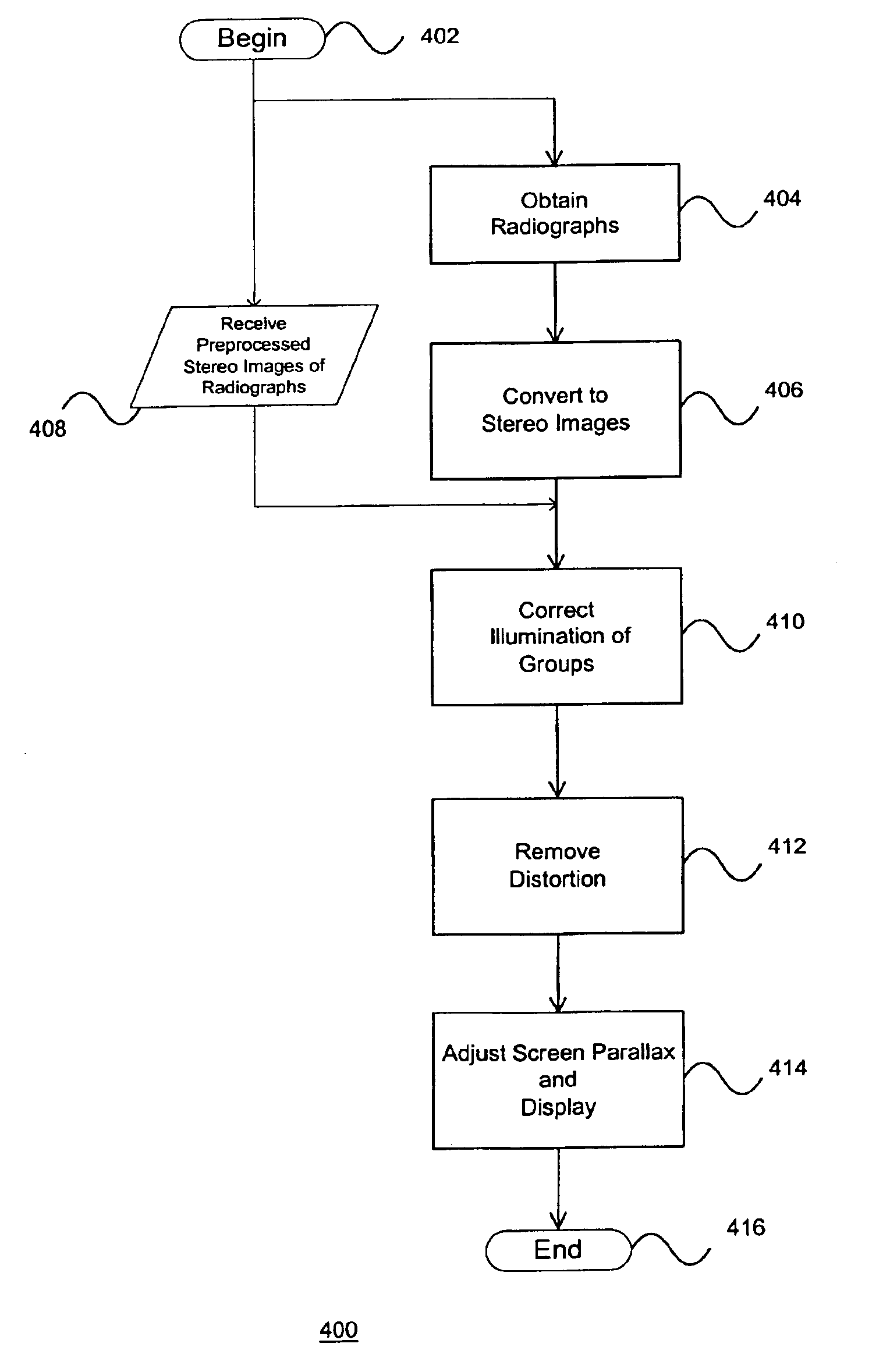
The present invention is directed to a system, method and computer readable medium for stereoscopic image processing that improves the image quality and depth perception of stereoscopic radiographs obtained with imaging devices. The method of the present invention includes a three step process. In the first step, the illumination of X-ray stereo pairs is adjusted to alleviate the illumination differences therebetween. In particular, the illumination of pixels in vertical columns for the pair of images is adjusted by equalizing the mean and standard deviation of these pixels. That is, upon calculating the mean and standard deviation in the vertical columns of the pixels, histograms are then adjusted to equalize the illumination. With the second step, the method of the present invention converts the toed-in stereoscopic X-ray images into a pair of images that are in the same plane, that is, both images being parallel with the surface of the display device and with the base line betwee...


PUM


Abstract

Pairs of stereo Xray radiographs are obtained from an X-ray imaging system and are digitized to form corresponding pairs of stereo images (602, 604). The pairs of stereo images (602, 604) are adjusted (410) to compensate for gray-scale illumination differences by grouping and processing pixel groups in each pair of images. Distortion in the nature of depth plane curvature and Keystone distortion due to the toed-in configuration of the X-ray imaging system are eliminated (412). A screen parallax for the pair of stereo images is adjusted (414) to minimize depth range so as to enable a maximum number of users to view the stereoscopic image, and particular features of interest.

Description

BACKGROUND OF THE INVENTION1. Field of the InventionThe present invention relates generally to the field of three-dimensional imaging and analysis, and more particularly, to a system, method and computer readable medium for adjusting stereo X-ray images in order to correct errors and distortion resulting from illumination disparities, depth plane curvature, keystone distortion, and depth range.2. Description of Background ArtFor both real-world and computer-generated imaging applications, there is a growing need for display techniques that enable determination of relative spatial locations between objects in an image. Once the spatial relations are established, a user may move through the display space and manipulate objects easily and accurately. There are a variety of techniques for collecting as much information as possible about the object by analyzing the image.One area of interest pertains to computer object recognition of binocular stereo, wherein three-dimensional shapes of ...

Claims


Application Information

Patent Timeline
IPC IPC(8): G06K9/00
CPCG06T5/008G06K2209/05G06K2209/40H04N13/0022G06T2207/10116G06T2207/10012H04N13/0025H04N13/128H04N13/133G06V2201/12G06V2201/03
Inventor BERESTOV, ALEXANDER
Owner CANON KK
Why Eureka
Try Eureka