
Method for combining proton beam irradiation and magnetic resonance imaging

a technology of magnetic resonance imaging and proton beam irradiation, which is applied in the field of system for localization and radiotherapy of various cell lines, can solve the problems that tumors and tissue located in organs subject to significant physiologic motion cannot be treated without significant collateral

Inactive Publication Date: 2005-03-01
VIEWRAY TECH
View PDF24 Cites 162 Cited by
  • Summary
  • Abstract
  • Description
  • Claims
  • Application Information

AI Technical Summary

Benefits of technology

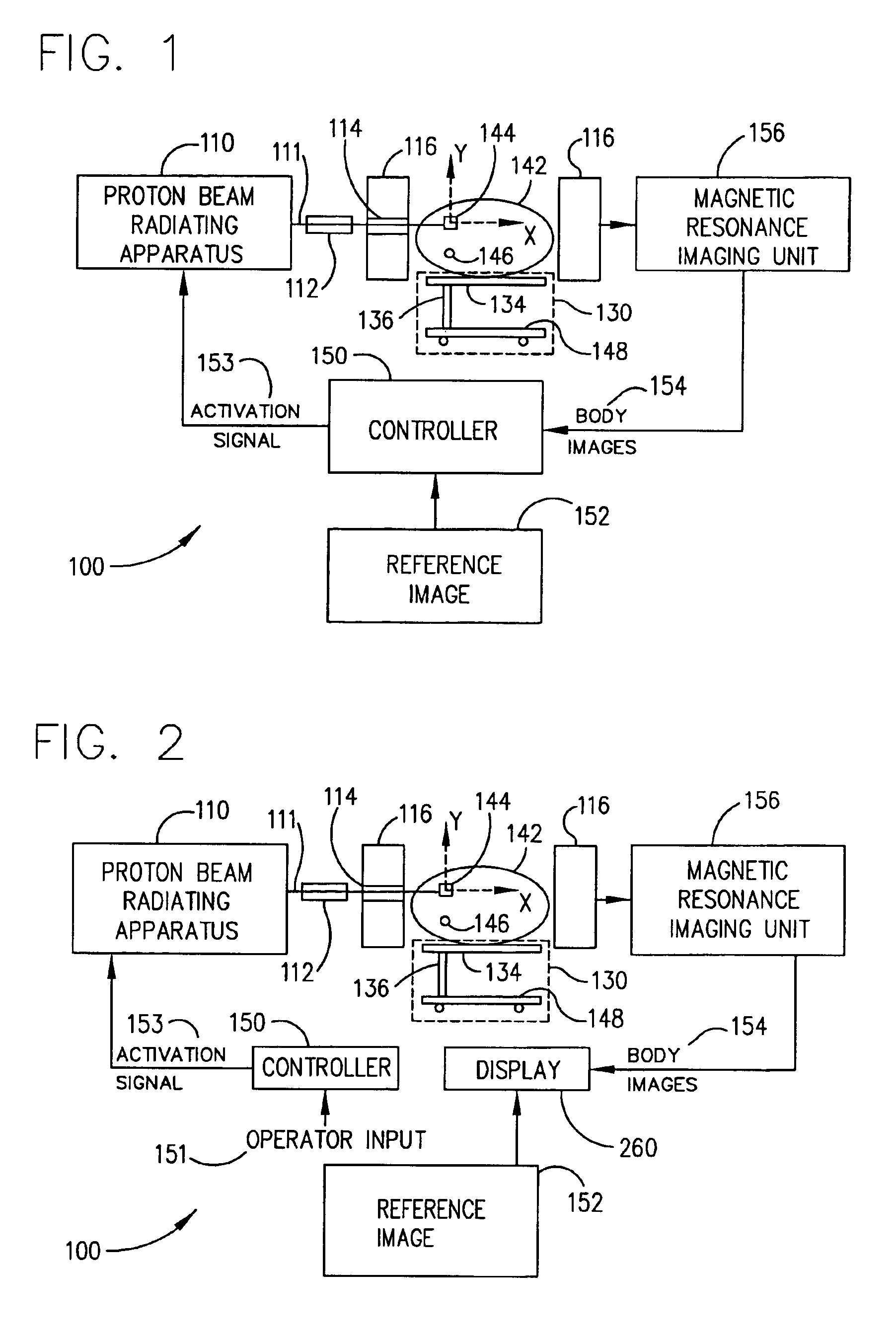
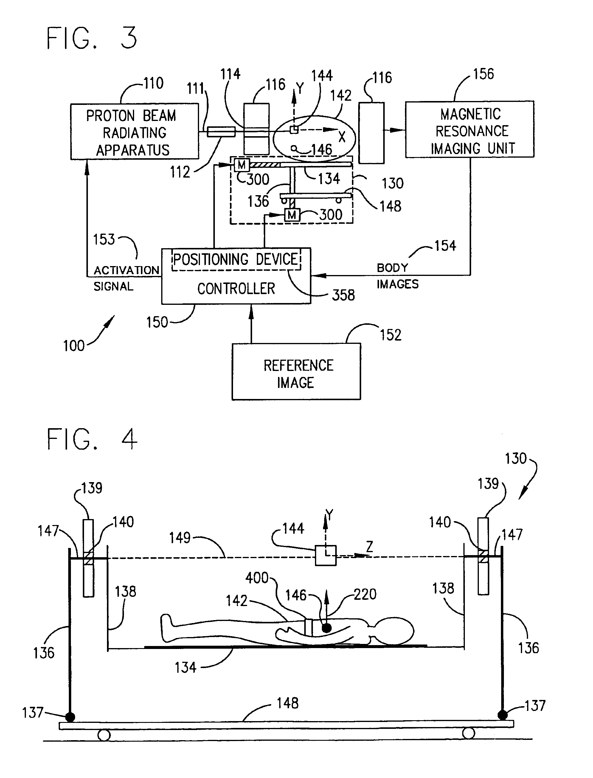
It is another object of this invention to provide a system which treats a target within a body of a patient by proton beam therapy by allowing irradiation of the target at multiple angles according to a treatment plan while minimizing radiation of surrounding tissues during the physiologic motion of the target.

Problems solved by technology

However, tumors and tissue located in organs subject to significant physiologic motion cannot be treated without significant collateral radiotoxicity.

Method used


Image

  • Method for combining proton beam irradiation and magnetic resonance imaging
  • Method for combining proton beam irradiation and magnetic resonance imaging
  • Method for combining proton beam irradiation and magnetic resonance imaging

Examples


Embodiment Construction

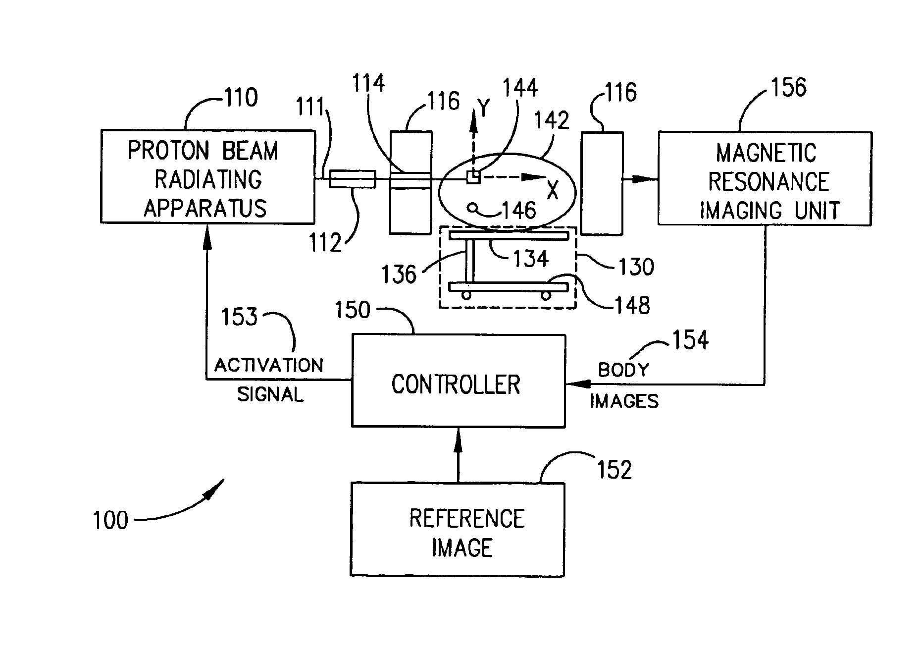
In general, the system 100 of the invention is a near-simultaneous, noninvasive localization and radiotherapy device capable of detecting and treating malignancies, benign tumors, and normal tissues of various cell lines in any anatomic location within the body.

Magnetic resonance imaging (MRI) is a noninvasive diagnostic imaging technique which uses powerful magnetic fields and rapidly changing radio-frequency energy pulses to tomographically represent the varied fat and water content within living tissues. Using standard, cylindrical, closed-bore magnet designs, MRI is extremely useful for the detection and localization of tumors, some of which may not be detected using X-ray computed tomography (CT). The open-architecture design of newer MRI units (e.g., units 156) has extended the utility of standard MRI by permitting the imaging of claustrophobic patients, and by permitting intra-operative imaging in real time during surgery. Proton beam therapy can be combined with MRI or other...


PUM


Abstract

A method which coordinates proton beam irradiation with an open magnetic resonance imaging (MRI) unit to achieve near-simultaneous, noninvasive localization and radiotherapy of various cell lines in various anatomic locations. A reference image of the target aids in determining a treatment plan and repositioning the patient within the MRI unit for later treatments. The patient is located within the MRI unit so that the target and the proton beam are coincident. MRI monitors the location of the target. Target irradiation occurs when the target and the proton beam are coincident as indicated by the MRI monitoring. The patient rotates relative to the radiation source. The target again undergoes monitoring and selective irradiation. The rotation and selective irradiation during MRI monitoring repeats according to the treatment plan.

Description

BACKGROUND OF THE INVENTION1. Field of the InventionThe present invention relates to systems for localization and radiotherapy of various cell lines in various anatomic locations. In particular, this invention relates to a system which coordinates proton beam irradiation with an open magnetic resonance imaging (MRI) unit to achieve near-simultaneous, noninvasive localization and radiotherapy of various cell lines in various anatomic locations by maintaining coincidence between the target and the proton beam.2. Description of the Prior ArtProton beam irradiation therapy treats tumors found in selected locations that are not subject to significant physiologic motion. Examples of such tumors include prostatic cancer, spinal chordomas, and certain retinal or orbital tumors. The proton beam generated by a medical cyclotron has similar biological activity for the destruction of tumors as standard radiation therapy techniques to target a fixed tumor site with minimal radiotoxicity to the s...

Claims


Application Information

Patent Timeline
IPC IPC(8): A61N5/10
CPCA61N5/1049A61B5/0555A61N5/1037A61N2005/1087A61N5/1067A61N5/107A61N2005/1055A61N5/1064A61B5/055A61B5/704
Inventor BUCHOLZ, RICHARD D.MILLER, D. DOUGLAS
Owner VIEWRAY TECH
Learn More