Methods of making medical devices

a technology of medical devices and materials, applied in the field of medical devices making, can solve the problems of waste, cost, and stents being more susceptible to failure, and achieve the effects of reducing the amount of stent material

Inactive Publication Date: 2005-03-15
BOSTON SCI SCIMED INC
View PDF22 Cites 215 Cited by
  • Summary
  • Abstract
  • Description
  • Claims
  • Application Information

AI Technical Summary

Benefits of technology

The invention features a method of manufacturing an endoprosthesis, such as a stent, that includes using sacrificial material that covers one or more portions of a tube of stent material. During manufacturing, particularly during removal of recast material, the sacrificial material protects the stent material from material loss. As a result, although the tube of stent material can be formed oversized, the tube does not need to be formed oversized, thereby reducing the amount of stent material used. When the stent material includes precious materials, such as gold or platinum, reducing the amount of stent material reduces cost. The sacrificial material may also be capable of reacting with the stent material to form a product, such as an alloy, that is relatively easy to remove, e.g., compared to the pure stent material. As a result, less hazardous chemicals may be used during recast material removal.

Problems solved by technology

The recast material can make a stent more susceptible to failure (e.g., cracking or fracture) during manufacturing or in use.
To compensate for loss of material during the removal step(s), the metal tube can be made oversized, which can be wasteful and costly, particularly if the tube includes precious metals.

Method used


Image

  • Methods of making medical devices
  • Methods of making medical devices
  • Methods of making medical devices

Examples


Embodiment Construction

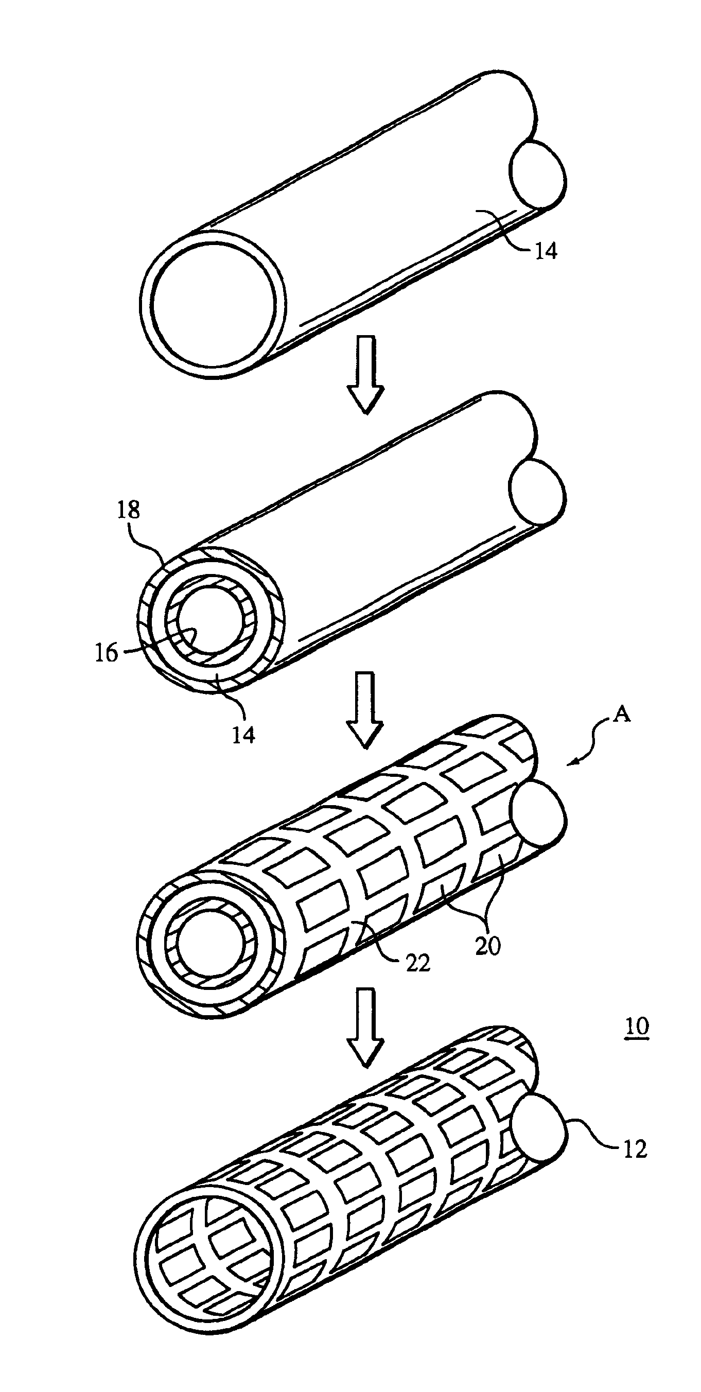
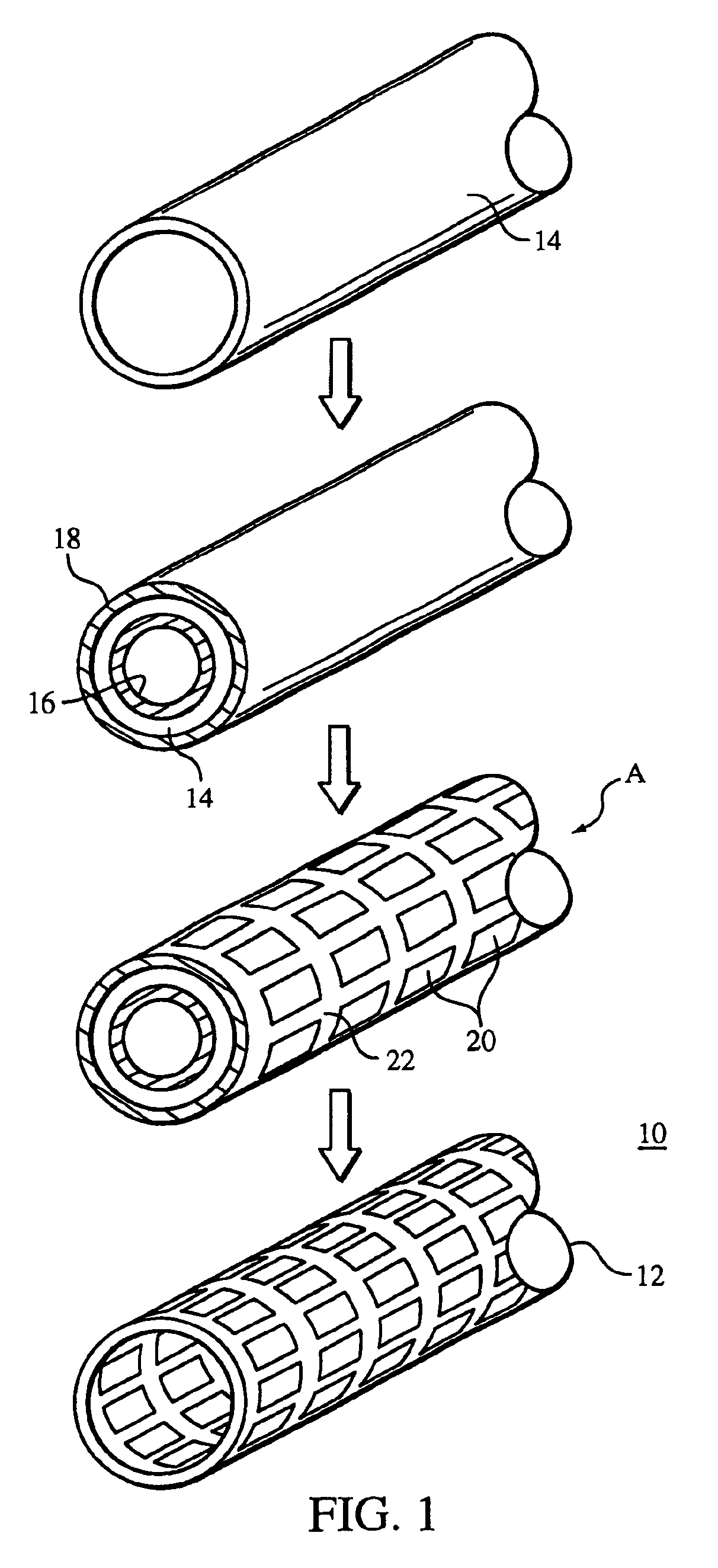
Referring to FIG. 1, a method 10 of making a stent 12 is shown. Method 10 generally includes providing a tubular member 14 that ultimately becomes stent 12, and forming an inner layer 16 and an outer layer 18 on the tubular member. Tubular member 14 is made of a stent material, e.g., platinum, and layers 16 and 18 can be made of, e.g., carbon steel. Portions of tubular member 14 and layers 16 and 18 are then removed, e.g., by laser cutting, to form openings 20 and struts 22 of stent 12. Subsequently, layers 16 and 18 are removed from tubular member 14 to yield stent 12.

Layers 16 and 18 serve as sacrificial layers that are ultimately removed at the end of method 10. Here, rather than starting with an oversized tubular member to compensate for loss of material that may occur during removal of recast material, a relatively smaller tubular member 14 can be used with layers 16 and 18. For example, tubular member 14 and layers 16 and 18 can have a total dimension similar to that of an ove...


PUM

PropertyMeasurementUnit
thickaaaaaaaaaa
thickaaaaaaaaaa
diameteraaaaaaaaaa

Abstract

A method of making a stent includes providing a tubular member having a first layer, the first layer and the tubular member having different compositions, removing a portion of the tubular member, and removing a portion of the first layer from the tubular member.

Description

TECHNICAL FIELDThe invention relates to methods of making medical devices, such as, for example, endoprostheses.BACKGROUNDThe body includes various passageways such as arteries, other blood vessels, and other body lumens. These passageways sometimes become occluded or weakened. For example, the passageways can be occluded by a tumor, restricted by plaque, or weakened by an aneurysm. When this occurs, the passageway can be reopened or reinforced, or even replaced, with a medical endoprosthesis. An endoprosthesis is typically a tubular member that is placed in a lumen in the body. Examples of endoprosthesis include stents and covered stents, sometimes called “stent-grafts”.An endoprosthesis can be delivered inside the body by a catheter that supports the endoprosthesis in a compacted or reduced-size form as the endoprosthesis is transported to a desired site. Upon reaching the site, the endoprosthesis is expanded, for example, so that it can contact the walls of the lumen.The expansio...

Claims


Application Information

Patent Timeline
Patent Type & Authority Patents(United States)
IPC IPC(8): A61F2/06A61F2/00A61F2/90B23K26/18
CPCA61F2/91B23K26/18B23K26/4065B23K26/0661A61F2240/001A61F2250/0067B23K2203/02Y10T29/49885A61F2210/0076Y10T29/49982Y10T29/49996Y10T29/49821Y10T29/496B23K2203/04B23K2103/02B23K2103/04B23K2103/05B23K2103/42B23K2103/50
Inventor STINSON, JONATHAN S.
Owner BOSTON SCI SCIMED INC
Why Eureka
Try Eureka