
Monocyte-specific particulate delivery vehicle
- Summary
- Abstract
- Description
- Claims
- Application Information
AI Technical Summary
Benefits of technology
Problems solved by technology
Method used
Image

Examples
example 1
This example demonstrates the construction of a typical bead vector.
An exemplary bead vector comprises adenovirus which has been engineered to contain the gene encoding Green Fluorescent Protein (GFP) attached to a Dynabead (Ad-GFP-bead). To make the AD-GFP-bead, first, Dynabeads (Dynal, prod. No. 115.19) were thoroughly resuspended and the desired amount of beads was transferred to a tube suitable for the magnetic device. The beads were washed three times with 1-2 ml buffer (RPMI with 1% FCS) with magnetic assistance. The beads were resuspended back into the original buffer volume. Next, in order to coat the beads with with anti-adenovirus fiber protein antibody AB-4 (Neomarker, cat. No. MS-1027), 2 micrograms mouse IgG were added per 107 beads. After rotating the bead / antibody mixture for 30 minutes at room temperature, the beads were washed 3 times with buffer (PBS plus 1% BSA) using magnetic assistance. The coated beads were then resuspended back into the original volume of buff...
example 2
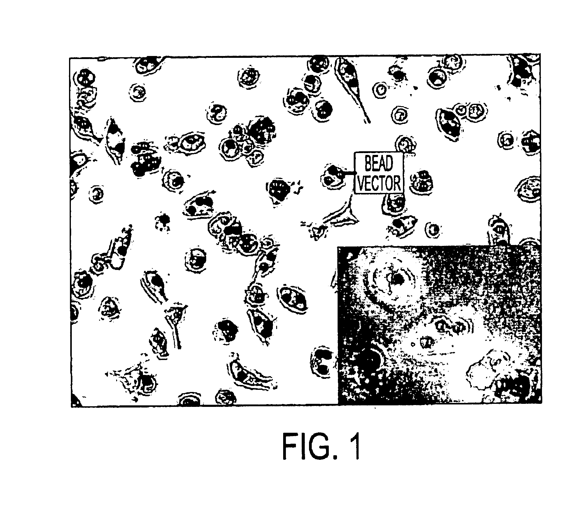
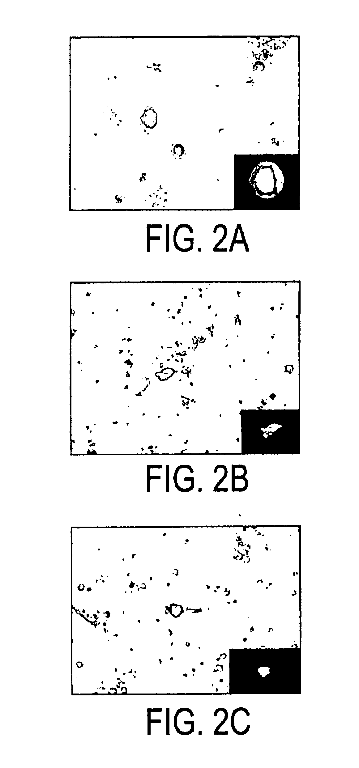
This example demonstrates that monocytic cells specifically take up the inventive bead vector in vitro. In this experiment, the Ad-GFP vector of Example 1 was incubated with a mixed culture containing monocytes. Examination by microscopy revealed high levels of GFP expression only in monocytic cells.
In this example, 50-100 ml of blood was obtained from a human donor and placed in sodium heparin tubes. The blood was diluted 1:1 with 1×PBS. 8 ml of the diluted blood was layered over 4 mls of room temperature Ficoll-Paque Plus in a 15 ml centrifuge tube. The tubes were centrifuged at 400 g at room temperature for 30 minutes. The PBMC layer was carefully removed from the Ficoll gradient using a pasteur pipet and put into a clean 15 or 50 ml centrifuge tube. 4 volumes of 1×PBS were added to the tubes and the tubes were inverted to mix. The tubes were centrifuged at 100 g at room temperature for 10 minutes. 10 mls of 1×PBS was added and the tubes were inverted to mix the cells. The tubes ...
example 3
This example demonstrates that monocytic cells specifically take up the inventive bead vector in vivo and that the cells that took up the vector migrate to the lymph nodes which mimics the normal immune response. This is proof of principle that the monocytic cells, once they phagocytize the vector, actively migrate to the lymphatic system, where the immune response is generated. The vector of Example 1 was injected IP into mice at the midline. After 24 hours, the axillary lymph nodes were removed and examined microscopically. Monocytes expressing high levels of GFP were observed.
PUM
| Property | Measurement | Unit |
|---|---|---|
| Length | aaaaa | aaaaa |
| Composition | aaaaa | aaaaa |
| Interaction | aaaaa | aaaaa |
Abstract
Description
Claims
Application Information