Device and method for cytology slide preparation

a technology of cytology and slides, applied in the field of preparation of cytology slides, can solve the problems of inability to accurately or read, inability to effectively transfer specimens to slides, and inability to accurately and accurately detect and treat precancerous lesions, etc., and achieve the effect of effectively transferring specimens to slides, preparing quickly and reliably

Inactive Publication Date: 2005-05-10
ABBOTT MOLECULAR INC
View PDF8 Cites 47 Cited by
  • Summary
  • Abstract
  • Description
  • Claims
  • Application Information

AI Technical Summary

Benefits of technology

[0017]In another aspect, the invention is directed to a method of preparing cytology slides. The method comprises combining a cellular sample with a liquid-based medium to create a solution, mixing the solution, extracting the sample or an aliquot of the sample from the solution and providing a slide preparation device. The device comprises a first cover having an inside surface, a second cover having an inside surface and an interposed spine. The first and second cover are pivotably secured to the spine so as to be foldable into a book form. An absorbent material is mounted to the inside surface of the first cover and a filter overlays the absorbent material. A slide is removeably mounted to the inside of the second cover so that the filter contacts the slide when the device is in the closed position. The method further comprises applying the sample to the filter, closing the book form so that the filter containing the sample contacts the slide, and applying a pressure to the contact surface to effectively transfer the specimen to the slide. The range of pressures appropriate for the absorbent matter and the filter in the device is provided by the design of the device. In this way, a cytology slide containing an evenly distributed number of cells is prepared quickly and reliably.

Problems solved by technology

The reliability of these slides is a primary issue.
Often, the results are not accurate or are unreadable.
Accurate interpretation of up to 40% of conventional Pap smears are compromised by the presence of blood, mucous, obscuring inflammatory cells, scant cellular material and air-drying artifacts.2 The presence of these contaminants can obscure many of the cells, causing important precancerous lesions to be missed when the slide is reviewed at the lab or, alternatively, making the entire slide unreadable.
One of the problems with the conventional Pap smear is how quickly the sample dries out once it is smeared on the slide.
Another problem with the conventional Pap smear is the frequent inaccuracy of the test result.
A false positive result may require a woman to undergo unnecessary and costly medical procedures.
A false negative Pap test may delay the diagnosis and treatment of a precancerous or even a cancerous condition.
However, in both the ThinPrep® and the CytoRich® slide preparation systems, a time consuming and expensive procedure is followed to prepare a mono-dispersed layer of cells on a cytology slide.
Although each of these systems reduces significantly the false-negative rate of the traditional Pap smears, the health care market has been slow in adopting these systems because of their cost and preparation time compared to the conventional Pap smears.

Method used


Image

  • Device and method for cytology slide preparation
  • Device and method for cytology slide preparation
  • Device and method for cytology slide preparation

Examples


example

Comparison with Conventional Pap Smear

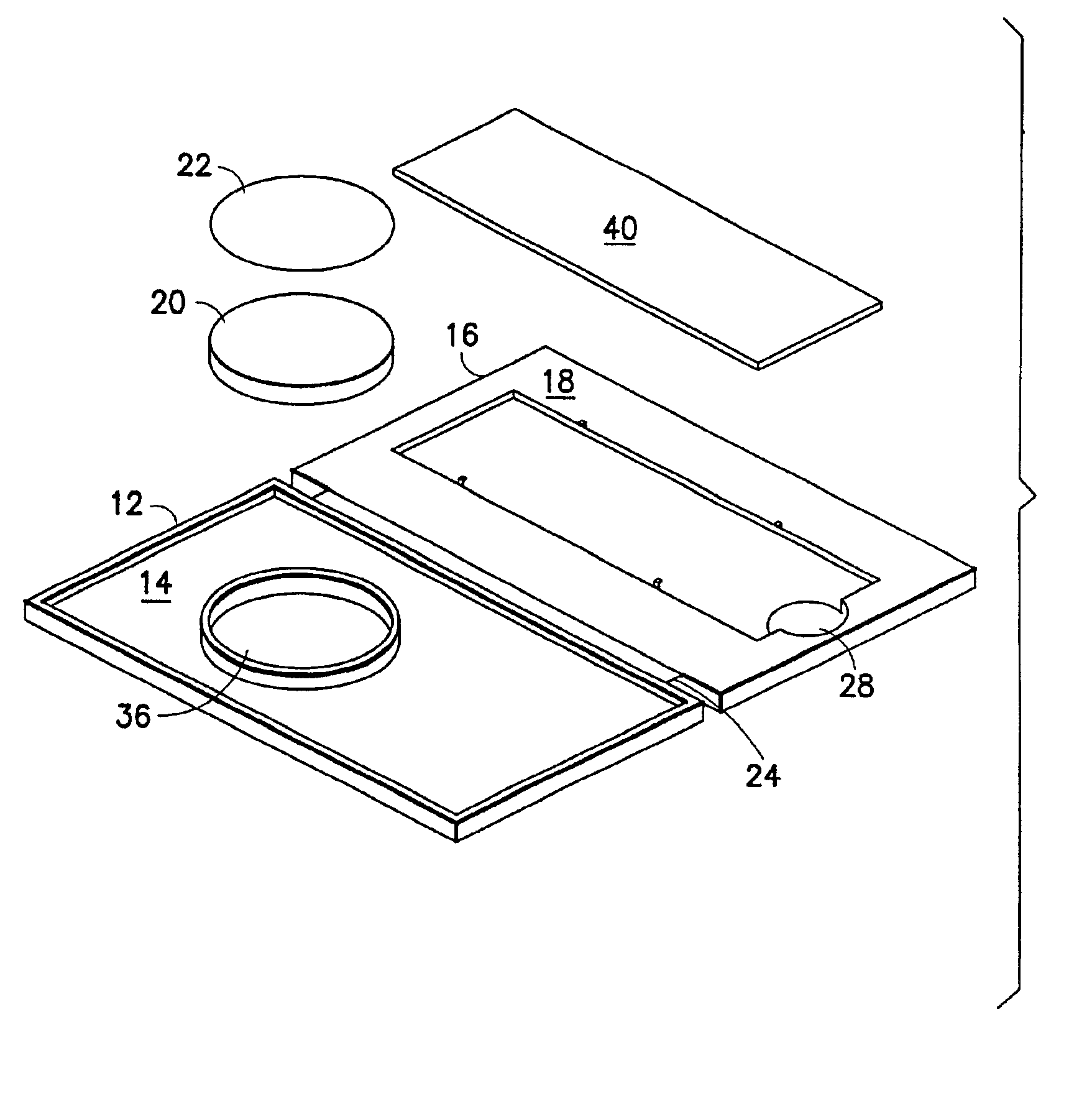
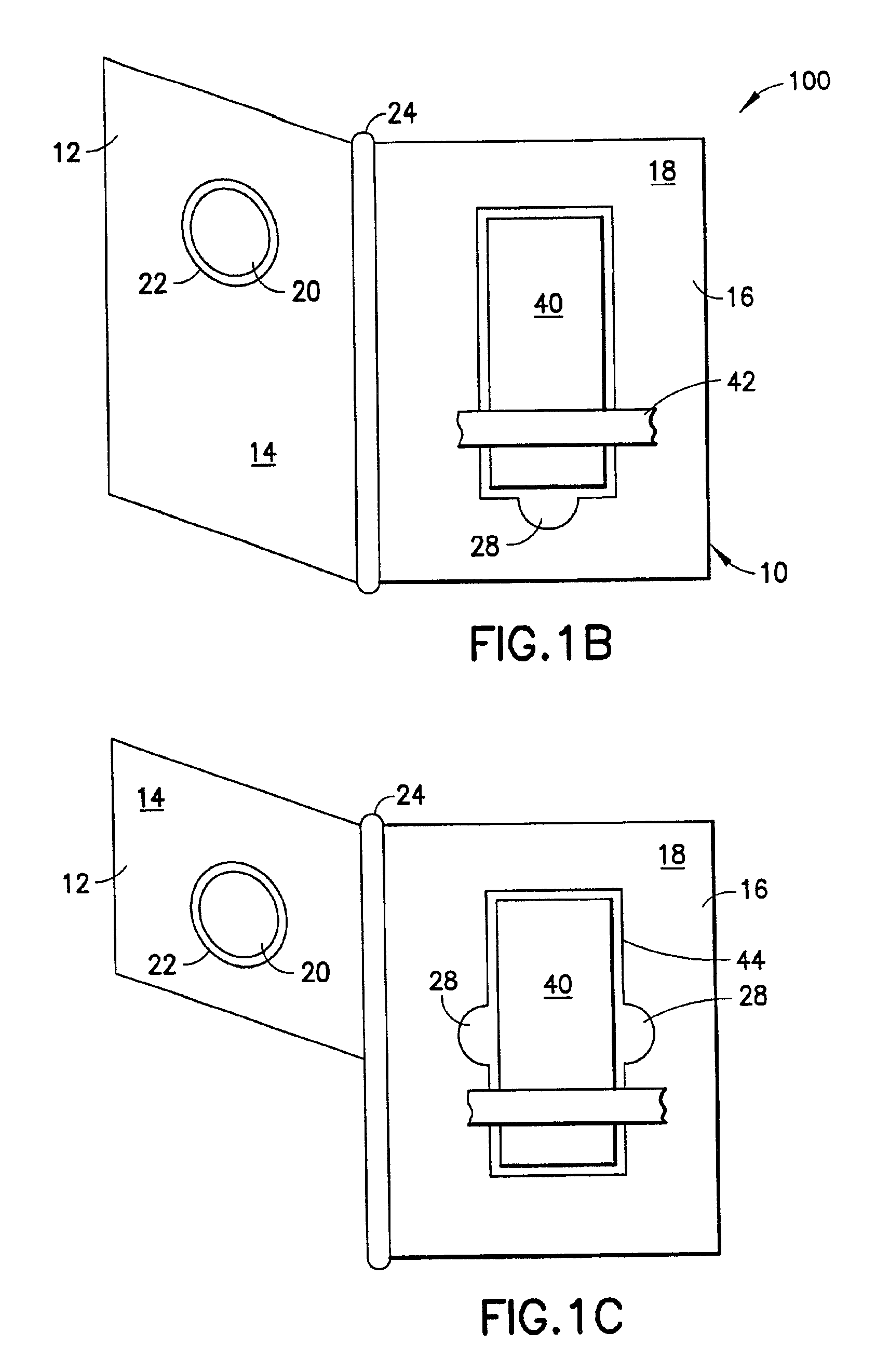
[0059]A cervical sample was taken from a female patient and placed in a one milliliter of Universal Collection Medium.7 An aliquot of 200 microliters is extracted from the solution and applied to the surface of the filter 22 of the book-form device 10. The book-form device 10 was closed for approximately 15 seconds while a controlled amount of pressure was applied. The slide 40 was then removed from the book-form device 10 and the sample was fixed, stained and placed under a light microscope. FIG. 6A shows the morphology of cervical cells prepared by the method of the present invention.

[0060]FIG. 6B was taken from archived routine Pap smears to show the morphology, distribution of cells and staining of stored samples fixed to slides for comparison. The morphology of the cells prepared by the method of the present invention shows better quality and more evenly distributed cells with less interference from debris than the conventional Pap smear.

[0...


PUM

PropertyMeasurementUnit
pressureaaaaaaaaaa
light microscopeaaaaaaaaaa
frequencyaaaaaaaaaa

Abstract

This invention provides a novel device and method for preparing cytology slides. The device comprises a book-like form including an absorbent material and filter attached to the inside surface of a front cover and a cytology slide removeably attached to an inside surface of a back cover. A sample is removed from the body of a patient, placed in a liquid-based solution, and then on the filter. When the book-like form is closed, the sample is effectively transferred to the slide.

Description

[0001]This is a divisional of application Ser. No. 09 / 542,746 filed Apr. 4, 2000 issued as U.S. Pat. No. 6,436,662 B1 on Aug. 20, 2002.FIELD OF INVENTION[0002]The present invention relates generally to the preparation of cytology slides and, more particularly, to a device and associated method for preparing cytology slides.BACKGROUND OF THE INVENTION[0003]Cytology slides are prepared to screen and diagnose cellular samples taken from, for example, tissue samples, samples from the uterine cervix, urine, sputum, blood, fine needle aspiration biopsy, urethral, bronchial brushings and washings, cerebral spinal fluid, and other body fluids. The reliability and efficacy of the screening methods of these slides are measured by their ability to diagnose infections, precancerous lesions or cancerous lesions while at the same time avoiding false positive or negative diagnosis. The reliability of these slides is a primary issue. Often, the results are not accurate or are unreadable. Thus, ther...

Claims


Application Information

Patent Timeline
Patent Type & Authority Patents(United States)
IPC IPC(8): G01N33/543G01N33/48G01N1/28
CPCG01N33/54386Y10S435/962Y10S435/97Y10S435/973Y10S436/81Y10T436/25Y10S435/922Y10S435/923Y10S435/924Y10T436/25375Y10S435/921
Inventor MIELZYNSKA, IWONAPAYNE, JAYLAZAR, JAMES
Owner ABBOTT MOLECULAR INC
Why Eureka
Try Eureka