Method and apparatus for three-dimensional imaging of a moving examination subject, particularly for heart imaging

a three-dimensional imaging and examination subject technology, applied in the field of three-dimensional imaging of moving examination subjects, can solve the problems of not being able to reconstruct the three-dimensional presentation of the diagnostic image, and the current inability to be don

Inactive Publication Date: 2005-06-21
SIEMENS HEALTHCARE GMBH
View PDF19 Cites 30 Cited by
  • Summary
  • Abstract
  • Description
  • Claims
  • Application Information

AI Technical Summary

Benefits of technology

[0005]An object of the present invention is to provide a method and apparatus for three-dimensional imaging of a moving examination subject, i.e. f

Problems solved by technology

The known possibilities, however, only allow the examination and three-dimensional presentation of examination regions from which adequate data has been obtained.
This is currently not possible, however, because too few projection images for the same heart phase can be re

Method used


Image

  • Method and apparatus for three-dimensional imaging of a moving examination subject, particularly for heart imaging
  • Method and apparatus for three-dimensional imaging of a moving examination subject, particularly for heart imaging
  • Method and apparatus for three-dimensional imaging of a moving examination subject, particularly for heart imaging

Examples


Embodiment Construction

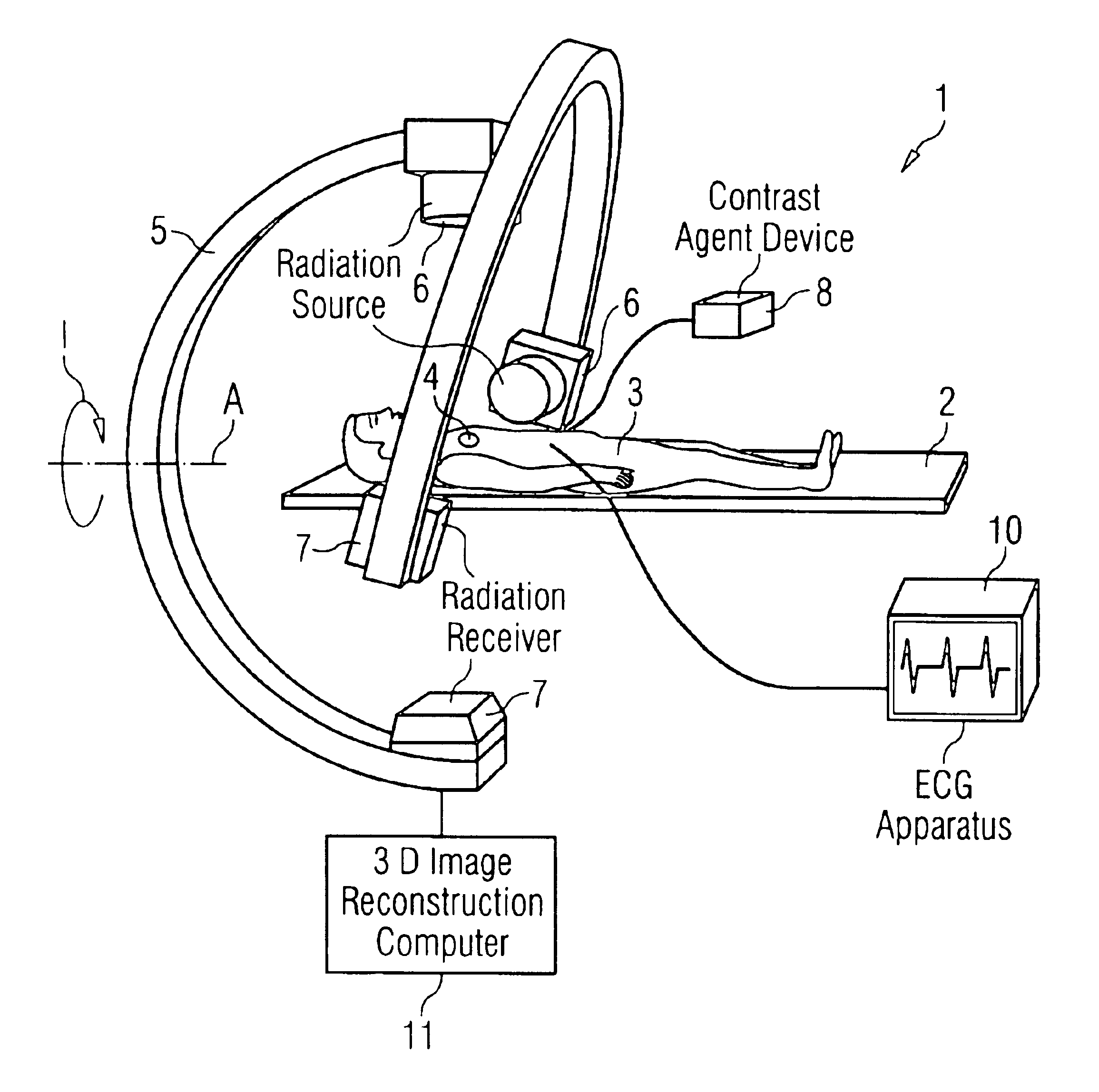
[0023]FIG. 1 shows an inventive medical examination apparatus 1 having a patient bed 2 on which a patient 3 whose heart 4 is to be examined lies in the illustrated exemplary embodiment. A C-arm 5 with a radiation source 6 and a radiation receiver 7 is provided for the examination, this being rotatable around the axis A, as indicated by the arrow 1. The radiation source 6 and the radiation receiver 7 thus rotate around the examination region, i.e. around the heart 4 in this case.

[0024]Contrast agent is administered to the patient with a contrast agent device 8 via a contrast agent line 9. This contrast agent is preferably arterially administered, so that it is already in the region of the heart 4 immediately after having been administered and is still present in adequate concentration. Further an ECG apparatus 10 is provided with which a ECG can be registered.

[0025]For the examination, the C-arm 5 rotates around the axis A during the time wherein the contrast agent is in the examinat...


PUM


Abstract

In a method and apparatus for three-dimensional imaging of a moving examination subject, particularly heart imaging with an examination apparatus having at least one C-arm with a radiation source and a radiation receiver, the C-arm rotates around the examination subject at least once through 180° plus the radiation fan angle during the time span in which a contrast agent is in the examination subject for the registration of the two-dimensional projection images, on the basis of which a three-dimensional image reconstruction ensues.

Description

BACKGROUND OF THE INVENTION[0001]1. Field of the Invention[0002]The present invention is directed to a method for three-dimensional imaging of a moving examination subject, particularly heart imaging with an examination apparatus having at least one C-arm with a radiation source and a radiation receiver.[0003]2. Description of the Prior Art[0004]A standard application for an examination apparatus or angiography system of the C-arm type is the registration of projection images from one or from a few different directions. Usually, the exposures are produced as two-dimensional projection images and are viewed as such. Three-dimensional reconstructions of the examination region for presenting a three-dimensional angiography image are used only in interventional imaging for treating diseases of the vascular system in the brain on the basis of the two-dimensional projection images that have been registered. The known possibilities, however, only allow the examination and three-dimensional...

Claims


Application Information

Patent Timeline
IPC IPC(8): A61B6/00A61B5/0456A61B5/0452A61B5/352
CPCA61B6/481A61B6/541A61B6/504A61B5/0456A61B6/503A61B5/352
Inventor BRUDER, HERBERTMERTELMEIER, THOMAS
Owner SIEMENS HEALTHCARE GMBH
Why Eureka
Try Eureka