Method and system for producing interactive three-dimensional renderings of selected body organs having hollow lumens to enable simulated movement through the lumen

a three-dimensional rendering and selected body technology, applied in the field of three-dimensional rendering of selected body organs having hollow lumens, can solve the problems of increasing the sensitivity of cancer detection, increasing the risk and cost, and easy to perform fecal occult blood tests, etc., to facilitate subsequent detection

Inactive Publication Date: 2005-06-21
WAKE FOREST UNIV HEALTH SCI INC
View PDF59 Cites 333 Cited by
  • Summary
  • Abstract
  • Description
  • Claims
  • Application Information

AI Technical Summary

Benefits of technology

[0013]A surface, or isosurface, of the air column representing the colon is then produced. A wireframe model of the isosurface is then generated using a selected image processing technique such as a marching cubes algorithm. From the wireframe model of the colon, a three-dimensional interactive rendering is produced that enables the user to rapidly view a series of three-dimensional images of the lumen of the colon for purpose of detection of pathological conditions. As such, a user has the perception of flying through the lumen and virtual reality environment is achieved.

Problems solved by technology

The fecal occult blood test is easy to perform but is plagued by many false positive and false negative results.
Examination of the entire colon by a barium enema or conventional colonoscopy increases sensitivity for cancer detection but also increases the risks and costs.
A barium enema causes patient discomfort and / or embarrassment and exposes the patient to substantial radiation.
In addition, colonoscopy requires patient sedation, places the patient at risk for bowel perforation, and is comparatively expensive.
Furthermore, with the exception of fecal occult blood testing, all of these procedures meet with significant patient discomfort.
As can be expected, proper placement of the needle can be extremely difficult and to a small degree somewhat imprecise.

Method used


Image

  • Method and system for producing interactive three-dimensional renderings of selected body organs having hollow lumens to enable simulated movement through the lumen
  • Method and system for producing interactive three-dimensional renderings of selected body organs having hollow lumens to enable simulated movement through the lumen
  • Method and system for producing interactive three-dimensional renderings of selected body organs having hollow lumens to enable simulated movement through the lumen

Examples


Embodiment Construction

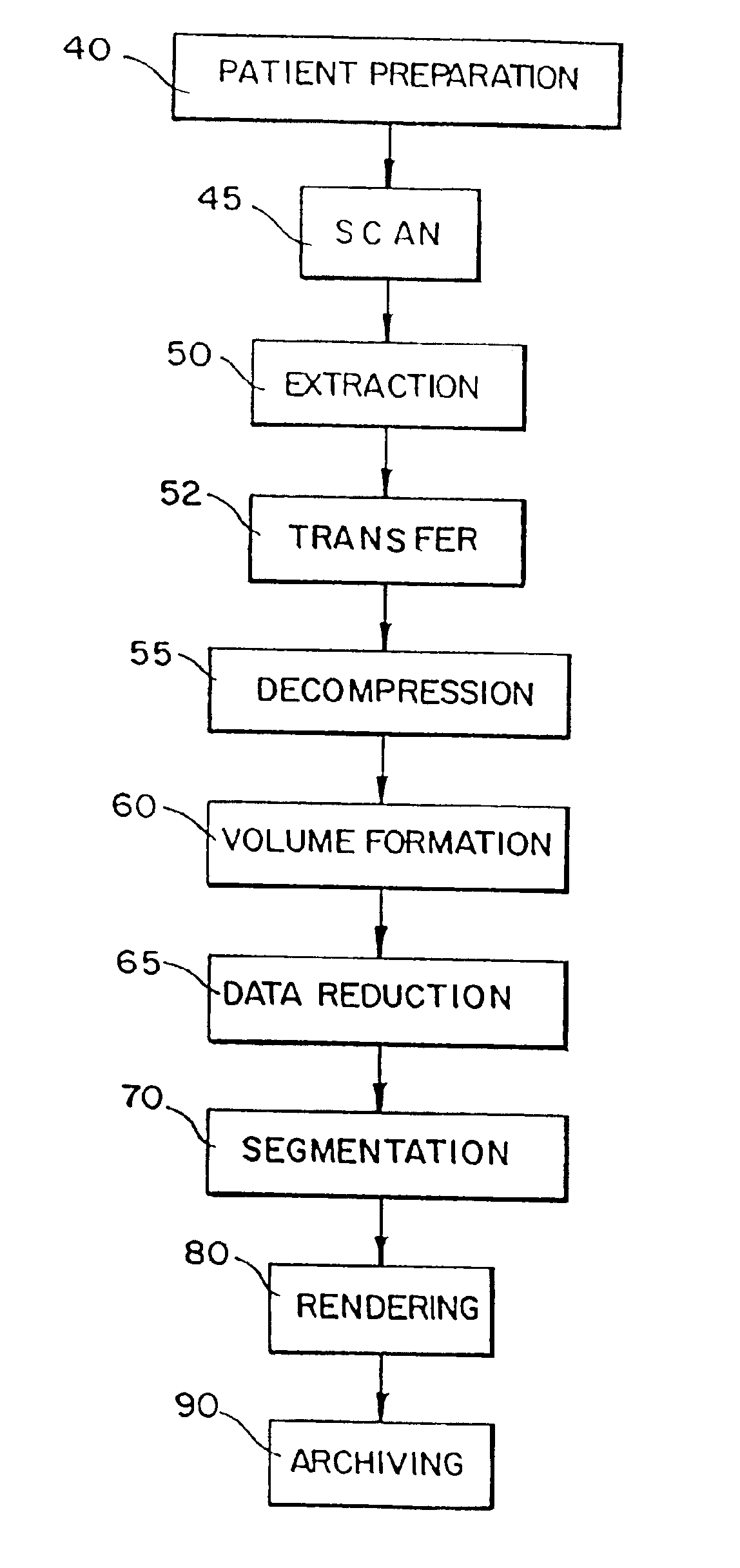
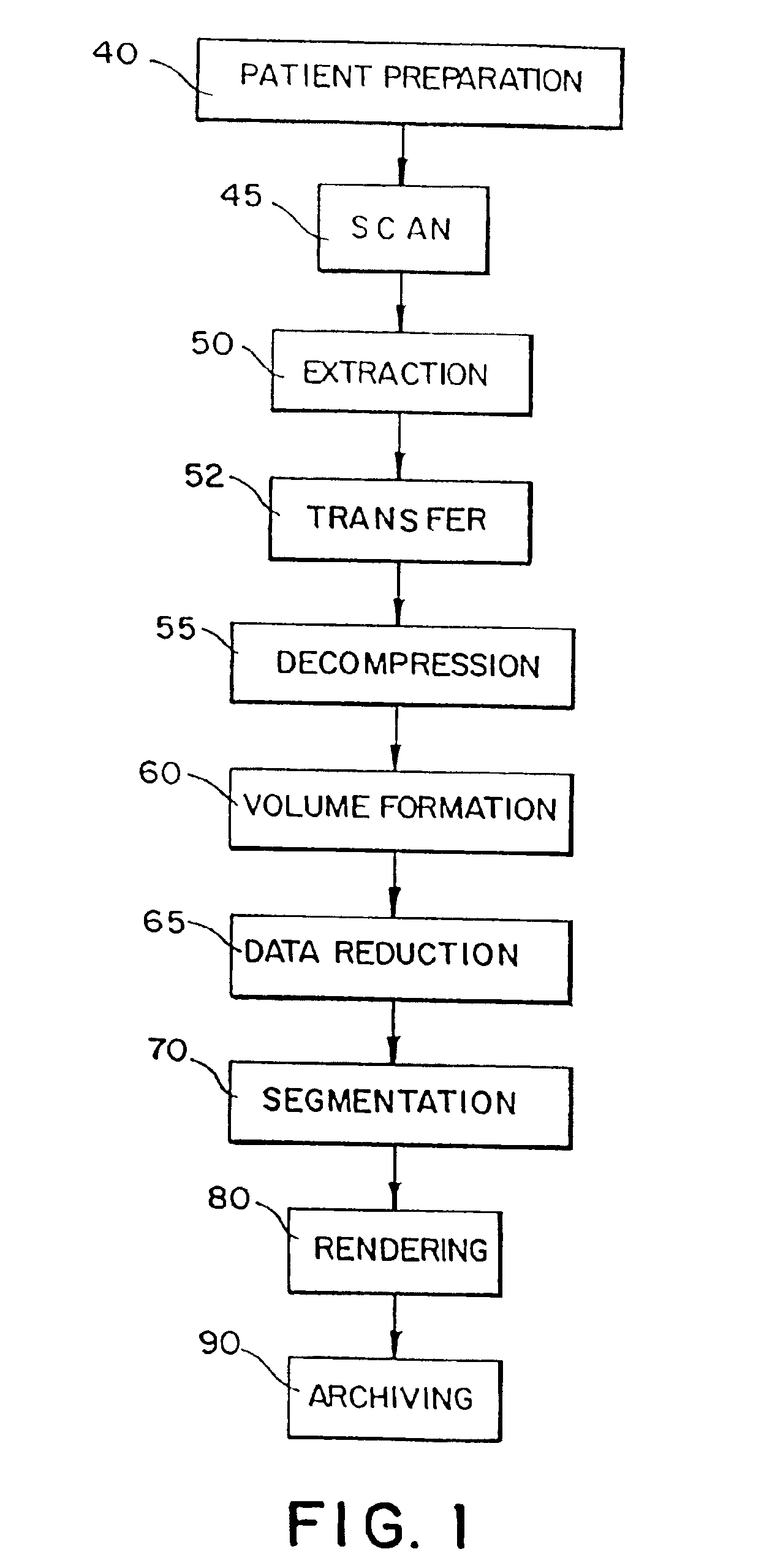
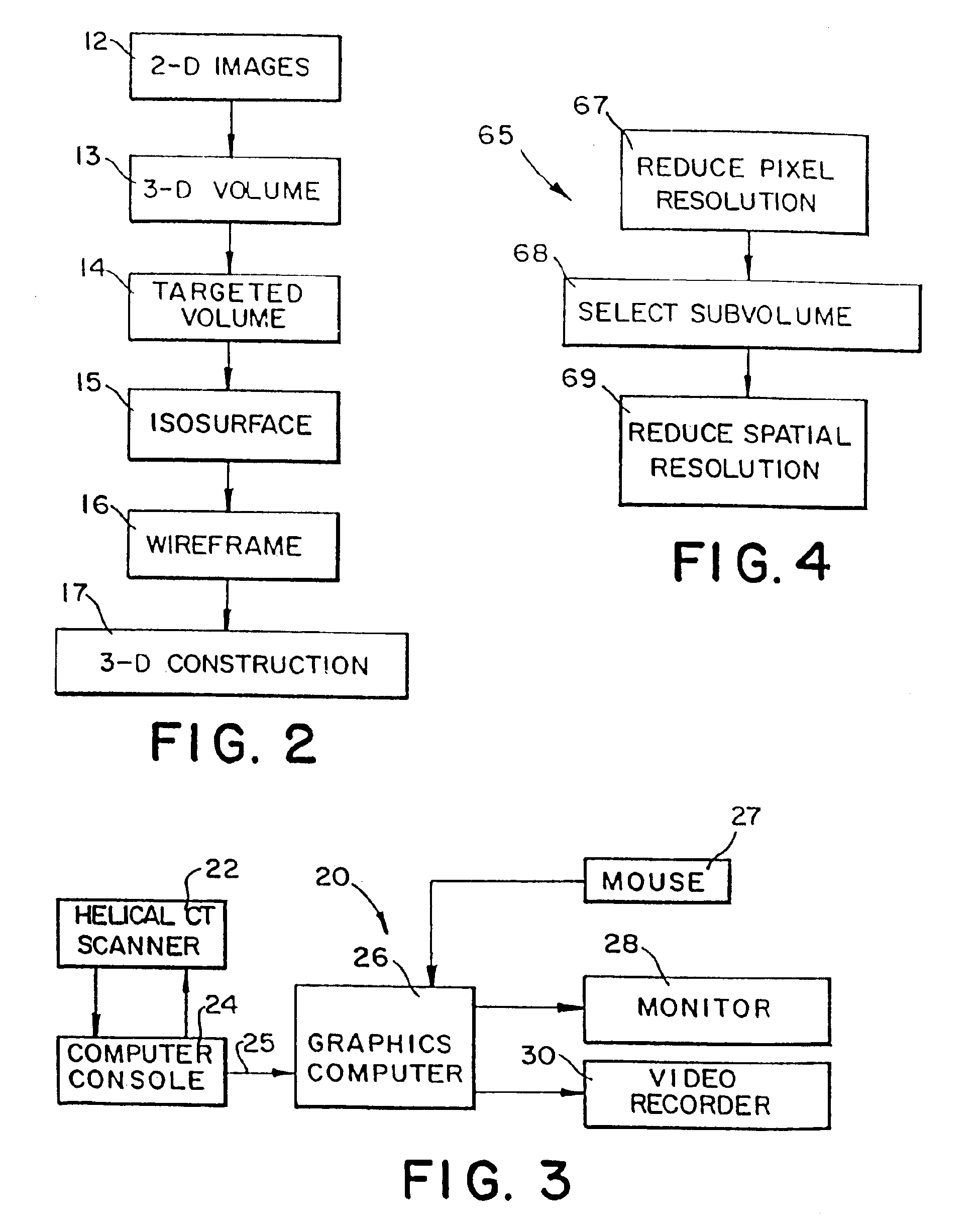
[0043]The present invention generally relates to a method and system, as schematically represented in FIGS. 1, 2, and 3, for generating and displaying interactive, three-dimensional structures. The three-dimensional structures are in the general form of selected regions of the body and, in particular, body organs with hollow lumens such as colons, tracheobronchial airways, blood vessels, and the like. In accordance with the method and system of the present invention, interactive, three-dimensional renderings of a selected body organ are generated from a series of acquired two-dimensional images.

[0044]As illustrated in FIG. 3, a scanner 22, such as a spiral or helical CT (Computed Tomography) scanner, operated by a computer console 24 is used to scan a selected three-dimensional structure, such as a selected anatomy, thereby generating a series of two-dimensional images 12 through that structure. A general procedure for converting or transforming the set of two-dimensional images 12 ...


PUM


Abstract

A method and system are provided for effecting interactive, three-dimensional renderings of selected body organs for purposes of medical observation and diagnosis. A series of CT images of the selected body organs are acquired. The series of CT images is stacked to form a three-dimensional volume file. To facilitate interactive three-dimensional rendering, the three-dimensional volume file may be subjected to an optional dataset reduction procedure to reduce pixel resolution and / or to divide the three-dimensional volume file into selected subvolumes. From a selected volume or subvolume, the image of a selected body organ is segmented or isolated. A wireframe model of the segmented organ image is then generated to enable interactive, three-dimensional rendering of the selected organ.

Description

[0001]This application is a continuation of application Ser. No. 08 / 734,427, entitled “Method and System for Producing Interactive Three-Dimensional renderings of Selected Body Organs Having Hollow Lumens to Enable Simulated Movement Through the Lumen”, filed Oct. 16, 1996, now U.S. Pat. No. 6,272,366, which is a continuation of issued patent application Ser. No. 08 / 331,352, entitled “Method and System for Producing Interactive, Three-Dimensional Renderings of Selected Body Organs Having Hollow Lumens to Enable Simulated Movement Through the Lumen”, filed on Oct. 27, 1994, now U.S. Pat. No. 5,782,762, which are incorporated herein by reference.BACKGROUND OF THE INVENTION[0002]For many forms of cancer, early detection is essential for a favorable prognosis. The cancerous growth must be detected at an early stage before the cancer is allowed to grow and spread. Such is the case for colorectal and lung cancers. As a result, techniques have been developed to examine the colon and trache...

Claims


Application Information

Patent Timeline
Patent Type & Authority Patents(United States)
IPC IPC(8): G06T5/00G06T15/00G09B23/00G09B23/28H04N5/74A61B6/03G06T15/08G06T17/00G06T17/20G06T19/00
CPCG06T7/0012G06T7/0091G06T15/08G06T17/20G09B23/285G06T19/003G06T7/0081G06T2210/41G06T2207/10081G06T2207/10136G06T2207/30028G06T2207/30032G06T2207/30064H04N9/3141Y10S128/92A61B6/027G06T2210/08G06T7/11G06T7/155
Inventor VINING, DAVID J.
Owner WAKE FOREST UNIV HEALTH SCI INC
Why Eureka
Try Eureka