Endoscopic sheath assembly and associated method

a technology of endoscopy and endoscope, which is applied in the field of medical devices, can solve the problems of difficult to securely apply the sheath to the endoscope without ripping the sheath, the possibility of infection transmission from one patient to another, and the difficulty of cleaning an endoscope, particularly the biopsy channel, to achieve the effect of avoiding infection and avoiding infection

Inactive Publication Date: 2005-08-16
GRANIT MEDICAL INNOVATION
View PDF4 Cites 50 Cited by
  • Summary
  • Abstract
  • Description
  • Claims
  • Application Information

AI Technical Summary

Benefits of technology

[0004]It is a general object of the present invention to

Problems solved by technology

One problem in endoscopy is the potential for the transmission of infection from one patient to another because of remaining bioburden inside or on the endoscope.
However, it is difficult to adequately clean an endoscop

Method used


Image

  • Endoscopic sheath assembly and associated method
  • Endoscopic sheath assembly and associated method
  • Endoscopic sheath assembly and associated method

Examples


Embodiment Construction

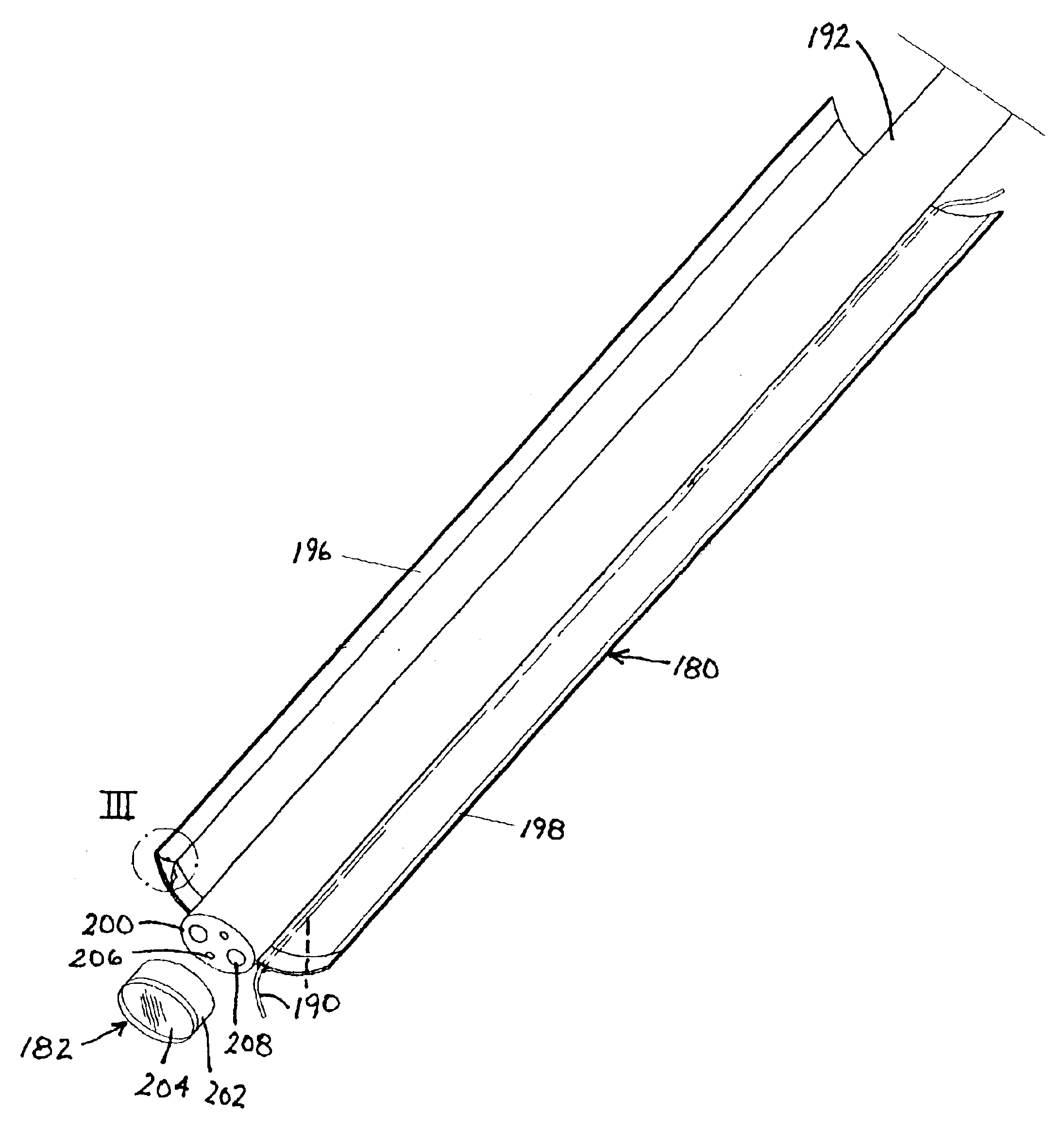
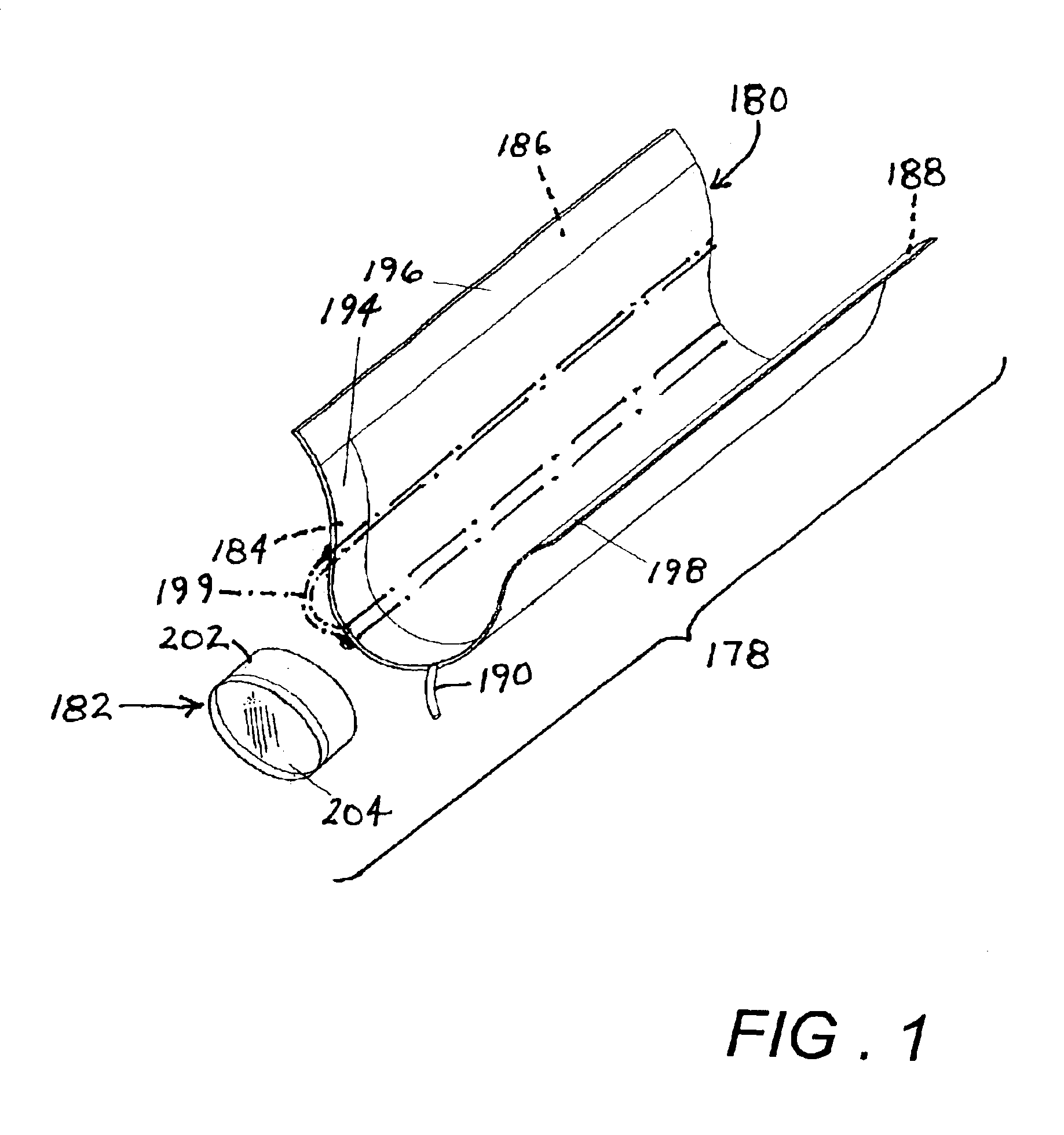
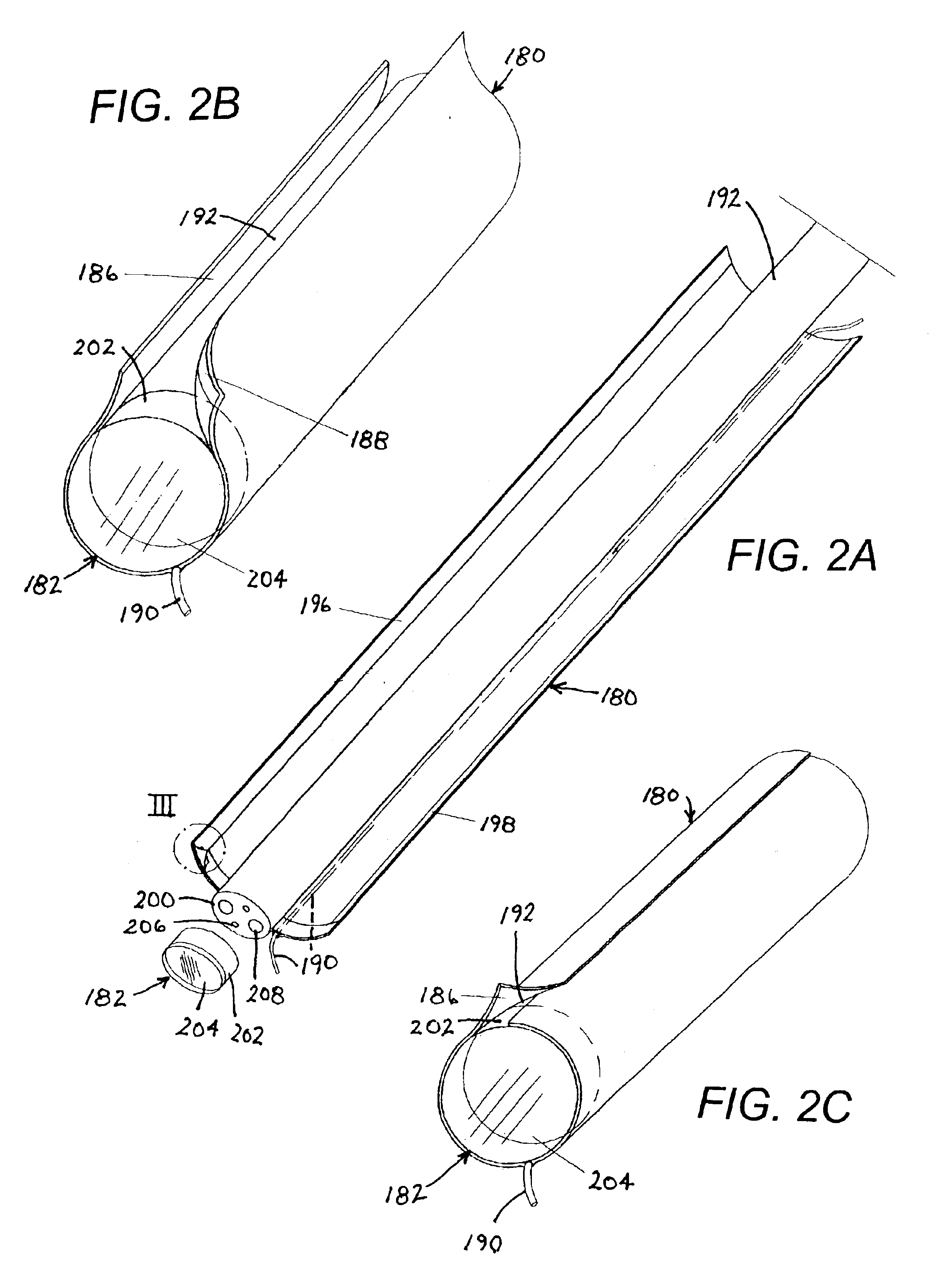
[0025]As depicted in FIG. 1, an endoscopic sheath assembly 178 includes an endoscope sheath 180 in the form of a web member or sheet and further includes an endoscope end cap 182. Sheath 180 is provided with a layer of adhesive material 184, 186, 188 along a forward end region, a first longitudinal edge region and a second longitudinal edge region, respectively (not separately enumerated). Sheath 180 is further provided along an elongate weakened (e.g., thinned or scored) region (not separately labeled) with an embedded tensile member 190 such as a tear string for enabling a severing of the sheath in preparation for removal of the sheath from an endoscope insertion member 192 (FIG. 2A). Releasable protective cover strips 194, 196, 198 are removably attached to the adhesive layers or regions 184, 186, 188. Preferably, the adhesive material of layers or regions 184 and 186 is a permanent adhesive, while the adhesive material of layer or region 188 forms a separable or releasable bond....


PUM


Abstract

An endoscope sheath includes a web member disposable in a tubular configuration in engagement with an endoscope insertion member, the member being provided with a tear string in the form of a tensile member. The tear string preferably extends longitudinally along the web member. In addition, the tear string preferably extends from one end of the web member to an opposite end thereof and is disposed along a weakened area of the web member. The web member is provided in a plurality of regions with a layer of adhesive material. The adhesive material of at least one of the regions is permanent adhesive, whereas the adhesive material of at least another of the regions forms a separable bond. These regions extend along opposing longitudinal edges of the web member.

Description

BACKGROUND OF THE INVENTION[0001]This invention relates to a medical device and to an associated medical technique. More particularly, this invention relates to an endoscopic sheath assembly and to an associated endoscopic method.[0002]Flexible endoscopes are inserted into the digestive tract for diagnostic and therapeutic purposes. Endoscopes generally include a light guide for transmitting optical-wavelength electromagnetic radiation into the patient. Images are captured, typically via lenses and an optical fiber bundle or a charge-coupled device, whereby a user can visually inspect the inner walls or surfaces of the digestive tract. One common objective of endoscopic investigations in the digestive tract is to detect the presence of polyps. Where a polyp is visually detected, particularly in the colon, it should be severed, captured, and removed from the patient. Alternatively, particularly where the polyp may be a malignant cancer, the polyp may be severed and captured for extra...

Claims


Application Information

Patent Timeline
IPC IPC(8): A61B1/00
CPCA61B1/00135A61B1/00142
Inventor NAKAO, NAOMI
Owner GRANIT MEDICAL INNOVATION
Why Eureka
Try Eureka