Minimally invasive method for implanting a sacral stimulation lead

a sacral stimulation and minimally invasive technology, applied in the field of sacral stimulation leads surgically implanted, can solve the problems of affecting the health and quality of life of millions of people with pelvic floor disorders, individuals with urinary control disorders often face debilitating challenges in their everyday lives, and becoming isolated and depressed

Inactive Publication Date: 2005-12-06
MEDTRONIC INC +1
View PDF21 Cites 145 Cited by
  • Summary
  • Abstract
  • Description
  • Claims
  • Application Information

AI Technical Summary

Problems solved by technology

Pelvic floor disorders adversely affect the health and quality of life of millions of people.
Individuals with urinary control disorders often face debilitating challenges in their everyday lives.
These individuals can be preoccupied with trips to the bathroom, fears of embarrassment, and sleepless nights.
Some suffers become so anxious that they become isolated and depressed.
Surgical treatments are the most invasive and are often considered after other therapies have proven ineffective.
Surgically implanting the stimulation lead in this manner near the patient's sacral nerve can cause patient complications, create significant patient's recovery time, and create a significant expense to the healthcare system.

Method used


Image

  • Minimally invasive method for implanting a sacral stimulation lead
  • Minimally invasive method for implanting a sacral stimulation lead
  • Minimally invasive method for implanting a sacral stimulation lead

Examples


embodiment

Fourth Minimally Invasive Method Embodiment

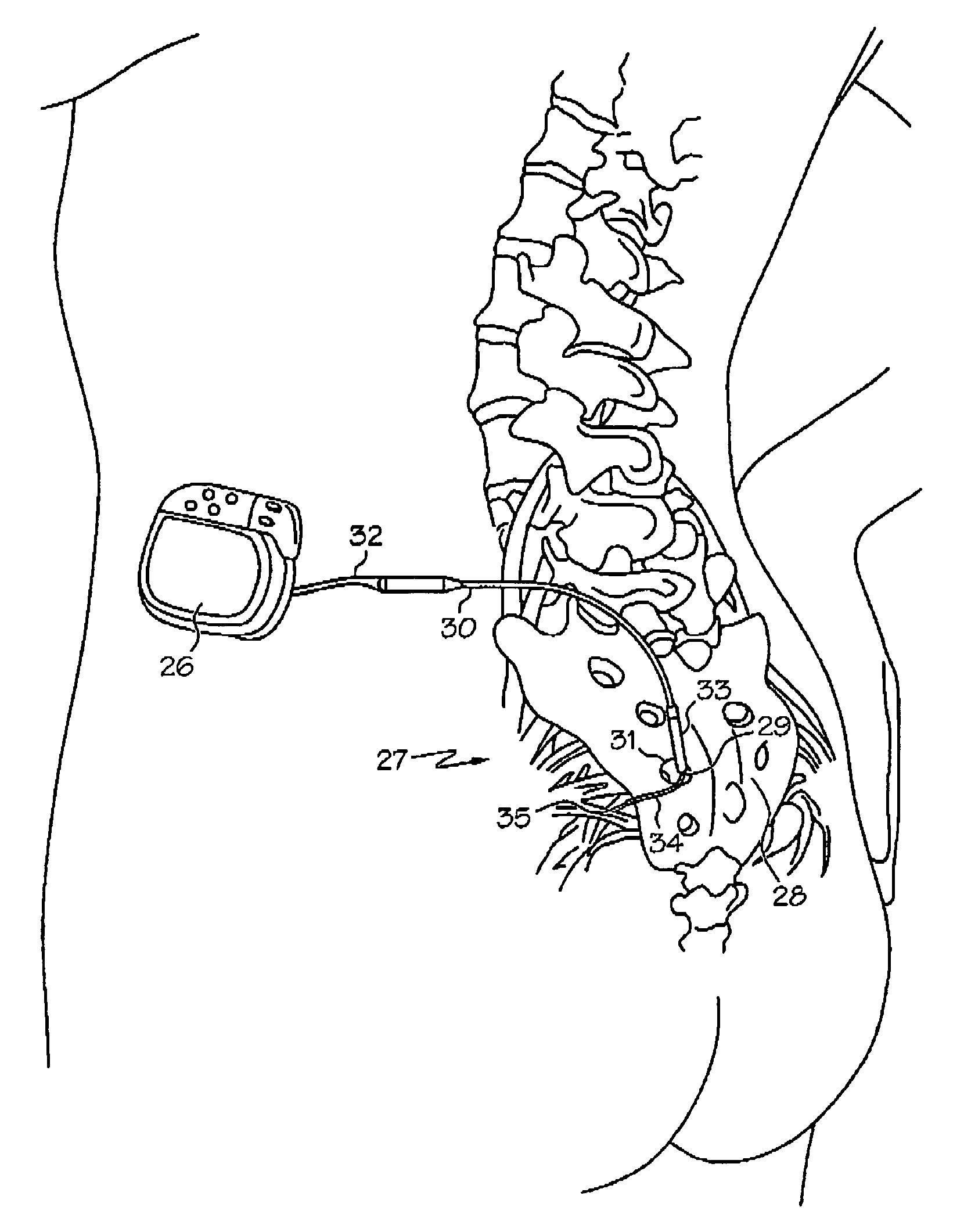
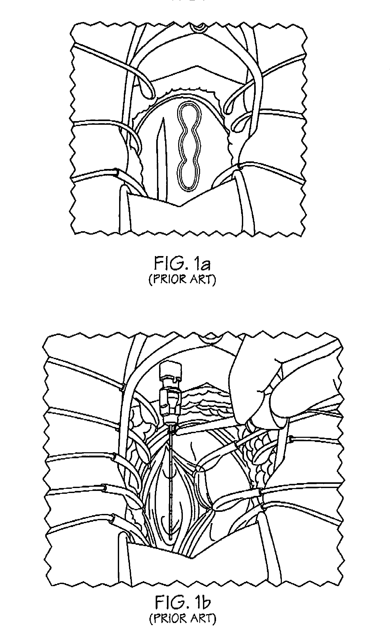
[0068]FIG. 7a shows a flowchart of a fourth minimally invasive implantation method embodiment 82, and FIGS. 7b–7g implementation element embodiments. The fourth minimally invasive method embodiment 82 is similar to the second minimally invasive method embodiment 74 with the exception that a guide wire 44 or stylet is inserted 78 to guide the stimulation lead 30 and the stimulation lead 30 functions as the dilator 42, so a separate dilator 42 is not used. More specifically, after the incision 68 is created 62, a guide wire 44 is inserted 78 into the needle. Once the guide wire 44 is in the desired location 35, the needle 36 is removed 56 from the insertion path 33. In one embodiment, the stimulation lead 30 is configured with a centrally located stylet lumen and a pointed tip, so the stimulation lead 30 can serve as the dilator 42. The stimulation lead 30 is inserted over the guide wire 44 to the desired location 35. In another embodiment, t...


PUM


Abstract

Method embodiments to implant a stimulation lead in a patient's sacrum to deliver neurostimulation therapy can reduce patient surgical complications, reduce patient recovery time, and reduce healthcare costs. A method embodiment begins by inserting a needle posterior to the sacrum through an entry point. The needle is guided into a foramen along an insertion path to a desired location. The insertion path is dilated with a dilator to a diameter sufficient for inserting a stimulation lead. The needle is removed from the insertion path. The stimulation lead is inserted to the desired location. The dilator is removed from the insertion path. Additionally if the clinician desires to separately anchor the stimulation lead, an incision is created through the entry point from an epidermis to a fascia layer. The stimulation lead is anchored to the fascia layer. After the stimulation lead has been anchored, the incision can be closed, or the stimulation lead proximal end can be tunneled to where an implantable neurostimulator is located and then the incision can be closed. A implanted sacral stimulation lead can be connected to the neurostimulator to delivery therapies to treat pelvic floor disorders such as urinary control disorders, fecal control disorders, sexual dysfunction, and pelvic pain.

Description

CROSS REFERENCE[0001]This disclosure is related to the following application entitled “Minimally Invasive Surgical Techniques For Implanting Devices That Deliver Stimulation To The Nervous System” by inventors Gerber et al. (application Ser. No. 09 / 489,544; filed Jan. 31, 2000 now U.S. Pat. No. 6,360,750), which is not admitted as prior art with respect to the present disclosure by its mention in this cross reference section.BACKGROUND OF THE INVENTION[0002]This disclosure relates to a method for surgically implanting an electric neurostimulation lead in the human sacrum.[0003]The medical device industry produces a wide variety of electronic and mechanical devices for treating patient medical conditions. Depending upon the medical condition, medical devices can be surgically implanted or connected externally to a patient receiving treatment. Clinicians use medical devices alone or in combination with drug therapies and surgery to treat patient medical conditions. For some medical co...

Claims


Application Information

Patent Timeline
Patent Type & Authority Patents(United States)
IPC IPC(8): A61N1/05A61N1/08
CPCA61N1/0551A61N1/08
Inventor MAMO, GEORGESPINELLI, MICHELE
Owner MEDTRONIC INC
Why Eureka
Try Eureka