Standoff holder and standoff pad for ultrasound probe

Inactive Publication Date: 2006-04-18
PMT
View PDF14 Cites 82 Cited by
  • Summary
  • Abstract
  • Description
  • Claims
  • Application Information

AI Technical Summary

Problems solved by technology

Without a standoff pad the ridged surface of a probe touching the rigid bony ankle causes gaping or spaces between the transducer and the ankle.
Such gaping yields artifacts and poor image quality during a diagnostic ultrasound exam.
At the same time, gaping reduces the healing benefits in therapeutic ultrasound due to poor contact.
Any space between the membrane and the probe surface will cause a lack of contact to occur and a failure to transmit ultrasound waves.
This means that the membrane interface itself can not move and therefore does allow for possibility of poor compliance using this cover.
Unfortunately not all ultrasound probes have collars as described.
This feature does pose inherent contact issues because the Marino described probe covers will not be able to snap onto the some transducers (probes).
An incorrect fit along with a fixed membrane will lead to poor contact with the surface of the probe.
This standoff adaptor is rather complex in that-it involves a two chamber device that is specifically made for a particular type of transducer shape.
This adaptor has inherent problems as it is described that can cause poor imaging and ultrasound waves not to be transmitted.
Some examples are improper filling of the chamber, gases or bubbles in the fluid within the chamber, and leaking fluid due to improper seals within the chamber.
Although the embodiment of the assembly serves as a coupling medium for ultrasound waves, it lacks the ability to serve as a standoff pad which shifts an image into the far field, one of the main attributes of a using a standoff pad.
Although these acoustic couplers provide an acoustical pathway for ultrasound waves, they are not intended to be disposable.
Such ultrasonic couplers are expensive to produce because each probe involves the production of a mold to be made before the standoff is produced.
A problem with using this standoff pad is that the operator of the ultrasound device needs to use two hands; one hand to hold the transducer, the other to hold the standoff pad.
Using both hands presents the sonographer with the difficulty of making adjustments to dials or controls on an ultrasound unit while performing an exam or treatment.
Another less obvious issue about the above stated standoff covers or assembles is that none allow for a variety of pad thickness.
Sill another problem with present molded or adaptor type standoff is the generation of artifacts within the ultrasound images.
These artifacts are caused by the aging and fatigue of the materials that make up the standoff pad itself which, are fixed to the above stated adaptors.
Although these methods are novel for their intended purposes all of the above described ultrasound couplers fail to provide a single coupling device that has all of the following properties.An ultrasound coupling pad that is intended to be disposable.An ultrasound coupling device that has a pad which is not molded or fixed to its holder.An ultrasound coupling device which uses a coupling pad that can be temporally fixed to transducers.An ultrasound coupling device that can accommodate various shapes and sizes of transducers.An ultrasound coupling device that allows for a gel pad that has variable thickness.An ultrasound coupling device that allows the operator to change the type of a gel pad being use anytime.An ultrasound coupling device which allows the operator of the ultrasound scanner to scan use one hand feeing the second hand to make adjustments to the controls on the ultrasound machine.An ultrasound coupling device that provides for free vertical movement of a gel pad within the standoff hold.

Method used


Image

  • Standoff holder and standoff pad for ultrasound probe
  • Standoff holder and standoff pad for ultrasound probe
  • Standoff holder and standoff pad for ultrasound probe

Examples


Embodiment Construction

—FIG. 1 TO 13

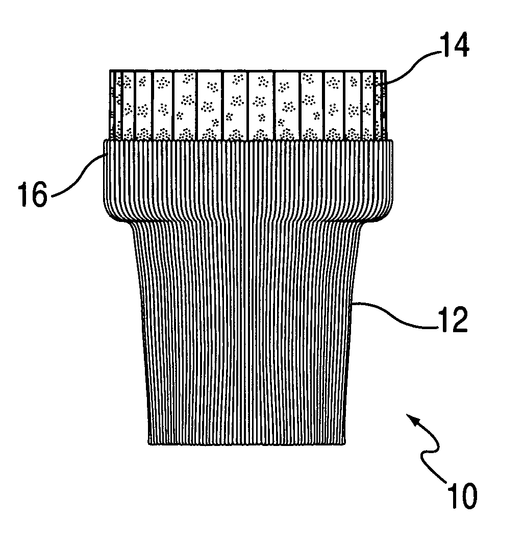
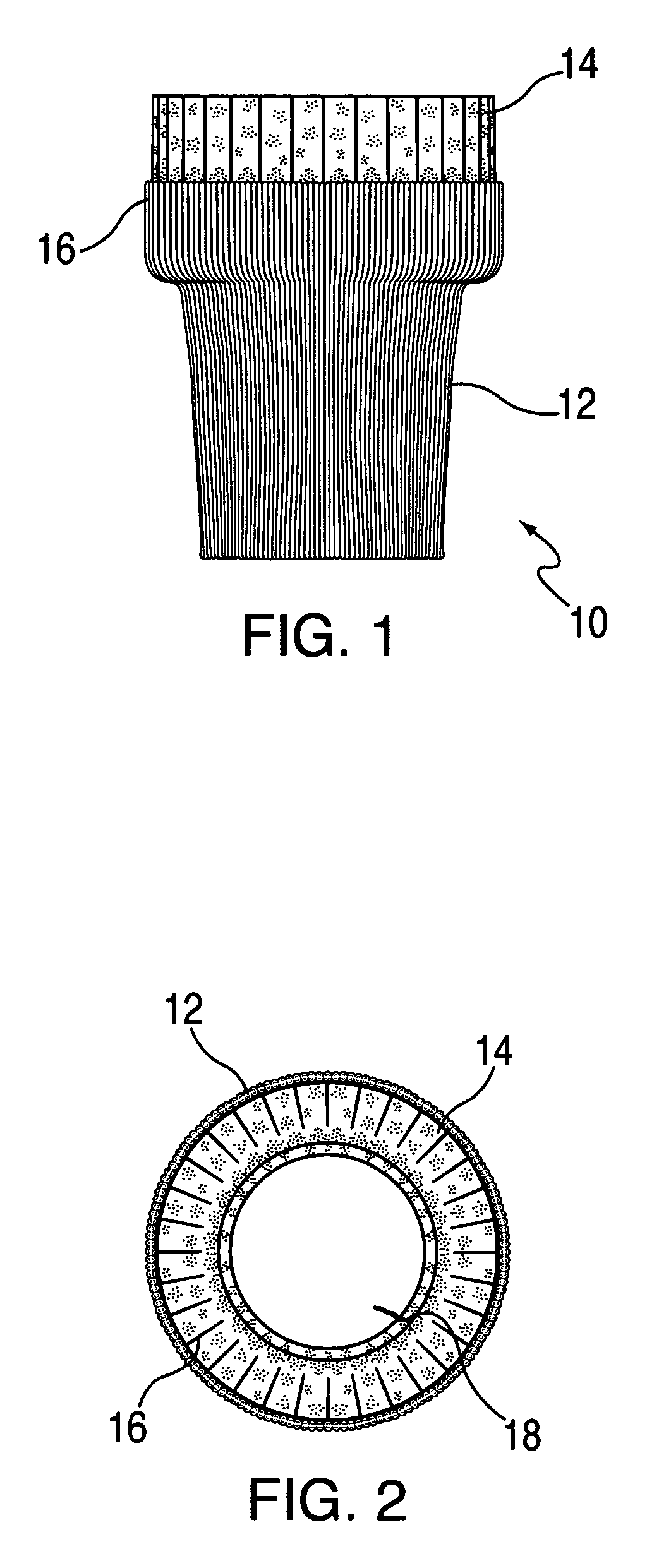
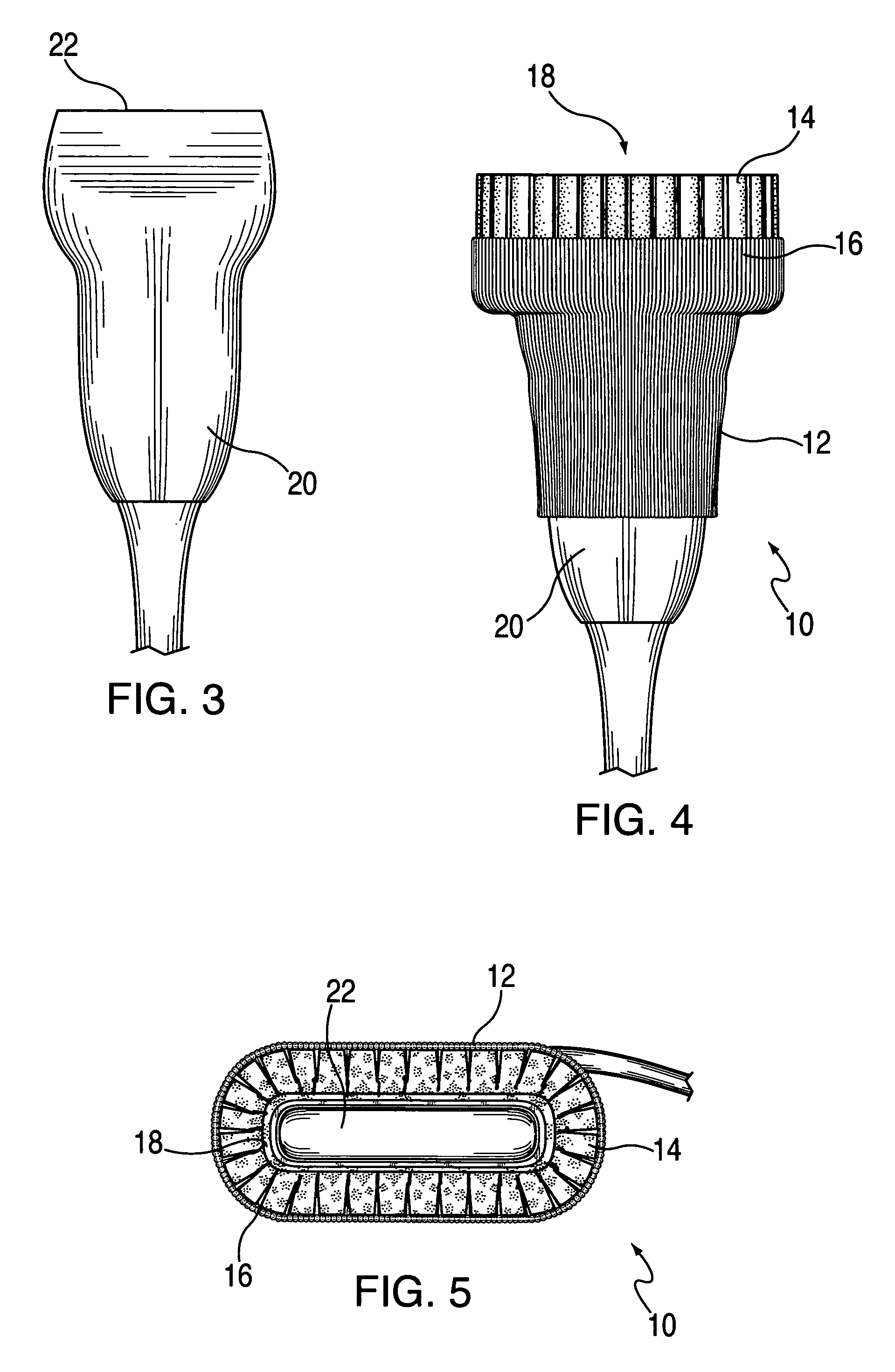
[0044]Referring to FIG. 1, a drawing of multi-ribbed standoff holder 10. Multi-ribbed standoff holder 10 is preferably based on sock 12 which is made of a soft pliable material such as an elastic cloth or rubber sock. Elastic sock 12 when stretched or expanded has memory which will cause it contract and to return to its normal shape when such tension is released. Elastic sock 12 on one end is comprised of solely elastic material and allows for great flexibility with little to no rigidity. As one travels along elastic sock 12 toward its opposite end, elastic sock 12 begins to loose flexibility and increases in rigidity. At the opposite end of elastic sock 12 an expansion collar 14 is comprised of a number of strips of rigid material such as plastic which are incorporated into elastic sock 12. Between these strips of plastic which make up expansion collar 14 areas of plain elastic sock exist providing and forming a number of expansion ribs 16. An expansion rib 16 is found...


PUM


Abstract

An accommodative standoff holder 10 which can be used for both diagnostic and therapeutic ultrasound is disclosed. The described standoff holder 10 has the ability to be mounted on and utilized with transducers 20 of different sizes and shapes. The standoff holder 10 includes a gel insert 26 which is removable and self adjusting assuring contact between a human body, an animal, or other object and the acoustic window 22 of a transducer 22.

Description

FIELD OF THE INVENTION[0001]The field of the invention relates to a temporary holder of a standoff pad for an ultrasound probe utilized during a diagnostic ultrasound exam or therapeutic ultrasound treatment.BRIEF DESCRIPTION OF PRIOR ART AND BACKGROUND OF THE INVENTION[0002]Diagnostic ultrasound is a medical procedure used to image both humans and animals in order to obtain images of a body. Images are taken of different organs or body systems and have been broken down into approximately 20 application areas as defined by the United States Food and Drug Administration (FDA). Some examples of the clinical applications are diagnostic ultrasound of musculoskeletal system, peripheral vascular system, and abdominal ultrasound. Therapeutic ultrasound is a medical treatment modality that is use to treat various abnormal conditions. The intent of this modality is to provide relief and cure the abnormal condition. An example therapeutic ultrasound would be to treat some inflammation of an i...

Claims


Application Information

Patent Timeline
Patent Type & Authority Patents(United States)
IPC IPC(8): A61B8/14A61B8/00
CPCA61B8/4281A61B8/4209
Inventor WENDELKEN, MARTIN EDMUNDPOPE, CHARLES
Owner PMT
Why Eureka
Try Eureka