
MR imaging method and MRI system

a technology of mr imaging and mri, applied in the direction of reradiation, measurement using nmr, instruments, etc., can solve the problems of inconvenient ssfp method, inability to separate water from fat, band artifacts in images constructed in the method employing fat suppressing pulses or images constructed according to femr techniques, etc., to achieve accurate and decisive verification, short scan time

Inactive Publication Date: 2006-06-27
GE MEDICAL SYST GLOBAL TECH CO LLC
View PDF15 Cites 13 Cited by
  • Summary
  • Abstract
  • Description
  • Claims
  • Application Information

AI Technical Summary

Benefits of technology

[0027]An object of the present invention is to provide an MR imaging method and an MRI system capable of shortening the time (scan time) elapsing from scanning to image construction, readily separating water from fat, and accurately constructing a water image and(or) a fat image.
[0030]According to the present invention, even a high-magnetic field strength system in which an in-phase time is short can construct images, which express water or fat separately from each other, according to the SSFP method.
[0031]Furthermore, according to the present invention, there is the advantage that when a repetition time TR is made equal to the product of the in-phase time by m (where m denotes a natural number) with spins excited in the SSFP, since both echoes induced by water and fat are affected by the inhomogeneity in a static magnetic field, an image unsusceptible to band artifact can be constructed.
[0032]Furthermore, according to the present invention, when an echo time TE is made equal to the product of the in-phase time by (m−1±1 / n) (where m denotes a natural number, n denotes a natural number equal to or larger than 3, and TE assumes a positive value), the single quadrature fat / water imaging method can be adapted. In short, since the present invention does not employ the phase cycling method presented in Japanese Unexamined Patent Application Publication No. 2001-414 and Thesis 1, an object can be scanned with 1 NEX. This leads to a shorter scan time.
[0033]When the method described in Thesis 2 is employed, since a phase difference between echoes induced by water and fat is π (radian) under the conditions of “m=1 and n=2,” a certain hypothesis is needed. According to the present invention, adjoining signals can be checked to see if they are induced by water or fat. Whether an echo is induced by water or fat can be accurately and decisively verified.

Problems solved by technology

On the other hand, the SSFP method confronts the problems that band artifact occurs and that it is hard to separate water from fat due to induction of an intense signal by fat.
However, when the distribution of static magnetic field strengths is not uniform, an image constructed in the method employing fat suppressing pulses or an image constructed according to the FEMR technique suffers from band artifact.
Furthermore, fat suppression to be achieved using fat suppressing pulses disorders the steady state of a magnetic field, and is therefore unsuitable for the SSFP method.
According to the method, since echoes are acquired using RF pulses exhibiting two kinds of phases, a scan time is long and a signal processing time is long.
Attempts have been made to compensate for the inhomogeneity in static magnetic field strength in terms of facilities, but have confronted limitations.
However, since the Dixon method requires production of two image data items, a scan time increases.
This poses a problem in that the Dixon method cannot be adapted to a system offering a low magnetic field.
The technology described in Japanese Unexamined Patent Application Publication No. 2001-414 and Thesis 1 has the drawback that the repetition time TR must equal the product of the in-phase time by 2 / n and the echo time TE must equal the quotient of the in-phase time by n (where n denotes an integer equal to or larger than 3).
Besides, the repetition time TR is so short that a system for realizing the technology will be large in scale and include special components.
Furthermore, since at least two scans must be performed, a scan time is long.
At this time, there is a possibility that the phases of the water and fat signals may be identified inversely and that incorrect judgment may be invited.

Method used


Image

  • MR imaging method and MRI system
  • MR imaging method and MRI system
  • MR imaging method and MRI system

Examples


first embodiment

Advantages of First Embodiment

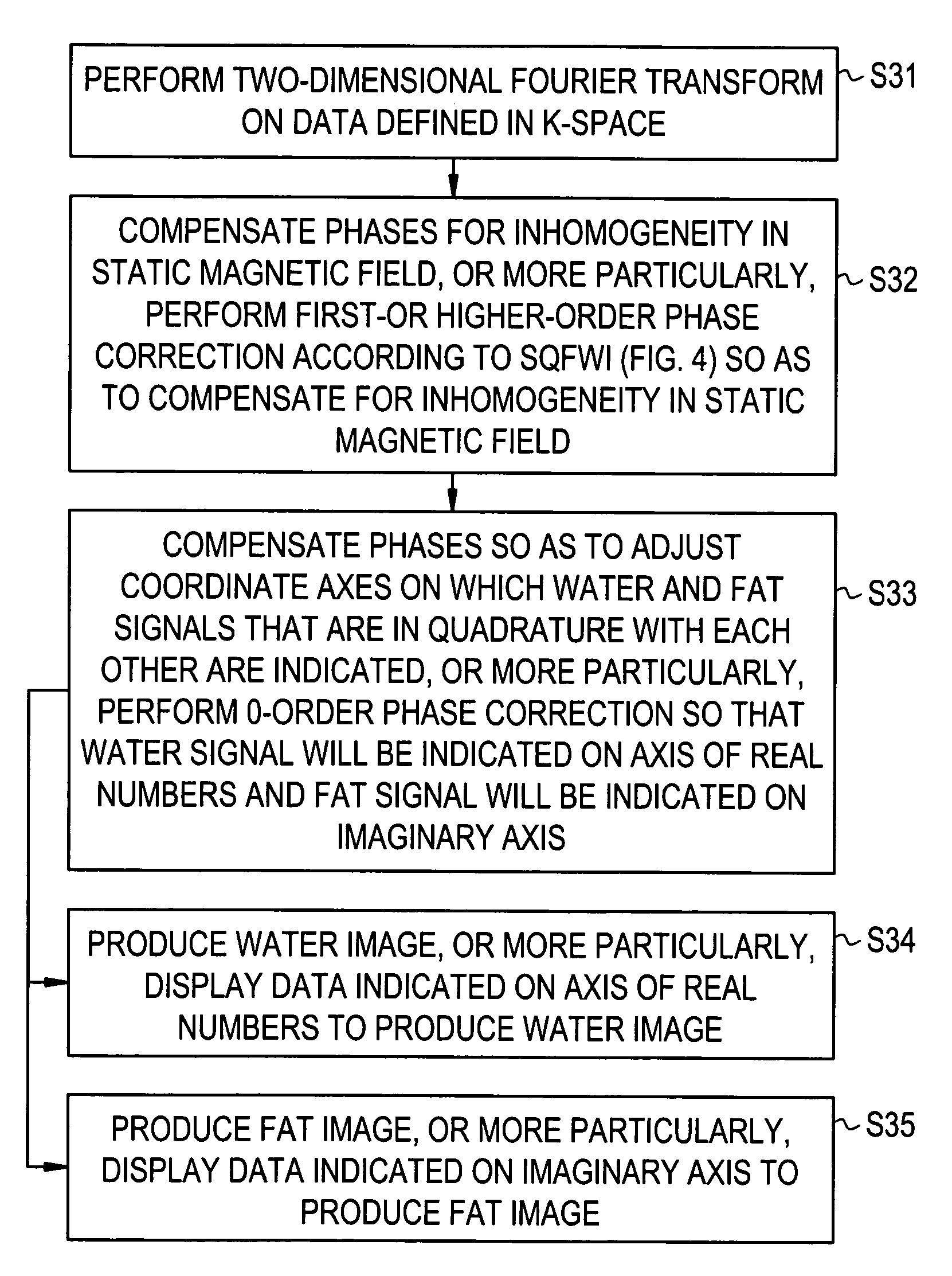
[0174]Since the SQFWI technique is adapted to the SSFP method that is implemented based on the contents of the PSD, after the spins in water and fat are excited in the SSFP method, an image whose water and fat components can be separated from each other can be constructed.

[0175]According to the contents of the PSD employed in the present embodiment, the repetition time TR is set to the product of the in-phase time by m (where m denotes a natural number) in order to implement the SSFP method. Consequently, a signal induced by water and a signal induced by fat alike are affected by the inhomogeneity in a static magnetic field. Eventually, the adverse effect of the inhomogeneity in a static magnetic field can be canceled.

[0176]Moreover, in the present embodiment, unlike the method described in Japanese Unexamined Patent Application Publication No. 2001-414, that is, the phase cycling SSFT fat / water image construction method, it is unnecessary to change the...

second embodiment

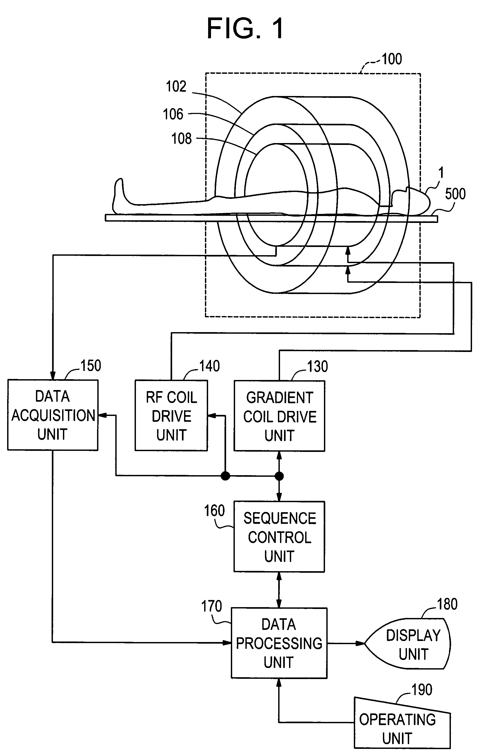
[0178]FIG. 7 shows the configuration of an MRI system in accordance with the second embodiment of the present invention.

[0179]The MRI system shown in FIG. 7 has the same components as the MRI system shown in FIG. 1 except a magnet system 100′. The magnet system 100′ will be mainly described below.

[0180]The magnet system 100′ includes a main field magnet unit 102′, a gradient coil unit 106′, and an RF coil unit 108′.

[0181]Each of the main field magnet unit 102′ and coil units comprises a pair of magnets or coils opposed to each other with a space between them. Moreover, the magnets and coils have a substantially disk-like shape and share the same center axis. The object of imaging 1 lying down on the cradle 500 is carried into the bore of the magnet system 100′ by means of a carrying means that is not shown.

[0182]The main field magnet unit 102′ produces a static magnetic field in the bore of the magnet system 100′. The direction of the static magnetic field is substantially orthogona...


PUM


Abstract

An MR imaging includes acquiring echoes from the object by implementing a pulse sequence which specifies the conditions which should be established with spins excited in the steady-state free precession (SSFP) method, so that the object can be scanned with an echo induced by water contained in the object and an echo induced by fat contained therein in single quadrature with each other or with a phase difference of 90° between the echoes induced by water and fat, constructing a tomographic image by performing frequency transformation on the acquired echoes, compensating the transformed data for the inhomogeneity in a static magnetic field and reconstructing an image, of which water and fat components are separated from each other, according to the result of the compensation.

Description

CROSS REFERENCE TO RELATED APPLICATIONS[0001]This application claims the benefit of Japanese Application No. 2004-026894 filed Feb. 3, 2004.BACKGROUND OF THE INVENTION[0002]The present invention relates to an MR imaging method and an MRI system.[0003]More particularly, the present invention is concerned with an MR imaging method and an MRI system that clearly separates water in an object from fat therein for a short period of time so as to construct an image.[0004]MR imaging methods and MRI systems are demanded to construct an accurate image for a short period of time.[0005]Scanning an object whose spins are excited in the steady-state free precession (SSFP) method has advantage of producing an intense signal, which represents a high contrast, for a short scan time. On the other hand, the SSFP method confronts the problems that band artifact occurs and that it is hard to separate water from fat due to induction of an intense signal by fat.[0006]As a technique for suppressing a magne...

Claims


Application Information

Patent Timeline
Patent Type & Authority Patents(United States)
IPC IPC(8): G01V3/00A61B5/055G01R33/32G01R33/54G01R33/561G01R33/565
CPCG01R33/4828G01R33/5613G01R33/56563G01R33/56527A01G9/021A01G9/024A01G9/12A01G9/083
Inventor MIYOSHI, MITSUHARU
Owner GE MEDICAL SYST GLOBAL TECH CO LLC
Learn More