
Ophthalmic drug delivery device

a drug delivery and ophthalmology technology, applied in the field of biocompatible implants, can solve the problems of progressive damage to the retina, impracticality, and difficult or impossible reading, driving,

Inactive Publication Date: 2006-08-22
NOVARTIS AG
View PDF79 Cites 80 Cited by
  • Summary
  • Abstract
  • Description
  • Claims
  • Application Information

AI Technical Summary

Benefits of technology

The present invention is an eye drop device that can deliver medicine to the eye. It has a needle for injecting the medicine, a reservoir for holding the medicine, and a sleeve for placing the device on the eye. The sleeve is designed to fit tightly around the device, creating a ring-shaped shape on the eye. This design helps to keep the device in place and prevents it from moving around. The technical effect of this invention is to provide a more effective and reliable way to deliver medicine to the eye.

Problems solved by technology

ARMD attacks the center of vision and blurs it, making reading, driving, and other detailed tasks difficult or impossible.
In wet ARMD, newly formed choroidal blood vessels (choroidal neovascularization (CNV)) leak fluid and cause progressive damage to the retina.
However, photocoagulation can be harmful to the retina and is impractical when the CNV is near the fovea.
Furthermore, over time, photocoagulation often results in recurrent CNV.
However, due to drug-specific metabolic restrictions, systemic administration usually provides sub-therapeutic drug levels to the eye.
Therefore, to achieve effective intraocular drug concentrations, either an unacceptably high dose or repetitive conventional doses are required.
Periocular injections of these compounds often result in the drug being quickly washed out and depleted from the eye, via periocular vasculature and soft tissue, into the general circulation.
Repetitive sub-Tenon's capsule injections of these compounds carry the potential risk of penetrating the globe and the severe, often blinding, complications of retinal detachment and endophthalmitis.
In addition, it is difficult to perform such injections in a reproduceable manner, and each injection may result in a different distribution of drug along the scleral surface.
Furthermore, many attempts to inject drug below the Tenon's capsule actually result in injections into the Tenon's capsule itself or the surrounding tissue, which is not desirable.
Repetitive intraocular injections may also result in retinal detachment and endophthalmitis.
Consequently, these requirements generally increase the complexity and cost of such implants.

Method used


Image

  • Ophthalmic drug delivery device
  • Ophthalmic drug delivery device
  • Ophthalmic drug delivery device

Examples


Embodiment Construction

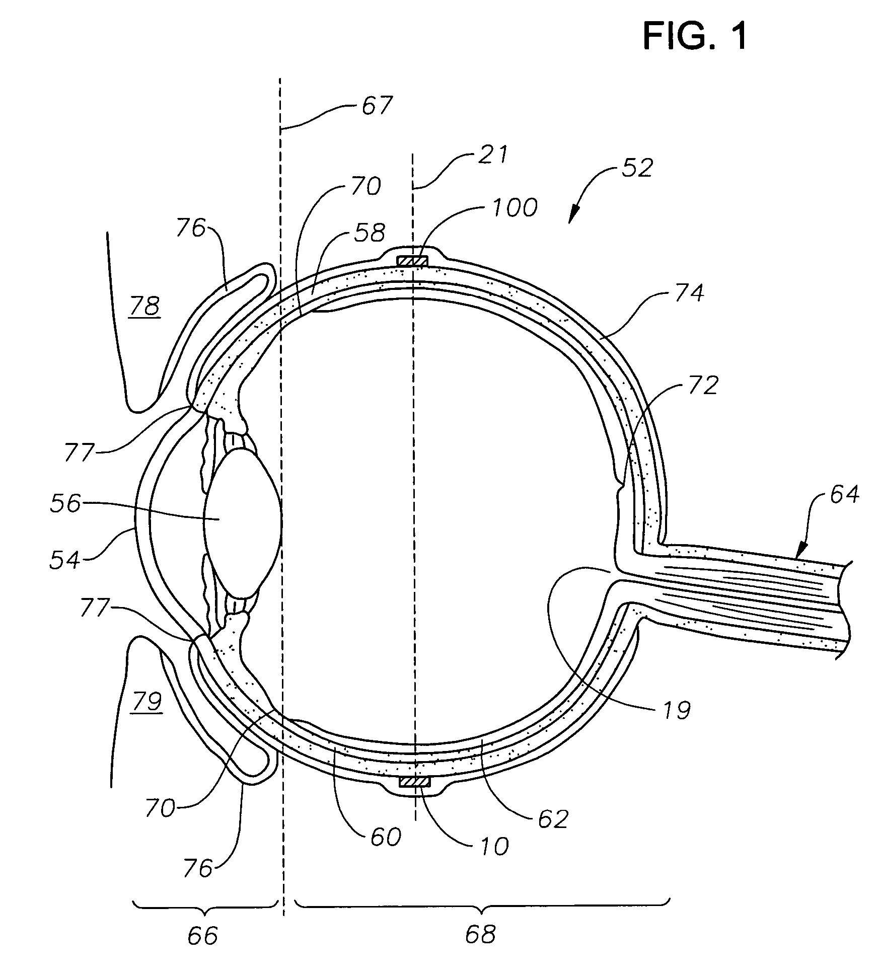
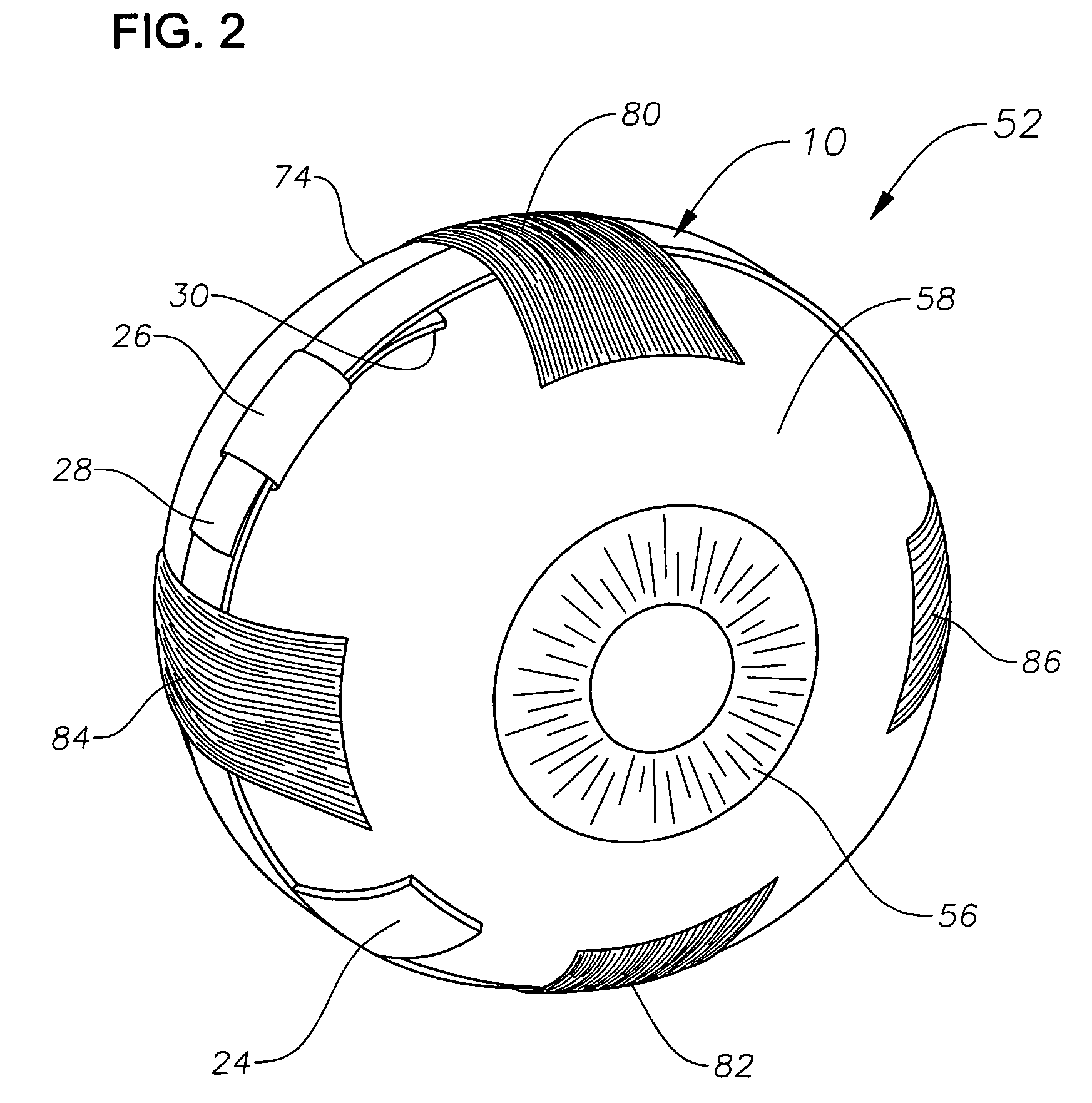
[0019]The preferred embodiments of the present invention and their advantages are best understood by referring to FIGS. 1–4 of the drawings, like numerals being used for like and corresponding parts of the various drawings.

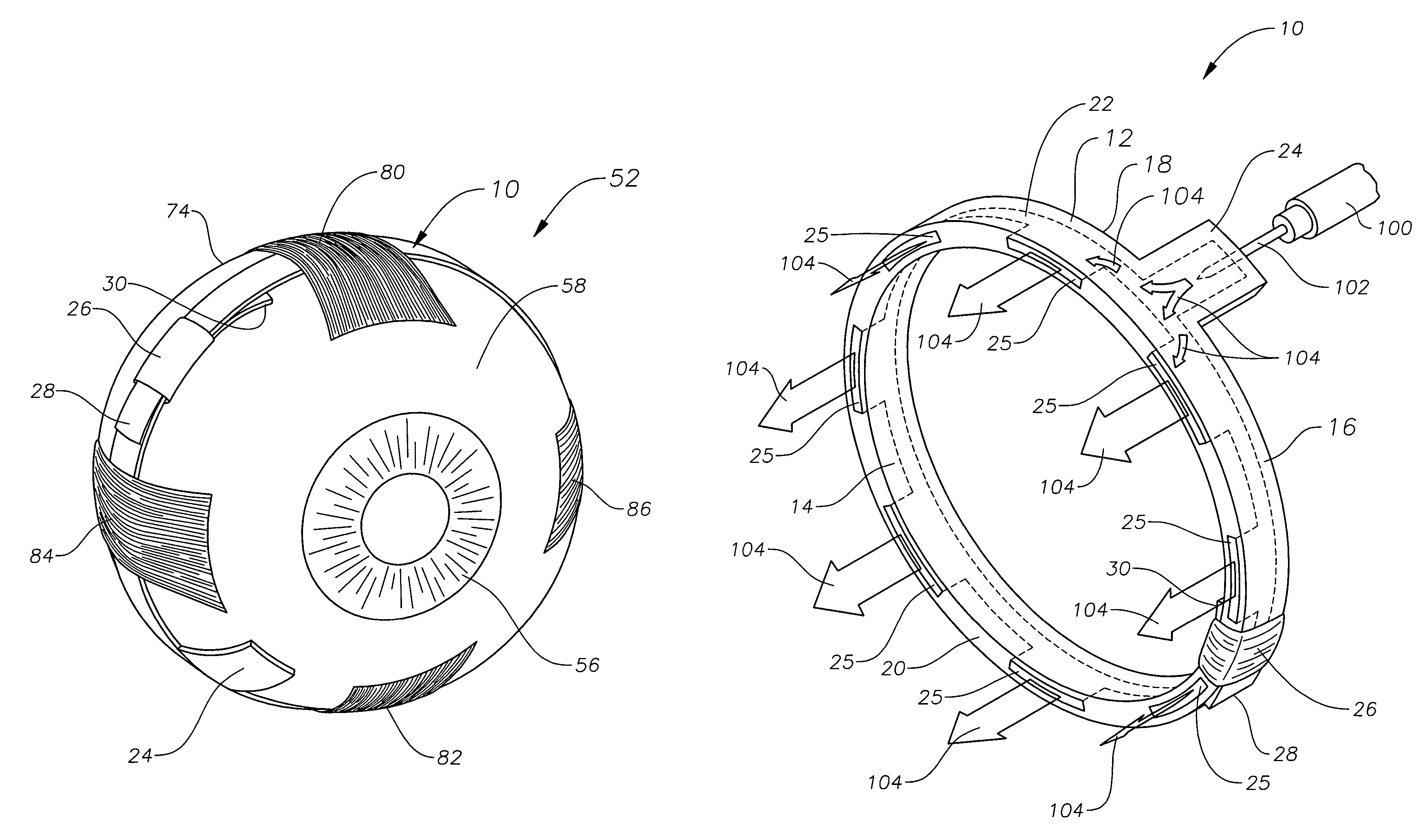
[0020]FIGS. 1–4 schematically illustrate an ophthalmic drug delivery device 10 according to a preferred embodiment of the present invention. Device 10 may be used in any case where delivery of a pharmaceutically active agent to the eye is required. Device 10 is particularly useful for delivery of active agents to the posterior segment of the eye. A preferred use for device 10 is the delivery of pharmaceutically active agents to the retina for treating ARMD, choroidial neovascularization (CNV), retinopathies, retinitis, uveitis, macular edema, and glaucoma.

[0021]Referring to FIGS. 1–2, a human eye 52 is schematically illustrated. Eye 52 has a cornea 54, a lens 56, a sclera 58, a choroid 60, a retina 62, and an optic nerve 64. An anterior segment 66 of eye 52 genera...


PUM


Abstract

An ophthalmic drug delivery device having a first end and a second end, an injection port, a reservoir, and a sleeve is disclosed. The injection port is for sealingly engaging a needle of a syringe, which is for providing a fluid comprising a pharmaceutically active agent. The reservoir is disposed within the device, is fluidly coupled to the injection port, and has an opening for communicating the fluid to an outer surface of a sclera of an eye. The sleeve is for engaging the device proximate overlapping portions of the first end and the second end for forming a generally ring-shaped three-dimensional geometry upon implantation of the device on the outer surface of the sclera. The device is useful for the treatment of a disease of the posterior segment of the eye.

Description

[0001]This application is a continuation of PCT / US02 / 23048 filed Jul. 22, 2002 entitled “Ophthalmic Drug Delivery Device,” which claims priority from U.S. Provisional Application No. 60 / 307,284 filed Jul. 23, 2001. This application is related to U.S. Pat. Nos. 6,413,540 and 6,416,777, both of which are incorporated herein in their entirety by this reference.FIELD OF THE INVENTION[0002]The present invention generally pertains to biocompatible implants for delivery of pharmaceutically active agents to the eye. More particularly, but not by way of limitation, the present invention pertains to biocompatible implants for delivery of pharmaceutically active agents to the posterior segment of the eye.DESCRIPTION OF THE RELATED ART[0003]Several diseases and conditions of the posterior segment of the eye threaten vision. Age related macular degeneration (ARMD), choroidal neovascularization (CNV), retinopathies (e.g., diabetic retinopathy, vitreoretinopathy), retinitis (e.g., cytomegalovirus ...

Claims


Application Information

Patent Timeline
Patent Type & Authority Patents(United States)
IPC IPC(8): A61M35/00A61M5/14A61F9/00A61F9/007A61M37/00
CPCA61F9/0017
Inventor YAACOBI, YOSEPH
Owner NOVARTIS AG
Learn More