

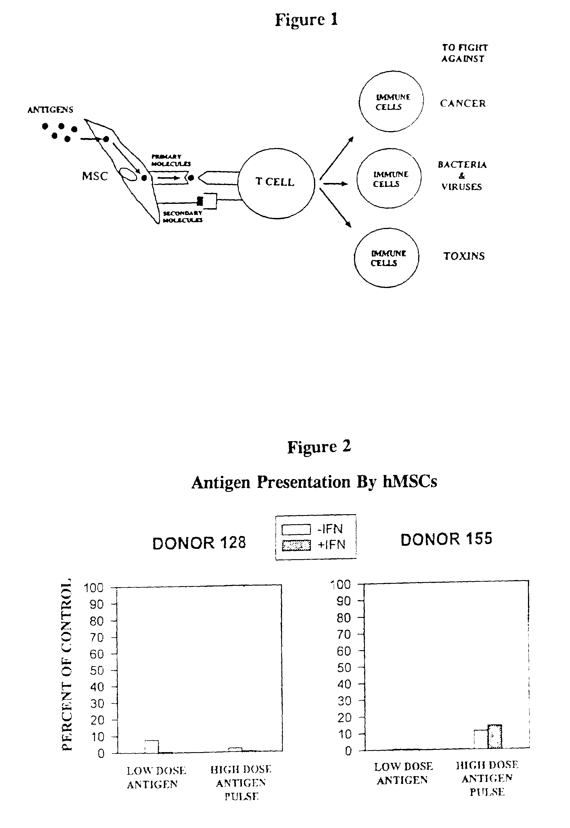
Antigen presenting mesenchymal stem cells
a mesenchymal stem cell and antigen technology, applied in the field of antigen-presenting mesenchymal stem cells, can solve the problems of receptors not being activated, eliciting an immune response, and not being able to do so
- Summary
- Abstract
- Description
- Claims
- Application Information
AI Technical Summary
Benefits of technology
Problems solved by technology
Method used
Image

Examples
example 1
Tetanus Antigen Presentation by hMSCs
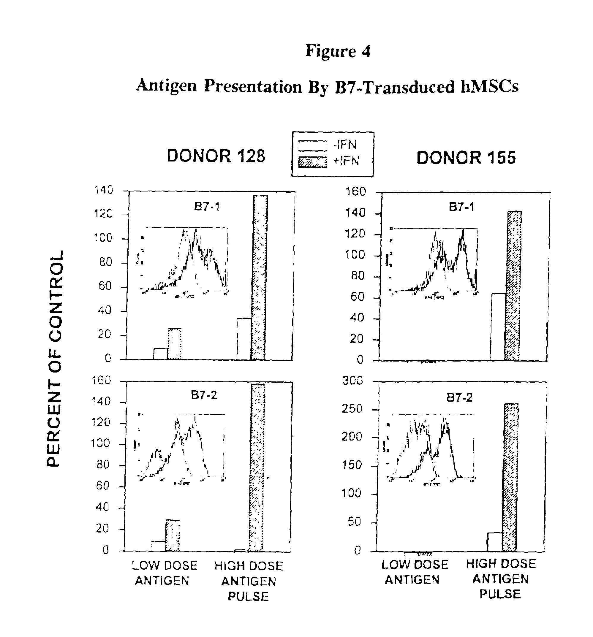
[0072]The principles of this invention are demonstrated by experiments using unmodified and B7-1 / B7-2 transduced MSCs, co-cultured with CD4+ tetanus toxoid enriched T-lymphocytes (Tet-T).
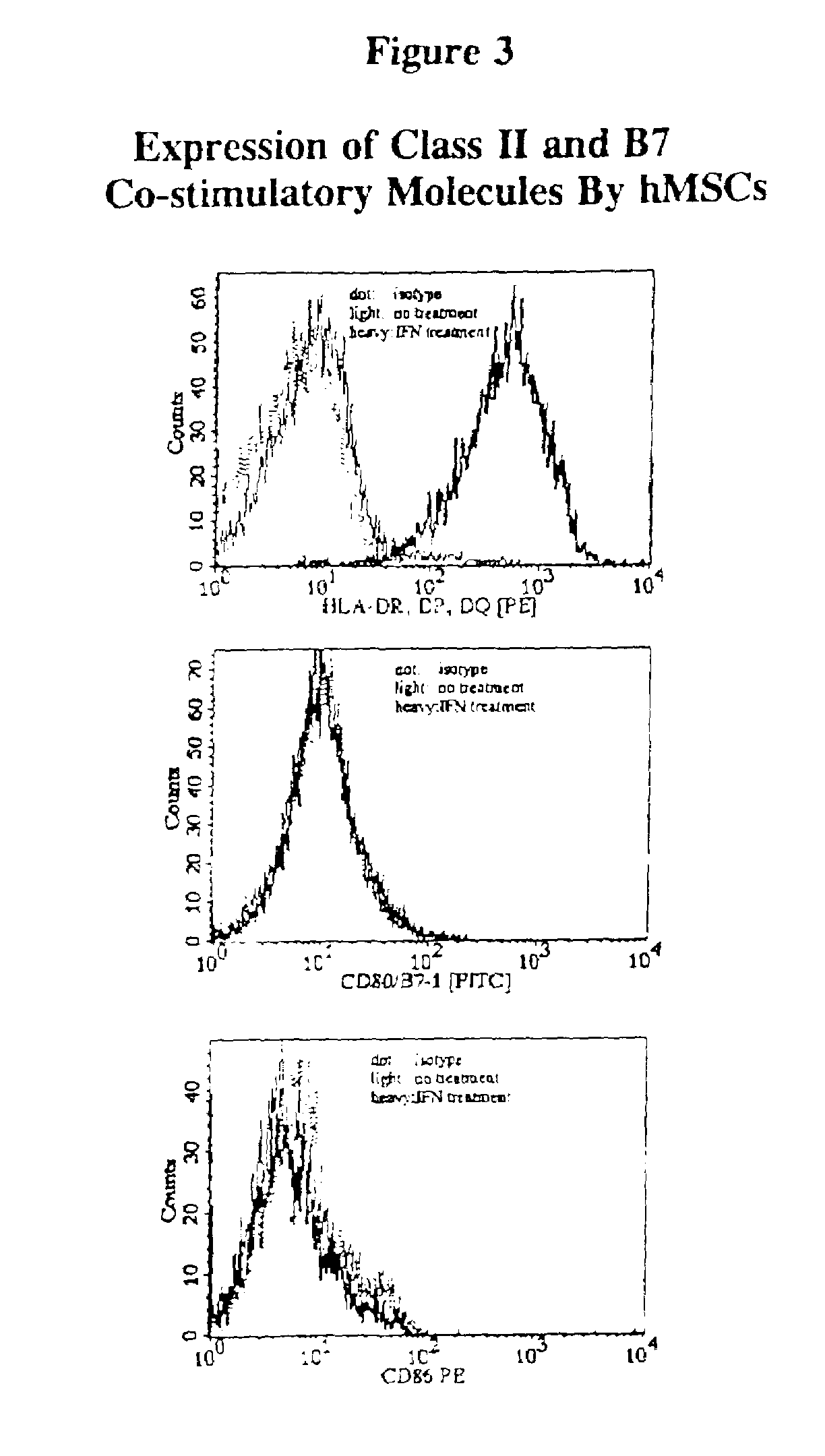
[0073]Human MSCs were evaluated for expression of MHC class II molecules and the co-stimulatory molecules, B7-1 and B7-2, to determine whether they were capable of antigen presentation resulting in activation of CD4+ T cells. Evaluation by immunofluorescence staining showed that hMSCs did not express any of these molecules on their surface; however, after induction with IFN-γ, hMSCs expressed cell surface class II molecules, but not B7-1 nor B7-2. hMSCs from two individuals were evaluated for their ability to present tetanus toxoid to autologous CD4+ T cell lines specific for this antigen. There was little or no stimulation of the T cell lines, as measured by lymphoproliferation, regardless of IFN-γ treatment. All of these results were confirmed by RT-PCR studies...
example 2
Expression of Class II MHC Molecules by Adipocytes
[0078]Post confluent hMSCs were differentiated with adipocyte differentiation medium (MDI+I) containing DMEM-High glucose / 10% FBS, insulin 10 μg / ml, antibiotic / antimycotic, dexamethasone 10−6 M, methylisobutylxanthine (MIX) 0.5 mM, and indomethacin 200 μM. The cells were treated with MDI+I for 3 days, and adipocyte maintenance medium (AM) containing DMEM-High glucose / 10% FBS, 10 μg / ml insulin and an antibiotic / antimycotic for 1 day (1 cycle) for 1 to 3 cycles. Control cells (undifferentiated MSCs) were treated with hMSC medium (described in U.S. Pat. No. 5,486,359) or AM. The cells were treated with 50 U / ml of IFN-γ for 3 days and Class II MHC expression was measured at different times during adipogenesis. The cells were trypsinized and incubated with anti-human DR, DP, DQ-FITC conjugated antibody or matching isotype, and the median fluorescence intensity was measured by FACS analysis. The value for isotype control is given in parent...
example 3
Antigen Presentation by Adipocytes
Materials and Methods.
[0081]hMSCs transduced with human B7-1 (Donor 128-B7-1) were differentiated in T175 flasks. After adipogenesis, the cells were trypsinized and counted. 1×10⁴ MSCs (undifferentiated treated with MSC medium) were plated into 96 well plates and then the T-cell proliferation assay was performed.
[0082]Tetanus toxoid (TT) (Accurate Chemical & Scientific Corp. Westbury, N.Y.) was added to final concentrations of 100, 20, 4 or μ / ml.
[0083]All cultures were washed 5× with hMSC medium (last time in T cell medium) and donor-matched TT-specific T cells were added. TT-specific T cells were thawed from liquid N2, washed 1× in hMSC medium and 1× in T cell medium (IMDM, 5% Human AB serum, non-essential amino acids, sodium pyruvate, beta mercaptoethanol and penicillin, streptomycin, amphotericin mix (Gibco BRL, Frederick, Md.). 4.5×10⁵T cells were added in 1.15 ml. Controls included Undifferentiated or adipo-MSCs alone, or +T cells, no Ag. PBMCs...
PUM
| Property | Measurement | Unit |
|---|---|---|
| concentration | aaaaa | aaaaa |
| concentration | aaaaa | aaaaa |
| interferon-γ | aaaaa | aaaaa |
Abstract
Description
Claims
Application Information