Water-fat separation for fast spin echo imaging in an inhomogeneous field with progressive encoding

a spin echo and water-fat technology, applied in the field of magnetic resonance imaging, can solve the problems of inability to achieve high levels of main field homogeneity, inability to discriminate between water and lipid signals, and inability to achieve long rf pulse lengths,

Active Publication Date: 2006-11-28
TOSHIBA AMERICA MRI
View PDF16 Cites 20 Cited by
  • Summary
  • Abstract
  • Description
  • Claims
  • Application Information

AI Technical Summary

Problems solved by technology

Such high levels of main field homogeneity are not always achievable over the entire field of view (FOV) being imaged.
Furthermore, at mid and low magnetic field strengths, the actual frequency difference between water and lipid signals is so small that impractically long RF pulse lengths are needed to discriminate between the water and lipid signals.
A difficulty with STIR is the loss of signals from tissues that have longitudinal relaxation times (T1) which are similar to the T1 of the attenuated lipids.
Although lipids have the fastest relaxations, the recovery curve after an inversion pre-pulse at an evolution time of about 100 to 150 ms removes significant amounts of signal from the water components which reduces the overall conspicuity of many features of clinical importance.
In the past, identifying and using the water-lipid phase difference to attenuate lipid signals has been problematic.

Method used


Image

  • Water-fat separation for fast spin echo imaging in an inhomogeneous field with progressive encoding
  • Water-fat separation for fast spin echo imaging in an inhomogeneous field with progressive encoding
  • Water-fat separation for fast spin echo imaging in an inhomogeneous field with progressive encoding

Examples


Embodiment Construction

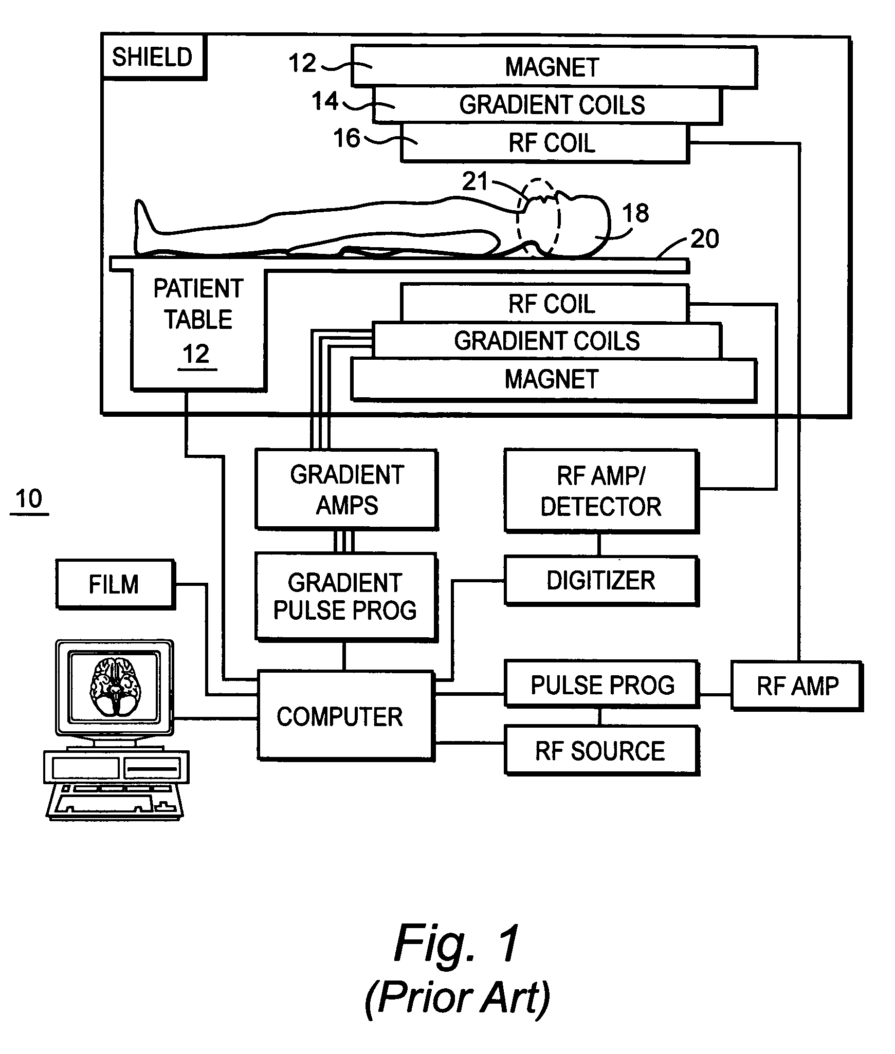
[0029]FIG. 1 shows a typical MR imaging system 10 including a magnet 12 to impose a static magnetic field (B0), gradient coils 14 for imposing spatially distributed gradient magnetic fields (Gx, Gy, and Gz) having gradients along three respective orthogonal coordinates, and RF coils 16 to transmit and receive RF signals to and from the selected nuclei of the body being imaged. The orthogonal gradients are applied to select a slice or other region of the body being imaged, to phase encode nuclei in the selected slice and to readout RF signals from the phase encoded nuclei. The patient 18 lies on a patient table 20 such that a portion of the patient to be imaged is in an “imaging volume”21 between the magnet and coils, which defines a field of view (FOV) of the MRI system.

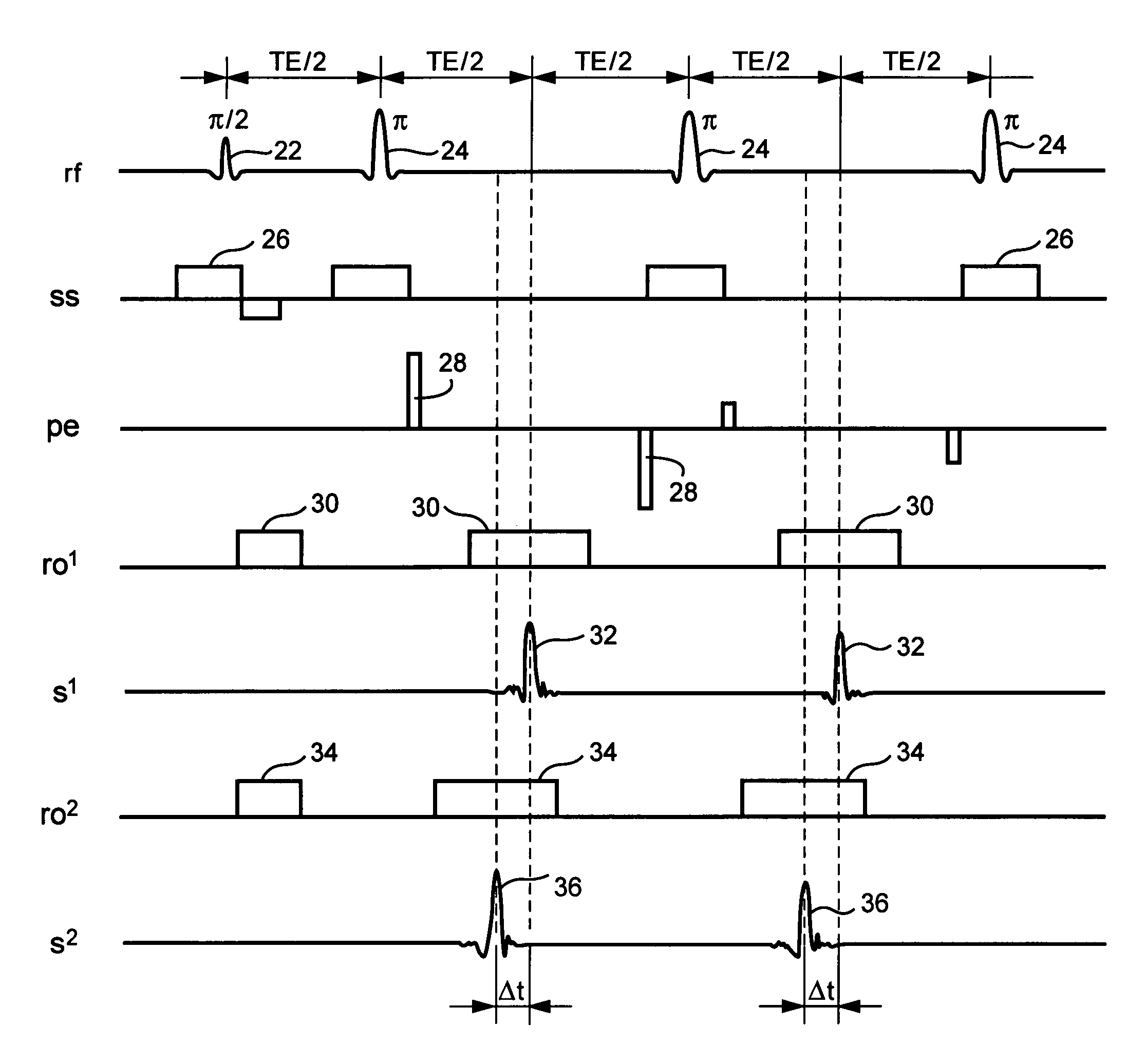
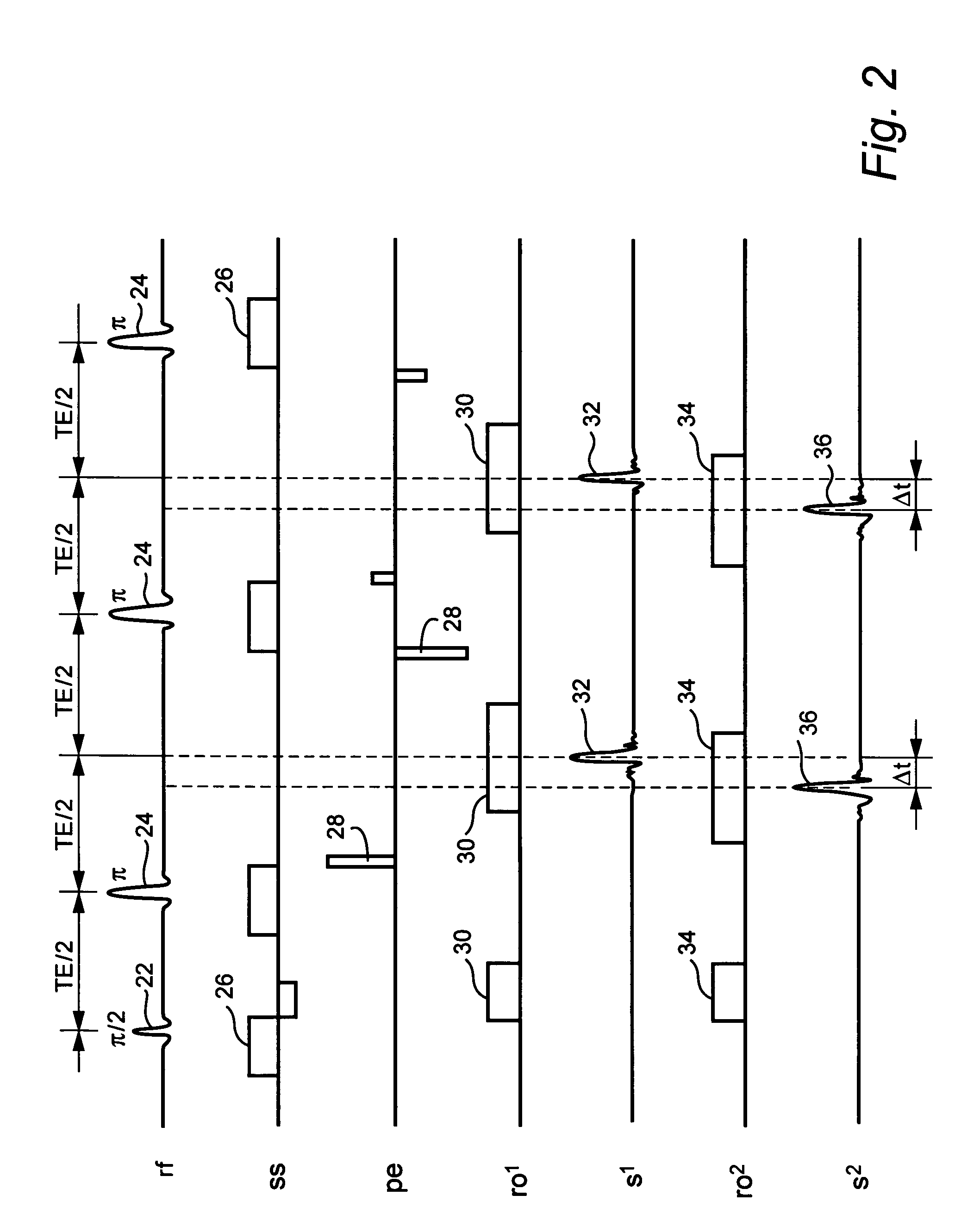
[0030]FIG. 2 shows a standard 2D fast spin echo (FSE) sequence (ro1, s1) and a shifted fast spin echo sequence (ro2, s2). Note that ro2 and s2 are shifted. As is conventional, a 90° (π / 2) initial phase RF pulse 22 is...


PUM


Abstract

A method to separate two chemically-shifted components of water (w) and fat (f) signals by: (i) estimating the field inhomogeneity β through a treatment of the residuals of the Moore-Penrose solution of the following equations: cos(αnβ)*w+cos [αn(1+β)]*f=real (In) and sin(αnβ)*w+sin [αn(1+β)]*f=imaginary (In), and (ii) using the estimated β to determine the resulting solution for w and f from the original input signals In.

Description

RELATED APPLICATION[0001]This application claims is a continuation of and claims priority of U.S. provisional application Ser. No. 60 / 520,321 filed on Nov. 17, 2003, the entirety of which is incorporated by reference.BACKGROUND OF THE INVENTION[0002]This invention generally relates to Magnetic Resonance Imaging (MRI) and, in particular, to a method and apparatus for water-fat image separation in MRI.[0003]Fast spin echo (FSE) imaging offers fast acquisition with long repetition times (TR). FSE can be used to improve contrast of long transverse relaxation time (T2) image components with respect to short transverse T2 image components. In clinical applications, where water signals are chiefly of interest, it is desirable to attenuate or eliminate MR signals from lipids (the terms “lipid” and “fat” are used interchangeably) which tend to reduce image contrast especially in the extremities and abdominal sections of a patient. The loss of water image contrast due to lipids is also exaspe...

Claims


Application Information

Patent Timeline
Patent Type & Authority Patents(United States)
IPC IPC(8): G01V3/00G01R33/48A61B5/055G01R33/54
CPCG01R33/4828
Inventor AVRAM, HECTOR C.SIMOVSKY, ILYA S.
Owner TOSHIBA AMERICA MRI
Why Eureka
Try Eureka