Method and system for the detection, comparison and volumetric quantification of pulmonary nodules on medical computed tomography scans

a computed tomography and volumetric technology, applied in the direction of material analysis using wave/particle radiation, instruments, nuclear engineering, etc., can solve the problems of time-consuming and tedious manual detection, time-consuming and human error-prone assessment of change on sequential chest cts, and none of the prior art registration approaches have compared two different ct studies of the chest to automatically, so as to reduce human error, reduce time spent reviewing images, and rapid and accurate quantitate tumor load

Inactive Publication Date: 2007-04-17
THE GENERAL HOSPITAL CORP
View PDF14 Cites 123 Cited by
  • Summary
  • Abstract
  • Description
  • Claims
  • Application Information

AI Technical Summary

Benefits of technology

[0030]In accordance with the present invention, a technique for the automatic detection of nodules within computed tomographic (CT) images of a lung includes processing a plurality of CT lung images, each of the images comprised of lung image data, taken from a patient in one CT study and relating the plurality of CT lung images taken from the patient in one CT study with a plurality of CT lung images taken from the same patient in another study. With this particular arrangement, a technique for automatically detecting nodules, quantitating their volume, and assessing for their change over time in sequential CT scans is provided. Such a technique is important for patient care and clinical productivity. In terms of patient care, such a system can be applied to two clinical scenarios in particular: 1) to quantitate tumor load and any change with treatment in oncology patients and 2) to screen high-risk populations for pulmonary nodules and lung cancer. With respect to clinical productivity, since the technique of the present invention can quantitate the number and volume of nodules and then calculate the overall tumor load in the lung parenchyma, the technique relieves the doctor from performing this task manually (i.e. by physically examining multiple CT images which is a very time consuming process subject to human error). Thus, the technique of the present invention can have significant impact on the treatment of oncology patients undergoing standard and experimental protocol treatments.
[0031]In accordance with a further aspect of the present invention, a system for automatically detecting nodules within computed tomographic (CT) images of a lung includes a CT image processor for processing a plurality of CT lung images, each of the CT lung images comprised of lung image data, taken from a patient in one CT study and a relationship processor for relating the plurality of CT lung images taken from the patient in one CT study with a plurality of CT lung im

Problems solved by technology

The manual detection, measurement of multiple pulmonary nodules, and assessment for change on sequential chest CTs can be time consuming and tedious.
Since such techniques are manual, however, they are time

Method used


Image

  • Method and system for the detection, comparison and volumetric quantification of pulmonary nodules on medical computed tomography scans
  • Method and system for the detection, comparison and volumetric quantification of pulmonary nodules on medical computed tomography scans
  • Method and system for the detection, comparison and volumetric quantification of pulmonary nodules on medical computed tomography scans

Examples


Embodiment Construction

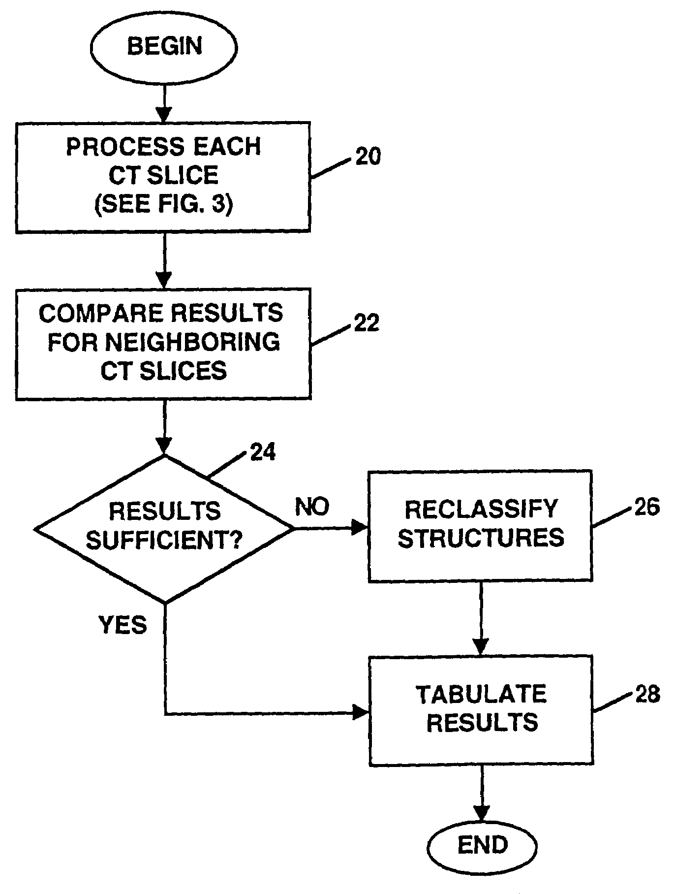
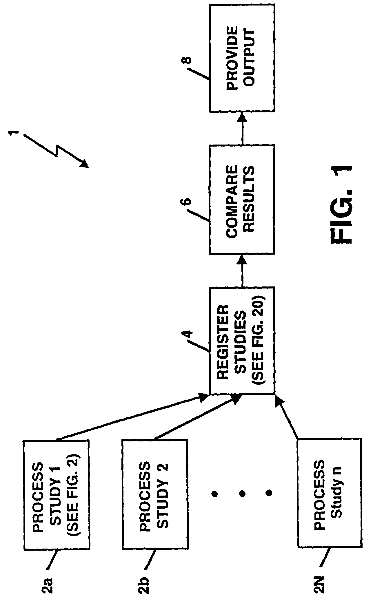
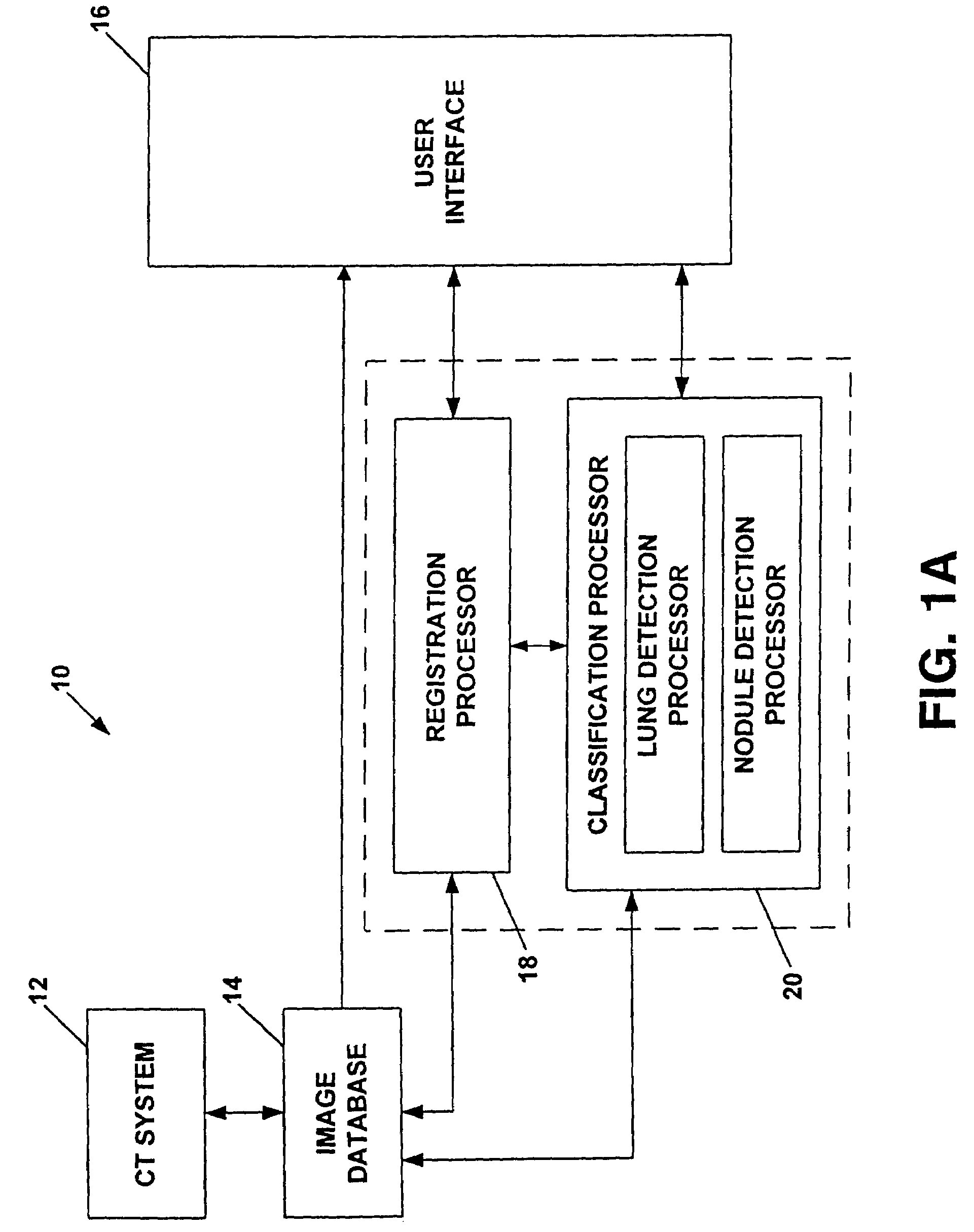
[0066]Before describing a system for the detection, comparison and volumetric quantification of pulmonary nodules on medical computed tomography scans and the techniques required to performed such operations some introductory concepts and terminology are explained.

[0067]A computed tomography (CT) system generates signals which can be stored as a matrix or array of digital values in a storage device of a computer or other digital processing device. Each of the numbers in the array correspond to a digital word (e.g. an eight-bit binary value) typically referred to as a “picture element” or a “pixel” or as “image data.” The array of digital values can be combined to display an image. Conversely, an image may be divided into a two dimensional array of pixels with each of the pixels represented by a digital word. Thus, a pixel represents a single sample which is located at specific spatial coordinates in the image.

[0068]The signals generated by the CT system are used to provide so-called...


PUM


Abstract

A method and system for the automated detection and analysis of pulmonary nodules in computed tomographic scans which uses data from computed tomography (CT) scans taken at different times is described. The method and system includes processing CT lung images to identify structures in the images for one patient in each of a plurality of different studies and then relating the plurality of CT lung images taken from the patient in one CT study with a plurality of CT lung images taken from the same patient in at least one other different study to detect changes in structures within the CT images.

Description

CROSS-REFERENCE TO RELATED APPLICATION[0001]This application is a continuation of U.S. patent application Ser. No. 09 / 812,021 filed on Mar. 19, 2001 now abandoned, which claims the benefit of U.S. Provisional Application No. 60 / 189,978 filed Mar. 17, 2000, under 35 U.S.C. §119(e) both of which are hereby incorporated herein by reference in their entirety.GOVERNMENT RIGHTS[0002]Not Applicable.FIELD OF THE INVENTION[0003]This invention relates generally to a method and system for automated processing of medical computer tomographic data and more particularly to an automated method and system for automated detection of pulmonary nodules and for volumetric assessment and quantification of nodule change over time in computed tomography (CT) images of the thorax.BACKGROUND OF THE INVENTION[0004]As is known in the art, each year a relatively large number of new cancer cases are diagnosed. Every one in four deaths in the United States is from cancer. The National Cancer Institute estimates ...

Claims


Application Information

Patent Timeline
IPC IPC(8): G06K9/36
CPCG06T7/0012
Inventor BETKE, MARGRITKO, JANE P.
Owner THE GENERAL HOSPITAL CORP
Why Eureka
Try Eureka