Method and apparatus for reducing x-ray dosage in CT imaging prescription

Active Publication Date: 2007-08-07
GENERAL ELECTRIC CO
View PDF20 Cites 32 Cited by
  • Summary
  • Abstract
  • Description
  • Claims
  • Application Information

AI Technical Summary

Benefits of technology

[0010]In accordance with a further aspect of the present invention, an electronic network includes at least one updatable database configured to store scan parameter values from one or more imaging sessions and at least one imager configured to acquire imaging data of a subject. The network further includes an ele

Problems solved by technology

Notwithstanding these highly sophisticated and developed protocols, it has been difficult to develop a protocol that provides the optimum scan parameters for each possible imaging condition.
As a result, it is difficult for imaging personnel to prescribe imaging sessions consistently or prescribe sessions similar to that prescribed by other system operators.

Method used


Image

  • Method and apparatus for reducing x-ray dosage in CT imaging prescription
  • Method and apparatus for reducing x-ray dosage in CT imaging prescription
  • Method and apparatus for reducing x-ray dosage in CT imaging prescription

Examples


Embodiment Construction

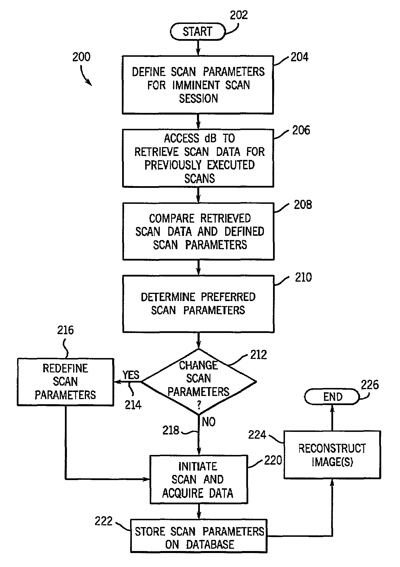
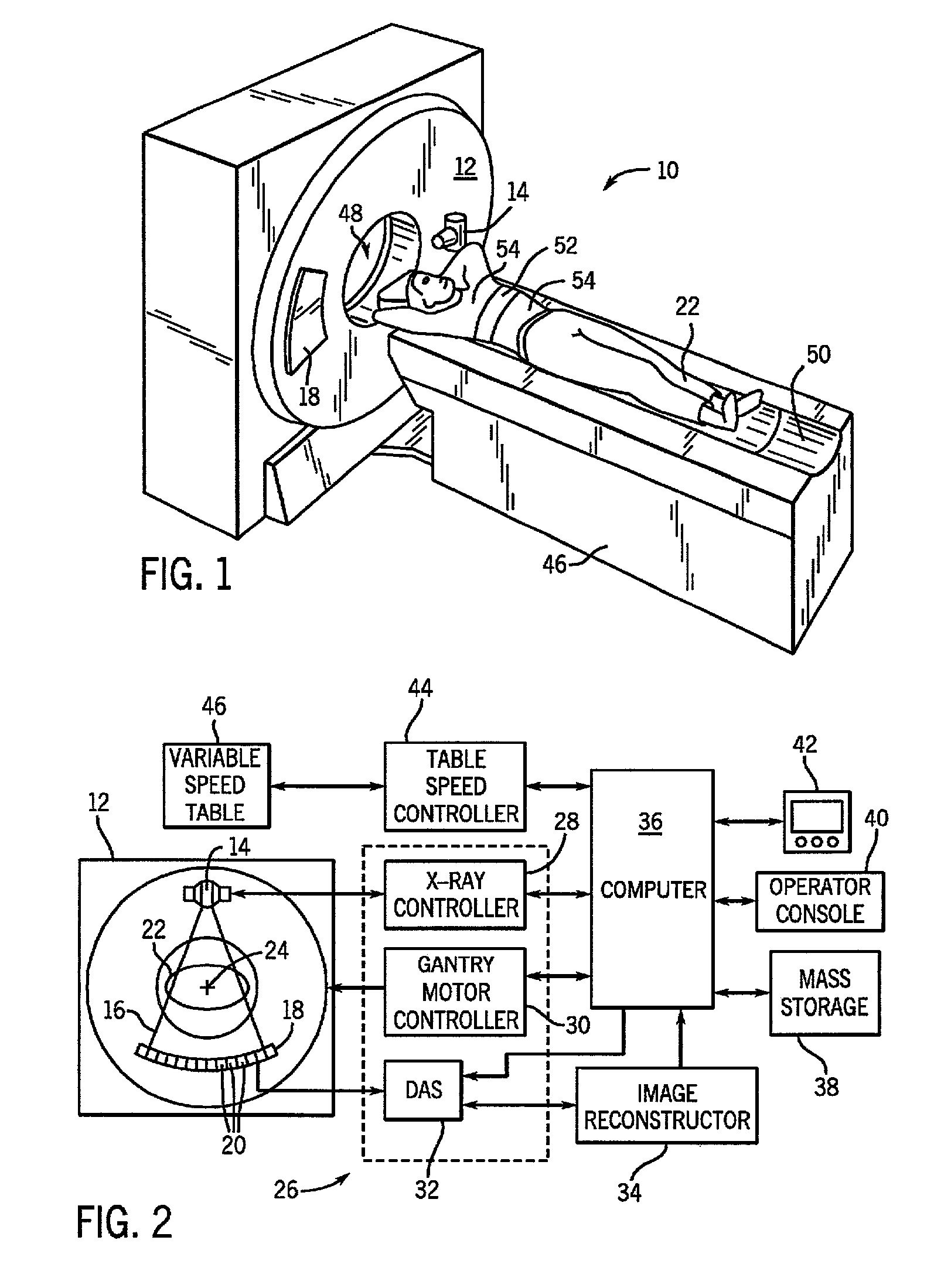
[0025]Referring to FIGS. 1 and 2, a “third generationCT imaging system 10 is shown as including a gantry 12. The present invention, however, is applicable with other CT systems as well as other imaging modalities such as x-ray and magnetic resonance. Gantry 12 has an x-ray source 14 that projects a beam of x-rays 16 through filter 15 toward a detector array 18 on the opposite side of the gantry 12. Detector array 18 is formed by a plurality of detectors 20 which together sense the projected x-rays that pass through a medical patient 22. Each detector 20 produces an electrical signal that represents the intensity of an impinging x-ray beam and hence the attenuated beam as it passes through the patient 22. During a scan to acquire x-ray projection data, gantry 12 and the components mounted thereon rotate about a center of rotation 24.

[0026]Rotation of gantry 12 and the operation of x-ray source 14 are governed by a control mechanism 26 of CT system 10. Control mechanism 26 includes ...


PUM


Abstract

The present invention is directed to a networked scanner environment wherein each scanner is communicatable with one or more databases configured to store data associated with previously executed imaging sessions. The one or more databases may be queried by a user to determine, based on a set of user inputs, an historical evaluation of the prior imaging sessions conducted in accordance with scan parameters similar to the scan parameters of an imminent imaging session. The present invention includes a global database that is accessible by a series of remotely located imaging systems as well as includes one or more databases particular to a specific treatment facility housing one or more imaging scanners. The present invention is also applicable with a stand-alone imaging system having a database local to that particular imaging system for storing and accessing data associated with imaging sessions conducted on that particular imaging system.

Description

BACKGROUND OF INVENTION[0001]The present invention relates generally to prescribing diagnostic imaging sessions and, more particularly, to a method and apparatus for prescribing an imaging session based on scan parameters of previously executed imaging sessions.[0002]Increasingly, diagnostic imaging systems are being used to non-invasively acquire diagnostic data of a subject such as a medical patient. For example, computer tomography, ultra-sound, and magnetic resonance imaging systems are increasingly being used to acquire medical diagnostic data to enable medical personnel to diagnose a number of medical conditions. Furthermore, x-ray and other systems are increasingly being used by airport security personnel to inspect luggage and other packages for contraband, guns, knives, explosives, etc.[0003]A number of protocols have been developed to assist personnel in prescribing an imaging session depending upon the diagnostic data sought and the modality of the imaging system being us...

Claims


Application Information

Patent Timeline
IPC IPC(8): G06F15/177G01R33/28A61B5/055A61B6/03G01R33/54G06Q50/00G06T1/00
CPCA61B6/032A61B6/4488A61B6/542A61B6/545A61B6/56G01R33/54Y10S707/99936Y10S707/99935
Inventor TOTH, THOMAS L.
Owner GENERAL ELECTRIC CO
Why Eureka
Try Eureka