Epicardial lead placement for bi-ventricular pacing using thoracoscopic approach

Inactive Publication Date: 2007-09-18
MEDTRONIC INC
View PDF32 Cites 139 Cited by
  • Summary
  • Abstract
  • Description
  • Claims
  • Application Information

AI Technical Summary

Benefits of technology

[0016]Once the lead has been anchored in place by the attachment member, the thoracoscope and introducer are removed and tests done to confirm the

Problems solved by technology

Despite considerable progress in the management of CHF, it remains a major health problem worldwide.
Despite significant advances in the treatment of CHF using various pharmacological therapies, the quality-of-life in patients with CHF remains poor as these patients are frequently hospitalized, and heart failure in these patients is a common cause of death.
In addition, there are significant long-term care costs associated with this problem.
The presence of LBBB delays left ventricular ejection due to delayed left ventricular activation.
Since the lead is advanced through the coronary sinus, the potential placement positions for the lead are severely limited.
Although bi-ventricular pacing has shown beneficial results, numerous problems are associated with this technique.
One such problem is the amount of time required for a physician to insert the lead into the desired location on the left ventricle.
Further, the placement of the lead on the left ventricle is limited to sites that provide adequate pacing and sensing signals.
Further, cannulation of the coronary sinus is often difficult due to either the rotation of the heart or the presence of an enlarged right atrium or Thebesian valve.
Further, the placement of a lead on the surface of the left ventricle though the coronary sinus cannot be carried out in some patients with prior bypass surgery or with coronary sinus stenosis.

Method used


Image

  • Epicardial lead placement for bi-ventricular pacing using thoracoscopic approach
  • Epicardial lead placement for bi-ventricular pacing using thoracoscopic approach
  • Epicardial lead placement for bi-ventricular pacing using thoracoscopic approach

Examples


Embodiment Construction

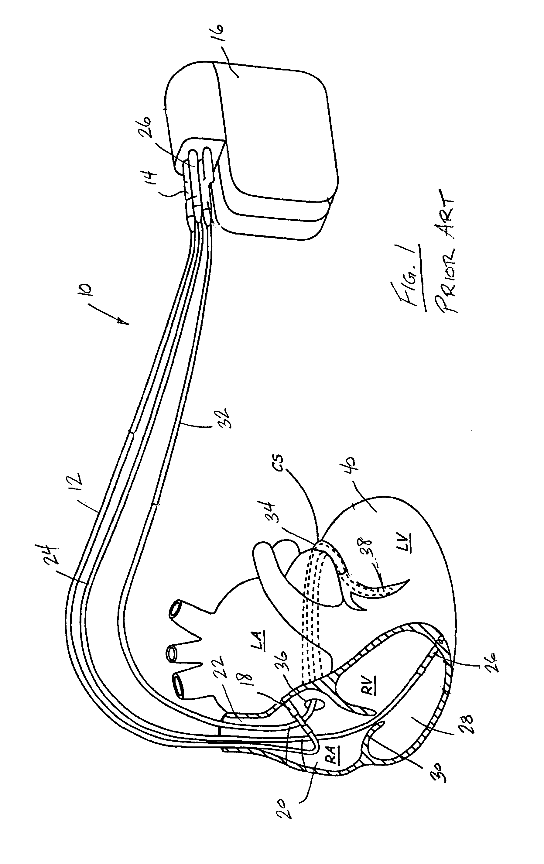
[0030]Referring first to FIG. 1, thereshown is a schematic illustration of a prior art bi-ventricular pacing method and apparatus 10 currently used. The bi-ventricular pacing system 10 shown in FIG. 1 utilizes a totally transvenous lead system. Specifically, a right atrial lead 12 has one end 14 connected to a pacemaker 16 and a distal end including an electrode 18 in contact with the inner wall of the right atrium 20. As illustrated in FIG. 1, the right atrial lead 12 is fed into the heart through the superior vena cava 22.

[0031]A second, right ventricular lead 24 is coupled at its end 26 to the pacemaker 16 and its distal end, including a electrode 26 is placed in contact with the inner wall of the right ventricle 28. The right ventricular lead 24 also passes through the vena cava 22 and passes through the tricuspid valve 30. The third lead 32 of the pacing system 10 passes through the vena cava 22 and enters into the coronary sinus 34 through the opening 36 in the right atrium 20...


PUM


Abstract

A method and apparatus for placing an epicardial lead over a desired, predetermined location on the left ventricle using a minimally invasive approach. A thoracoscope having a handle portion and a probe tube defining a central opening is placed through an incision in the patient generally aligned with the desired position for the pacing lead. An introducer and pacing lead are placed within the central opening of the thoracoscope and moved into contact with the pericardium at the desired lead location. An attachment member of the pacing lead attaches an electrode of the pacing lead to the pericardium. The pacing lead includes a mesh disk surrounding the electrode to aid in long term attachment of the electrode to the heart. Alternatively, the pericardium can be incised such that the lead is placed directly over the epicardial surface of the left ventricle.

Description

CROSS REFERENCE TO RELATED APPLICATION[0001]The present invention is based on and claims priority to U.S. Provisional Patent Application Ser. No. 60 / 364,311 filed on Mar. 14, 2002.FIELD OF THE INVENTION[0002]The present invention, called “stingray technique”, generally relates to a technique of inserting an epicardial lead in a patient with congestive heart failure (CHF) requiring bi-ventricular pacing. More specifically, the present invention is related to a thoracoscope design and technique of inserting a left ventricular lead using an thoracoscopic approach that places the lead at a specific, known location on the epicardial surface of the left ventricle.BACKGROUND OF THE INVENTION[0003]Despite considerable progress in the management of CHF, it remains a major health problem worldwide. It is estimated that there are 6-7 million people with CHF in the United States and Europe alone and approximately 1 million new patients are diagnosed with CHF every year.[0004]Despite significant...

Claims


Application Information

Patent Timeline
IPC IPC(8): A61B17/94A61N1/05
CPCA61N1/0587
Inventor SRA, JASBIR S.
Owner MEDTRONIC INC
Why Eureka
Try Eureka