
Illuminated retractor for use in connection with harvesting a blood vessel from the arm

a retractor and arm technology, applied in the field of medical devices, can solve the problems of inhibiting the ability of life-sustaining blood supply to the patient's heart, coronary bypass surgery is not warranted, and coronary bypass surgery is required, so as to facilitate safe, reliable and expeditious harvesting of the vessel

Inactive Publication Date: 2007-12-25
TELEFLEX MEDICAL INC
View PDF3 Cites 7 Cited by
  • Summary
  • Abstract
  • Description
  • Claims
  • Application Information

AI Technical Summary

Benefits of technology

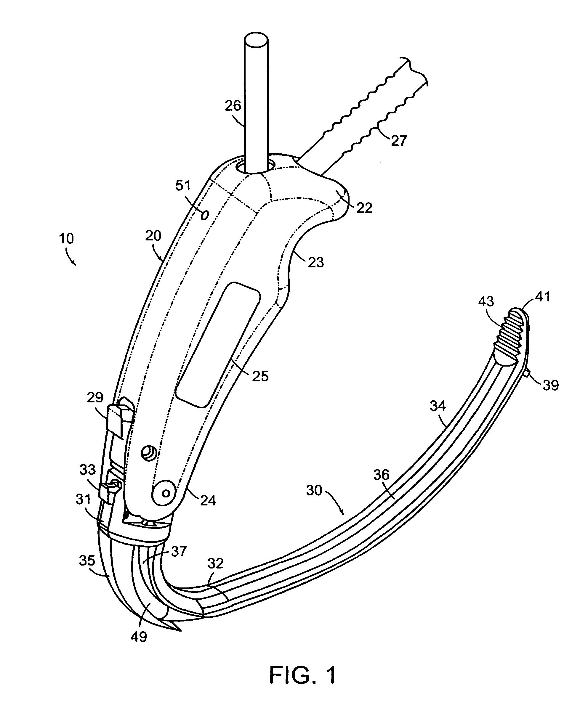
[0029]The present invention provides a surgical retractor that meets this, and other needs. As shown in the drawings, the present invention provides a retractor for defining, accessing and illuminating a subcutaneous working space near a blood vessel (e.g., the radial artery, the basilic vein) located within a patient's arm in order to facilitate safe, reliable and expeditious harvesting of the vessel.

Problems solved by technology

Once transplanted, the removed section (or entirety) of the vein / artery functions as a graft that replaces both the coronary arteries, which, as a result of aging and / or disease, have become blocked by plaque deposits, stenosis, or cholesterol, thus severely inhibiting their ability to supply life-sustaining blood to the patient's heart.
In some instances, these blockages can be treated with angioplasty, atherectomy or stent placement, and, therefore, coronary bypass surgery is not warranted.
Quite often, however, a coronary bypass is required because these treatment methods are either contraindicated, or have proven incapable of removing blockages from coronary arteries.
Making this “fillet-like” incision inherently presents a serious risk of injury to the medial lymph bundle and / or to nerves located within the leg, as well as a realistic risk of infection to the incision site.
Moreover, the healing process associated with this long, “fillet-like” incision is protracted (often more prolonged than the incision(s) to the patient's chest in furtherance of the coronary bypass) and very painful, especially if the patient has circulation problems in his / her extremities.
Ultimately, in fact, the incision often does not heal properly, thus requiring medical attention and / or invasive procedures (e.g., corrective surgery).
Unfortunately, despite this advancement, many patients are not ideal candidates for saphenous vein harvesting by virtue of having an unacceptably high risk of minor to extensive complications associated with even these smaller leg incisions.
Just as making a “fillet-like” incision causes significant risks in the context of saphenous vein harvesting, making an approximately 23 cm (i.e., 9 inch) continuous cut into a patient's arm to harvest the radial artery also presents serious risks, such as severing the delicate superficial radiac and / or lateral antebrachial cutaneous nerves.
There are several notable drawbacks associated with using endoscopic technology for visualization of the subcutaneous space in connection with radial artery harvesting.
First, the endoscope (or the display unit that depicts its image) provides a distorted visual perspective, which is a poor substitute for actual visualization of the surgical field by the naked eye.
Second, compounding the first drawback, use of an endoscope provides for visualization of only a small portion of the subcutaneous space, namely, the area that is immediately in front of the endoscope.
Third, illumination within the subcutaneous space created by this type of endoscope is also limited to the light emitted directly at the distal portion of the endoscope.
Because of these disadvantages, there is a significant learning curve to safely and efficiently practice this radial artery harvesting procedure.
Other drawbacks also are caused when attempting to use conventional equipment in connection with the skin bridging radial artery harvesting procedure.
For example, because the skin bridges between the transverse incisions are quite long, it is difficult to sufficiently illuminate the subcutaneous space, especially when conventional, non-illuminated retractors are implemented / utilized.
As such, the subcutaneous space within a patient's arm is likely comparatively more crowded with instruments during the harvesting process, thus complicating the process and further prolonging the learning curve for safely and efficiently practicing radial artery harvesting.

Method used


Image

  • Illuminated retractor for use in connection with harvesting a blood vessel from the arm
  • Illuminated retractor for use in connection with harvesting a blood vessel from the arm
  • Illuminated retractor for use in connection with harvesting a blood vessel from the arm

Examples


Embodiment Construction

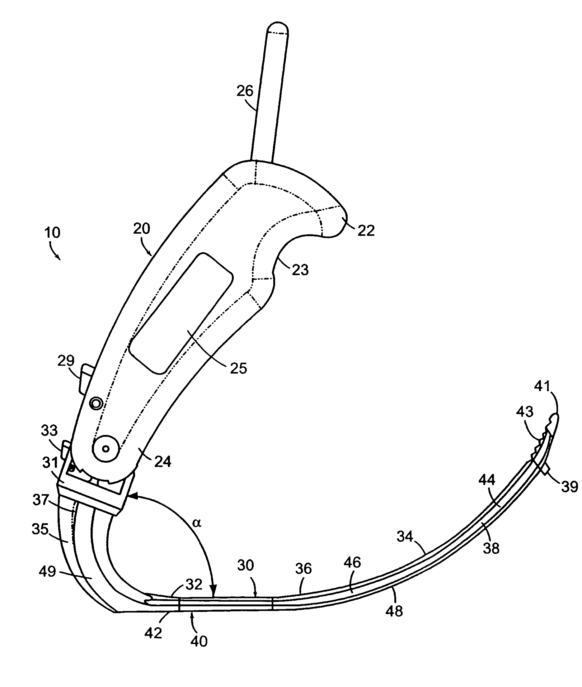
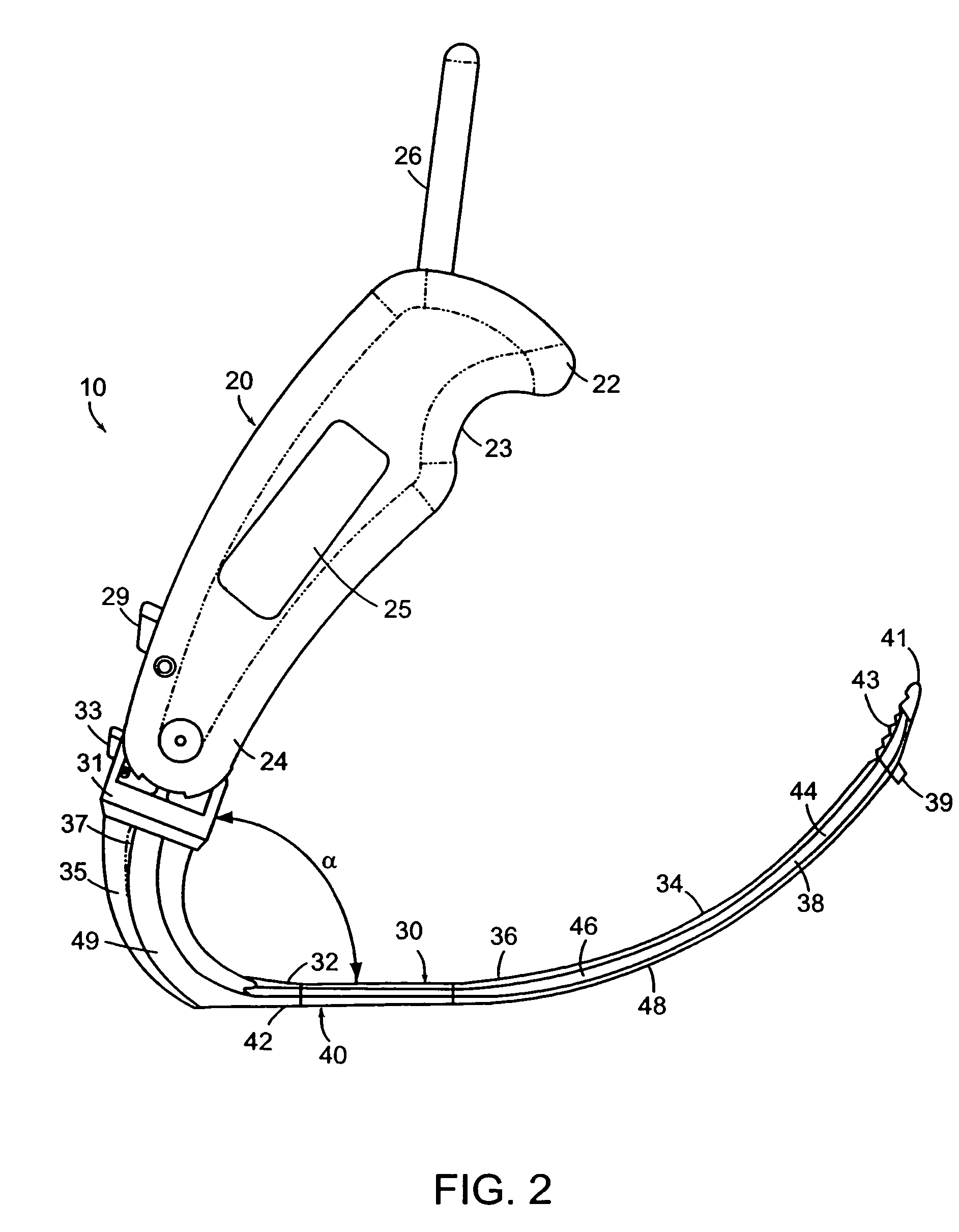
[0070]The present invention provides an illuminated retractor for assisting in accessing, defining and illuminating the subcutaneous space within a patient's arm in which a blood vessel (e.g., the radial artery, the basilic vein) is located, thus allowing for improved visualization of this space, and, in turn, facilitating a procedure wherein the vessel is harvested.

[0071]As shown in the drawings, the present invention provides an illuminated surgical retractor 10 having a handle member 20, a first elongate section 30, a second elongate section 40, and a twist connector 31.

[0072]The handle member 20 is an elongate and generally cylindrical member that has a first top handle member end portion 22 and a second bottom handle member end portion 24. The second handle member end portion 24 of the handle member 20 is connected to the first elongate section 30 of the retractor 10 at the shaft portion 35 distal to the distal end portion 32 of the first elongate section 30. This connection is...


PUM


Abstract

An illuminated surgical retractor for defining and illuminating a subcutaneous surgical field in the space near a vessel (such as the radial artery or the basilic vein) during a procedure for harvesting the vessel, wherein the illuminated surgical retractor includes a handle member pivotally connected at an acute angle to a first elongate section that has a non-liner shape, and includes a second elongate section releasably connected to the first elongate section, wherein a portion of the second elongate section defines an illumination input end portion, which is optically coupled to a light source to substantially illuminate the second elongate section, and further including an insertion area positioned on the proximal end portion of the first elongate section to allow the second elongate section to be inserted into the first elongate section.

Description

[0001]This application is a continuation of application Ser. No. 10 / 055,778 now U.S. Pat. No. 6,817,978, filed on Jan. 23, 2002.BACKGROUND OF THE INVENTION[0002]1. Field of the Invention[0003]The present invention relates to medical devices, and, in particular, to a retractor for harvesting a blood vessel, which, in turn, is used in connection with an ongoing or subsequent surgical procedure. More particularly, the present invention provides such a retractor that is capable of defining and illuminating a subcutaneous working space to ameliorate accessibility to, and to facilitate visualization and harvesting of one or more blood vessels (e.g., radial artery, basilic vein) for grafting / transplantation in connection with a surgical procedure (e.g., coronary bypass surgery).[0004]2. Description of Related Art[0005]In certain surgical procedures, it is necessary to remove a portion (or even the entirety) of a patient's blood vessel for use in another, often remotely located part of that...

Claims


Application Information

Patent Timeline
IPC IPC(8): A61B1/32A61B17/00A61B17/02A61B17/32A61B19/00
CPCA61B17/00008A61B17/02A61B1/32A61B2017/0046A61B1/07A61B2017/00969A61B2017/320044A61B2019/5206A61B2017/00469A61B2090/306
Inventor HOLLAND, DONNA D.POND, JR., JOHN D.EVANS, DOUGLAS G.
Owner TELEFLEX MEDICAL INC
Learn More