
Gene sets for glioma classification

a gene set and glioma technology, applied in the field of molecular biology and oncology, can solve the problems of small sample size, inability to apply the gene-based classification to gliomas, and limited number of human tissues for study, and the cost of such gene expression profiling projects

Inactive Publication Date: 2008-03-11
BOARD OF RGT THE UNIV OF TEXAS SYST +1
View PDF4 Cites 18 Cited by
  • Summary
  • Abstract
  • Description
  • Claims
  • Application Information

AI Technical Summary

Benefits of technology

[0020]In a further particular embodiment, the present invention provides a method of classifying a glioma comprising (a) obtaining a tumor sample; (b) obtaining from cells of the tumor sample information on the expression of one or more of the following gene combinations: Follitropin receptor and Thymosin β10; Cyclin D3 and SMARCA4; MRP and HSP70.1; MUC18, Clusterin and TNFSF5; MUC18, Transducin β1 and GRB2; MUC18, Transducin β1 and RXR-β; MUC18, Transducin β1 and Clusterin; MUC18, BCL-W and GRB2; MUC18, Transducin α1 and α1 catenin; MAP kinase 10, SMARCA4 and Neuronal aceytlcholine receptor α3; MUC18, GRB2 and Erythropoietin receptor; Clusterin, ISGF3γ and Erythropoietin receptor; TIE-2 and TNFSF5; IGFBP2 and EPHA1; IGFBP2 and CDK7; IGFBP2 and TNFSF5; Myc, IGFBP2 and CDK7; IGFBP2, TNSFS5 and CC chemokine receptor type 2; TIE-2, CXC chemokine receptor type 4 and EPHA1; TIE-2, CDK7 and JAK3; TIE-2, IGFBP2 and JAK3; TNFSF5 and DNase X; Prostaglandin E2 receptor E4 and DNase X; Prostaglandin E2 receptor E4 and TNFSF5; GNA13 and TNFSF5; ErbB4 and Prostaglandin E2 receptor E4; PKA C-α, TNFSF5 and Preprotachykinin β; DNase X, RκB DNA-binding and Preprotachykinin β; PKA C-α, DNA ligase IV and TNFSF5; DNA ligase IV, TNFSF5 and Acidic fibroblast growth factor; CREB1 and BCL2A1; CREB1, BCL2A1 and CD11B; CREB1, VEGFR2 and Thymosin β10; CREB1, VEGFR2 and CD11B antigen; CREB1, cyclin E and Thymosin β10; CREB1, BCL2A1 and Ednothel...

Problems solved by technology

The results are subjective and there can be disagreements among physicians as to what kind of tumor cell is involved.
To date, a successful application of gene-based classification has not been applied to gliomas.
Thus, the issue is how to best identify the “strong” feature genes that are closely linked to specific phenotypes from among the thousands of genes in gene expression profiles, and whether this information really aids classification of tumors more.
There are many technical challenges in the path to accomplishing the task of finding the key links.
However, a major roadblock is the small sample size issue inherent to microarray-based classification efforts (Dougherty, 2001).
Contributing to this are the limited numbers of human tissues for study and the cost of such gene expression profiling projects.

Method used


Image

  • Gene sets for glioma classification
  • Gene sets for glioma classification
  • Gene sets for glioma classification

Examples


example 1

Materials and Methods

[0231]Primary glioma tissues. All primary GM tissues were acquired from the Brain Tumor Center tissue bank of The University of Texas M. D. Anderson Cancer Center. The tumors were diagnosed according to two classifications: St. Anne-Mayo (Daumas-Duport et al., 1988) and the World Health Organization (Kleihues et al., 2000). Herein, gliomas are termed according to the St. Anne-Mayo nomenclature: low-grade OL, AO, AA, and GM. Tissue bank specimens were quick-frozen shortly after surgical removal and stored at −80° C. Hematoxylin-eosin (H&E)-stained frozen tissue sections are routinely prepared from all tissue bank specimens for screening. All tissue specimens for CDNA array analysis were screened by a neuropathologist (G.N.F.), and the diagnoses were independently confirmed by a second neuropathologist. The glioma tissue blocks were specifically selected for densest and purest tumor. All were comparatively and uniformly “pure” with minimal contamination by normal ...

example 2

Preliminary Results

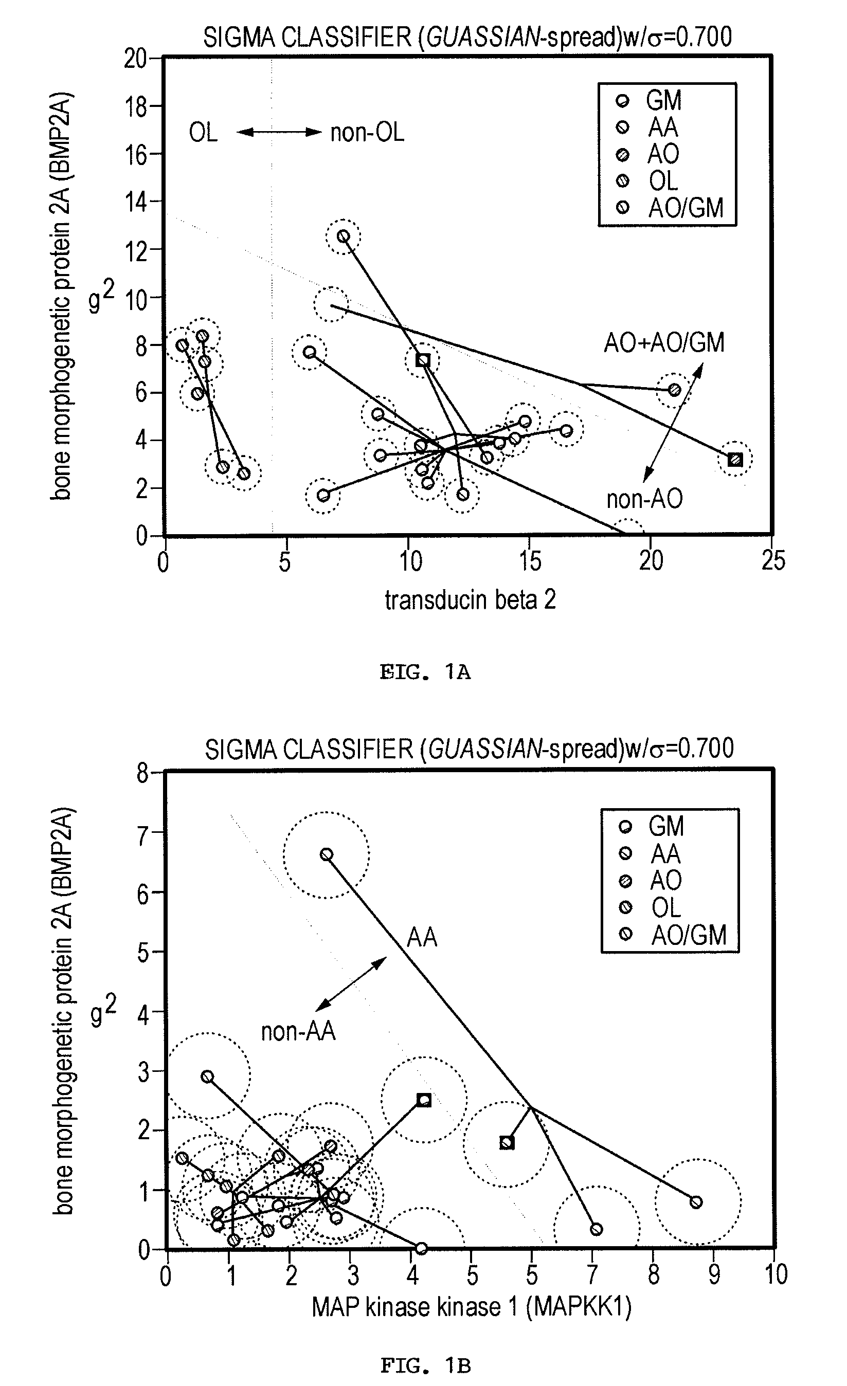
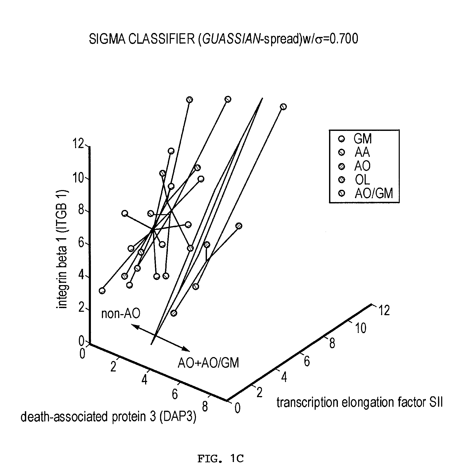
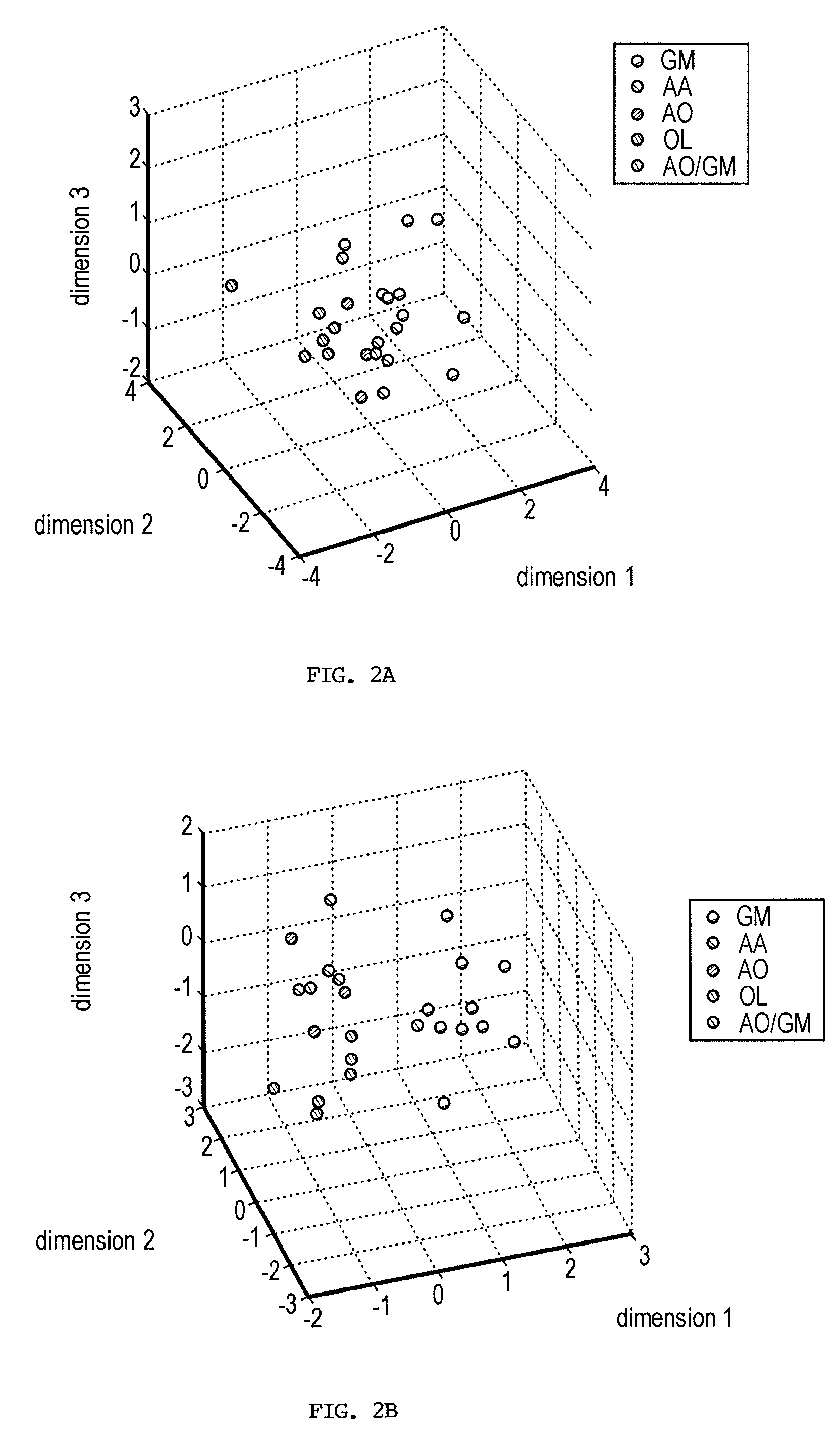
[0239]Current glioma classifications are based on morphologic feature assessment and remain subjective and problematic. For example, among the 25 glioma cases in this study, two cases could be classified as either AO or GM depending on the morphologic criteria used, and are thus designated AO / GM. A primary goal of functional genomics is to identify key genes and gene combinations, small in number, that are potentially useful for diagnosis or as candidate therapeutic targets (Golub et al., 1999; Alizadeh et al., 2000; Bittner et al., 2000; Hedenfalk et al., 2001; Perou et al., 2000; Ben-Dor et al., 2000; Khan et al., 2001). A new algorithm (Kim et al., 2002) designed to find robust gene sets from small samples has been applied to the gene expression profile data derived from 25 human glioma surgical specimens. This algorithm builds classifiers from a probability distribution resulting from spreading the mass of the sample points to make the classification more diff...

example 3

Discussion

[0255]Gliomas are complex cancers whose classification relies upon morphologically based tumor classification schemes that are frequently subjective. Gene expression profiling provides a promising objective approach to method to classify such cancers. Thus, the issue remains as to how to identify the relevant genes that are closely linked to specific disease phenotypes.

[0256]The inventors used a novel method to find both strong classifiers and strong features. This method is briefly described in the Materials and Methods section and is explicated in more detail in (Kim et al., 2002). This algorithm considers the inherently variable or “high-noise” nature of microarray measurements and mimics this fuzziness by adding noise to the sample sets. The basic idea is that if the data are deliberately made “worse” and classifier genes can still be identified, then these genes are very likely to be robust.

[0257]Using this algorithm, the inventors identified robust classifier gene se...


PUM

PropertyMeasurementUnit
Volumeaaaaaaaaaa
Fractionaaaaaaaaaa
Fractionaaaaaaaaaa

Abstract

The present invention provides a number of gene markers whose expression is altered in various gliomas. In particular, by examining the expression these markers, one can accurately classify a glioma as glioblastoma multiforme (GM), anaplastic astrocytoma (AA), anaplastic oligodendroglioma (AO) or oligodendroglioma (OL). The diagnosis may be performed on nucleic acids, for example, using a DNA microarray, or on protein, for example, using immunologic means. Also disclosed are methods of therapy.

Description

[0001]The present application claims priority to now abandoned U.S. Provisional Patent Application Ser. No. 60 / 364,608 filed on Mar. 15, 2002. The entire text of the above-referenced disclosure is specifically incorporated herein by reference without disclaimer.BACKGROUND OF THE INVENTION[0002]1. Field of the Invention[0003]The present invention relates generally to the fields of molecular biology and oncology. More particularly, it concerns the classification of gliomas based on the expression of various proteins identified as relevant to various glioma states.[0004]2. Description of Related Art[0005]Gliomas are complex cancers with different growth characteristics and involves different types of cells. Because the original clone of tumor cells may exist at any stage during the cell differentiation, the boundaries between cell lineages can be blurred. The current morphologically-based tumor classifications often mix cell lineage features with tumor growth characteristics. The resul...

Claims


Application Information

Patent Timeline
IPC IPC(8): C40B30/00C12NC12P19/34C12Q1/68G01N33/574
CPCC12Q1/6883G01N33/57407C12Q2600/158C12Q2600/118C12Q2600/112
Inventor ZHANG, WEIFULLER, GREGDOUGHERTY, EDHESS, KENNETH
Owner BOARD OF RGT THE UNIV OF TEXAS SYST
Learn More