Construction method and application of glioblastoma organ model

A technology of glioblastoma and organoids, which is applied in the field of construction of glioblastoma organoid models, which can solve the problems of invasion and so on

Active Publication Date: 2019-11-22
XIANGYA HOSPITAL CENT SOUTH UNIV
View PDF7 Cites 5 Cited by
  • Summary
  • Abstract
  • Description
  • Claims
  • Application Information

AI Technical Summary

Problems solved by technology

However, there is still a certain gap between this culture method and the characteristics of clinical tumor growth. Clinically, tumor gr

Method used


Image

  • Construction method and application of glioblastoma organ model
  • Construction method and application of glioblastoma organ model
  • Construction method and application of glioblastoma organ model

Examples


Embodiment 1

[0033] Embodiment 1, the culture of cerebral cortex organoid

[0034] Refer to "Generation of cerebral organoids from human pluripotent stemcells" (Lancaster MA, Knoblich JA. Nature protocols 2014; 9:2329-2340.) to culture cerebral cortical organoids.

[0035] Human induced pluripotent stem cells were purchased from System Biosciences (product number: SC101A-1). The culture process is as follows: human induced pluripotent stem cells were recovered and cultured, and the feeder layer cells were radiation-inactivated mouse fibroblasts, which were purchased from MTI-GlobalStem. First, resuscitate the human induced pluripotent stem cells in the liquid nitrogen tank, preheat the fresh medium in a 37°C constant temperature water tank in advance, and prepare sterile straws / centrifuge tubes / culture bottles. After the fresh medium returns to temperature, spray 70% alcohol and wipe it clean, and immediately move it into the sterile operating table. The principle of resuscitating cells ...

Embodiment 2

[0037] Example 2. Morphological Observation of Cerebral Cortical Organoids at Different Developmental Stages During Culture

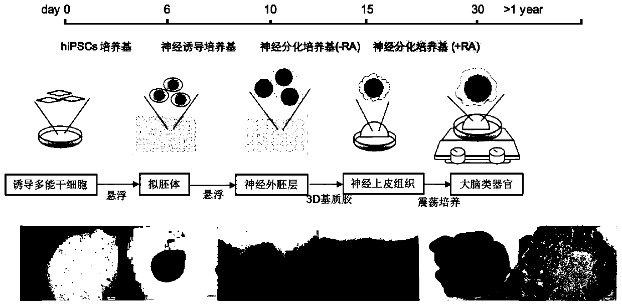
[0038] Cerebral cortical organoids have different characteristics in each differentiation process, and need to be observed regularly and ready for the next step at any time. Human induced pluripotent stem cells (hiPSCs) in the logarithmic growth phase were seeded on culture dishes pretreated with irradiation-inactivated mouse fibroblasts (iMEFs), and hiPSCs spontaneously formed after 6 days of low-adhesion suspension culture in whole stem cell medium Cell clusters of uniform size, that is, embryoid bodies. At this time, the tissue has three layers of germ layers: inner, middle, and outer; use neural induction medium to make it oriented to differentiate into neuroectoderm, and gradually differentiate into translucent on the 10th day. A round cell mass with a darker center and clear and smooth edges, indicating the formation of neuroectoderm; cultured in ...

Embodiment 3

[0040] Example 3. Staining and Identification of Cultured Cerebral Cortical Organoids

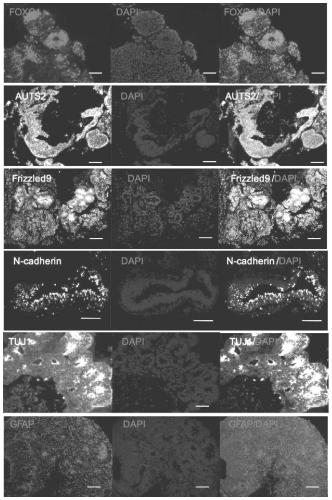
[0041] After tissue fixation, dehydration, clearing, paraffin immersion and paraffin embedding, the cerebral cortical organoids were sectioned for H&E staining, and the expression of different marker proteins in the cerebral cortical organoids was detected by immunofluorescence. The formation of cerebral cortical organoids was confirmed by detecting the marker proteins of different brain regions and nerve cells on the cerebral cortical organoids cultured for 30 days. Marker proteins include forebrain marker protein FOXG1 / EMX1, hippocampus marker protein Frizzled9 / Isl1, midbrain marker protein Otx2, cerebellum or hindbrain marker protein En2 / Islet-1 / Nell2, neural stem cell marker protein N-cadherin, forehead Cortical marker protein Auts2, neuron and cortical development marker protein Pax6, glial cell marker protein GFAP and nerve cell marker protein TUJ1, etc. See the results figure 2 . ...


PUM


Abstract

The invention belongs to the technical field of medicines, and discloses a construction method and application of a glioblastoma organ model. The method for constructing the glioblastoma organ model comprises the following steps: pretreating a glioblastoma cell line to obtain single glioblastoma cells, and transplanting the single glioblastoma cells into cerebral cortex organs cultured for 30 days. The method provided by the invention can simulate the growth characteristics of glioblastoma and the microenvironment of tumors clinically to realize simulation of the growth process of tumor cellsin the normal brain tissues. Furthermore, in order to observe the growth of glioblastoma cells in organoids, a red fluorescent dye is used for marking tumor cells instead of lentiviral transfection oftumor cells. The method is simple and feasible. The influence of lentivirus transfection labeling on the cell growth state is avoided. Meanwhile, the culture time of the system is shortened, the fluorescence labeling difficulty is reduced, and conditions are provided for rapidly applying a tumor sample to experimental researches clinically.

Description

technical field [0001] The invention belongs to the technical field of medicine, and in particular relates to a construction method and application of a glioblastoma organoid model. Background technique [0002] Glioblastoma (GBM) is a common malignant tumor of the central nervous system, which belongs to WHO grade IV, progresses rapidly, and has a poor prognosis. Therapeutic strategies of tumor sites and tumor microenvironment have become a new research direction. Currently commonly used GBM models are mainly tumor cell culture and animal models, but it is difficult for cell lines to simulate tumor heterogeneity, tumor microenvironment and oxygen concentration gradient, and its limited types are also difficult to use to study differences in individual genetic characteristics. Patient-derived tumor xenograft (PDX) models can reproduce the original tumor genome characteristics, but some copy number mutations may be lost after several passages, and it is time-consuming and co...

Claims


Application Information

Patent Timeline
IPC IPC(8): C12N5/09C12Q1/02
CPCC12N5/0693C12N2509/00G01N33/5011
Inventor 刘志雄刘方琨黄兢张李洋
Owner XIANGYA HOSPITAL CENT SOUTH UNIV
Why Eureka
Try Eureka