
Methods for treating malignancies expressing ICAM-1 using coxsackie a group viruses

a technology of coxsackie a group and malignancies, which is applied in the direction of viruses, drug compositions, biological testing, etc., can solve the problems of less successful control of more advanced forms, fewer deaths per year in that country alone, and increased risk of infection, so as to improve the ability of the virus to in

Inactive Publication Date: 2008-04-22
MERCK SHARP & DOHME CORP
View PDF77 Cites 6 Cited by
  • Summary
  • Abstract
  • Description
  • Claims
  • Application Information

AI Technical Summary

Benefits of technology

The present invention is about using a virus to treat abnormal cells, such as melanoma cells, that express certain proteins that the virus recognizes. The virus can infect and kill these cells, providing a new method for treating cancer. The invention also includes screening viruses to determine which ones are most effective at infecting and killing abnormal cells. Overall, the invention provides a way to target and kill cancer cells using a virus that recognizes specific proteins on the cells.

Problems solved by technology

Melanoma is a leading cause of morbidity in the human population.
It is predicted that one in thirty Australians have a form of melanoma resulting in the death of more than one thousand people per year in that country alone.
However, the control of more advanced forms is less successful and an area of intensive research.
Despite progress being made in the treatment of malignancies, the treatment of cancer including melanoma presents a major challenge for research and there remains the need for alternatives to existing therapy approaches.

Method used


Image

  • Methods for treating malignancies expressing ICAM-1 using coxsackie a group viruses
  • Methods for treating malignancies expressing ICAM-1 using coxsackie a group viruses
  • Methods for treating malignancies expressing ICAM-1 using coxsackie a group viruses

Examples


example 1

1.1. Cell Lines

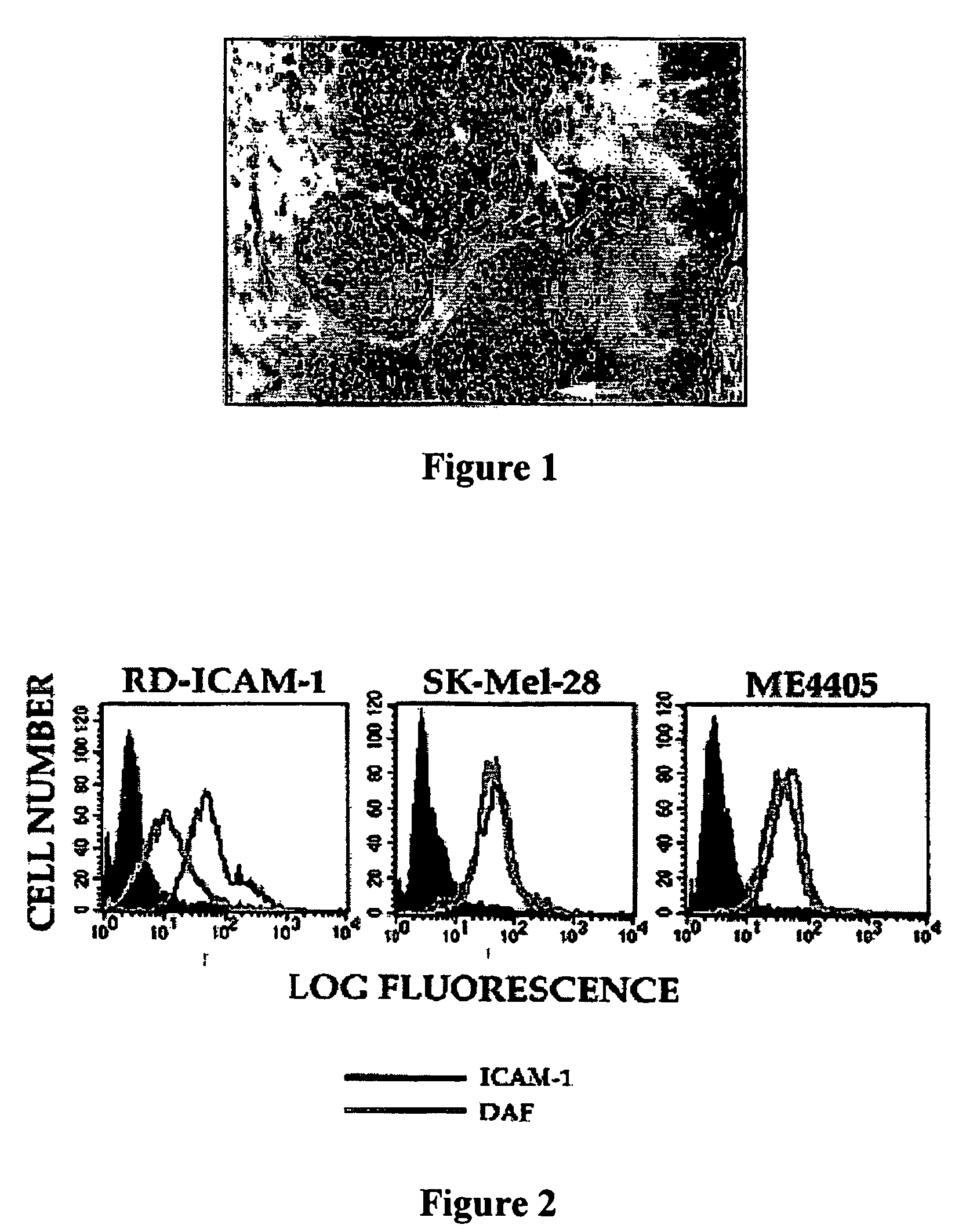
[0112]Continuous cultures of Rhabdomyosarcoma expressing ICAM-1 cells (RD-ICAM-1), HeLa-B cells, and human lung fibroblast cells (MRC5) were maintained in Dulbecco's Modified Eagle's Medium (DMEM) and 10% fetal calf serum (FCS). Two melanoma cell lines Sk-Mel-28 and ME4405 were obtained from Dr. Ralph (Department of Biochemistry and Molecular Biology, Monash University, Victoria, Australia) and Dr. Peter Hersey, Cancer Research Department, David Maddison Building Level 4, Royal Newcastle Hospital, Newcastle, New South Wales, Australia, respectively. The cell line Sk-Mel-28 is a metastatic melanoma cell line found to be resistant to chemotherapeutic drugs (56). The melanoma cell culture ME4405 was established from specimens of primary melanoma lesions (69). The two melanoma cell lines were maintained in DMEM containing 10% FCS. Rhabdomyosarcoma cells (RD) a heteroploid human embryonal cell line, and HeLa-B cells an aneuploid cell clone derived from human squamous epith...

example 2

2.1. Infection of Melanoma Cell Lines by CAV21

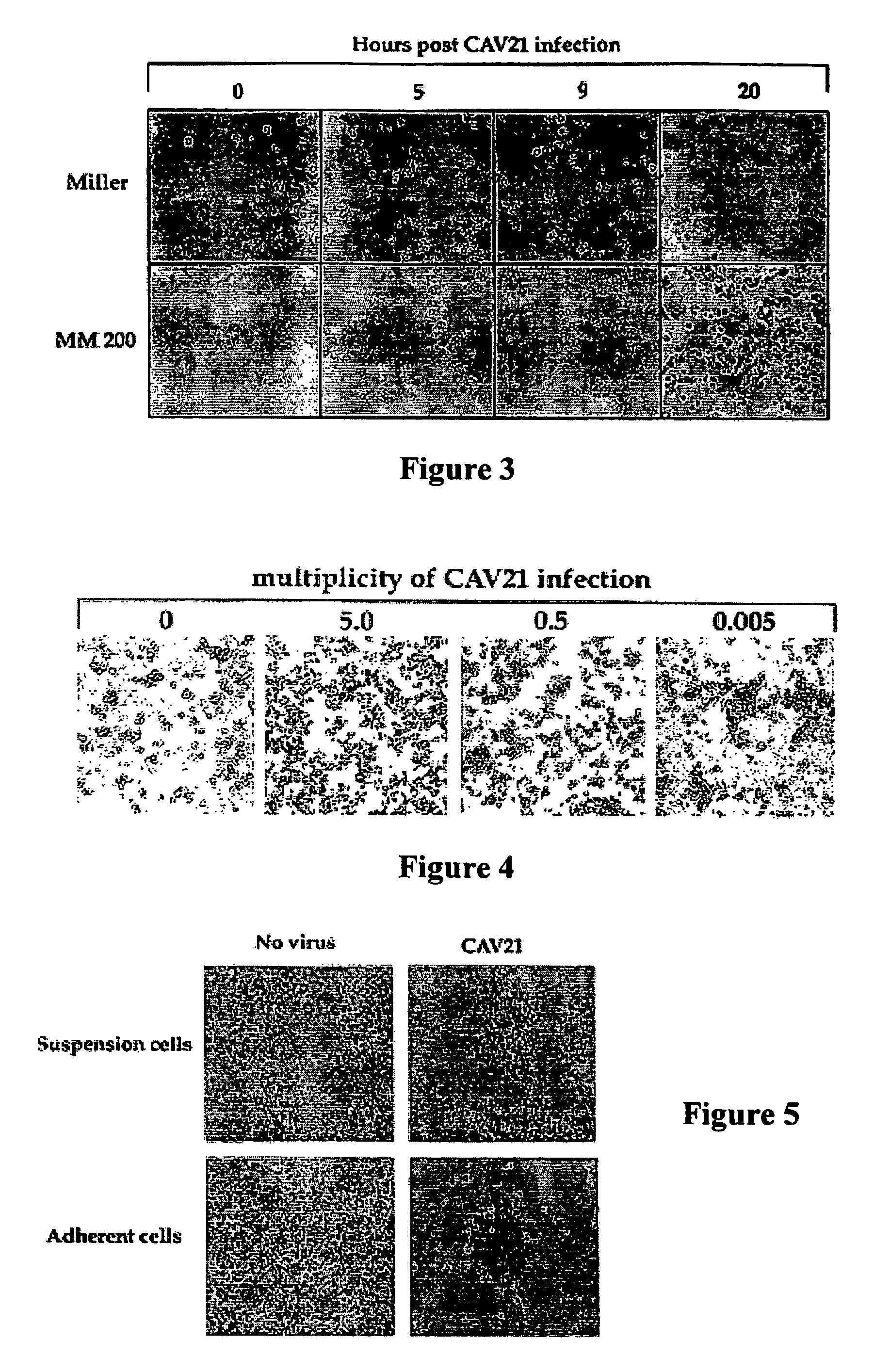
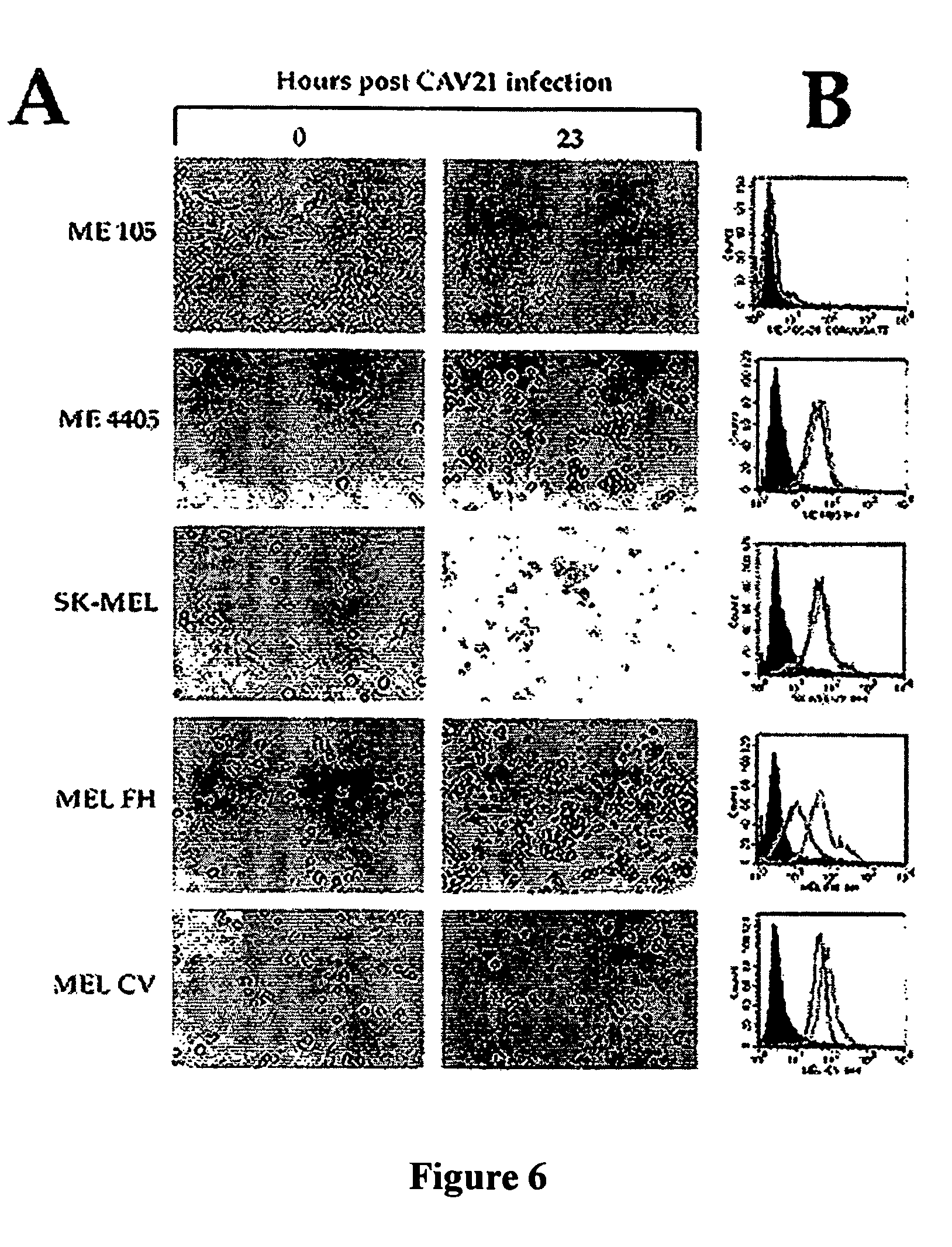
[0120]Monolayers of two culture-adapted melanoma cell lines Miller and MM200 were infected with CAV21 prepared in Example 1 at a multiplicity of infection of 1.0 for 1 hour prior to removal of the inoculum and the cells incubated in culture medium (DMEM containing 1% foetal calf serum and penicillin streptomycin) for 24 hours at 37° C. The results shown in FIG. 3 indicate that CAV21 was able to induce significant changes in the cellular cytopathology of both cell lines as early as five hours post infection (PI) and by nine hours PI almost complete killing of all the melanoma cells.

example 3

3.1. Infection of Melanoma Cells from Primary Melanoma by CAV21

[0121]Cells from a primary melanoma removed from a nude mouse that had been previously subcutaneously inoculated with human melanoma cells from cell line ME 4405 using conventional methods, were highly susceptible to CAV21 infection and killing, even at a challenge rate of 0.005 CAV21 particles per melanoma cell as shown in FIG. 4.


PUM

PropertyMeasurementUnit
wavelengthaaaaaaaaaa
final volumeaaaaaaaaaa
volumeaaaaaaaaaa

Abstract

There is disclosed a method of killing abnormal cells such as malignant cells including melanoma cells, using a virus recognizing at least one of a cell adhesion molecule and a complement regulatory protein. The virus may be a member of the Picornaviridae family. Coxsackie A-group viruses have been found to be particularly suitable. The cell adhesion molecule is desirably a member of the immunoglobulin (Ig) superfamily. Typically, the complement regulatory protein will be DAF.

Description

[0001]This application is a 35 U.S.C. § 371 of PCT / AU00 / 01461, filed Nov. 27, 2000, which claims benefit of Australian Patent Application PQ 4256, filed Nov. 25, 1999. The contents of each of these prior applications are incorporated by reference herein in their entirety.FIELD OF THE INVENTION[0002]The present invention relates to the killing of abnormal cells utilising a virus. There is also described a method of screening cells to ascertain whether they are susceptible to treatment with the virus, as well as pharmaceutical compositions incorporating the virus. The invention finds veterinary use as well as broad application in the human medical field.BACKGROUND OF THE INVENTION[0003]Melanoma is a leading cause of morbidity in the human population. Australia has the highest rate of melanoma in the world. Melanoma is an aggressive skin cancer and is the third most common cancer in Australia for both men and women. It is predicted that one in thirty Australians have a form of melanoma...

Claims


Application Information

Patent Timeline
Patent Type & Authority Patents(United States)
IPC IPC(8): A01N63/00C12N15/00C12N15/01C12N15/09G01N33/48A61K38/00A61K39/00A61K39/125A61P35/00C12Q1/02C12Q1/70G01N33/15G01N33/50
CPCG01N33/5011C12N7/00A61K35/768C12N2770/32332C12N2770/32371C12N2770/32333A61P35/00A61P35/04A61K9/0019C12Q1/025
Inventor SHAFREN, DARREN R.
Owner MERCK SHARP & DOHME CORP
Learn More