Diagnosis of blood clots using fibrin-binding proteins bound with contrast agents

a technology of fibrin-binding proteins and contrast agents, which is applied in the direction of peptidases, peptide/protein ingredients, enzymology, etc., can solve the problems of not being sensitive enough for clinical practice, and also storing and disposing of radioactive materials, so as to limit side effects and facilitate interpretation. , the effect of limiting side effects

Inactive Publication Date: 2008-12-16
BENFORD JACOB
View PDF5 Cites 2 Cited by
  • Summary
  • Abstract
  • Description
  • Claims
  • Application Information

AI Technical Summary

Benefits of technology

[0015]This invention describes the steps to modify a fibrin-binding protein and then combine this with a contrast agent. The contrast agent may be a single ion, or a molecule of a pre-formulated contrast agent. This compound is then given through an IV to a patient suspected of having a blood clot (see FIGS. 1-9). After waiting a sufficient amount of time for the compound to circulate and to bind the suspected blood clot, a radiographic study (plain x-ray or CT) is then obtained. A “hot spot” or collection of the contrast agent seen on the radiographic study would represent the presence and location of a blood clot (FIGS. 10-11). As blood vessels would not need to be filled with the contrast agent, relatively small amounts of this compound are used, thus limiting side effects. The results are also quickly and easily interpreted by a clinician, without the need for specialized personnel or equipment for administration and interpretation.

Problems solved by technology

Unfortunately all of these methods have limitations, such as high radiation load or nephrotoxicity, and many are not sensitive enough for clinical practice.
Problems also arise concerning storing and disposing radioactive material when using some tests.

Method used


Image

  • Diagnosis of blood clots using fibrin-binding proteins bound with contrast agents
  • Diagnosis of blood clots using fibrin-binding proteins bound with contrast agents
  • Diagnosis of blood clots using fibrin-binding proteins bound with contrast agents

Examples


example 1

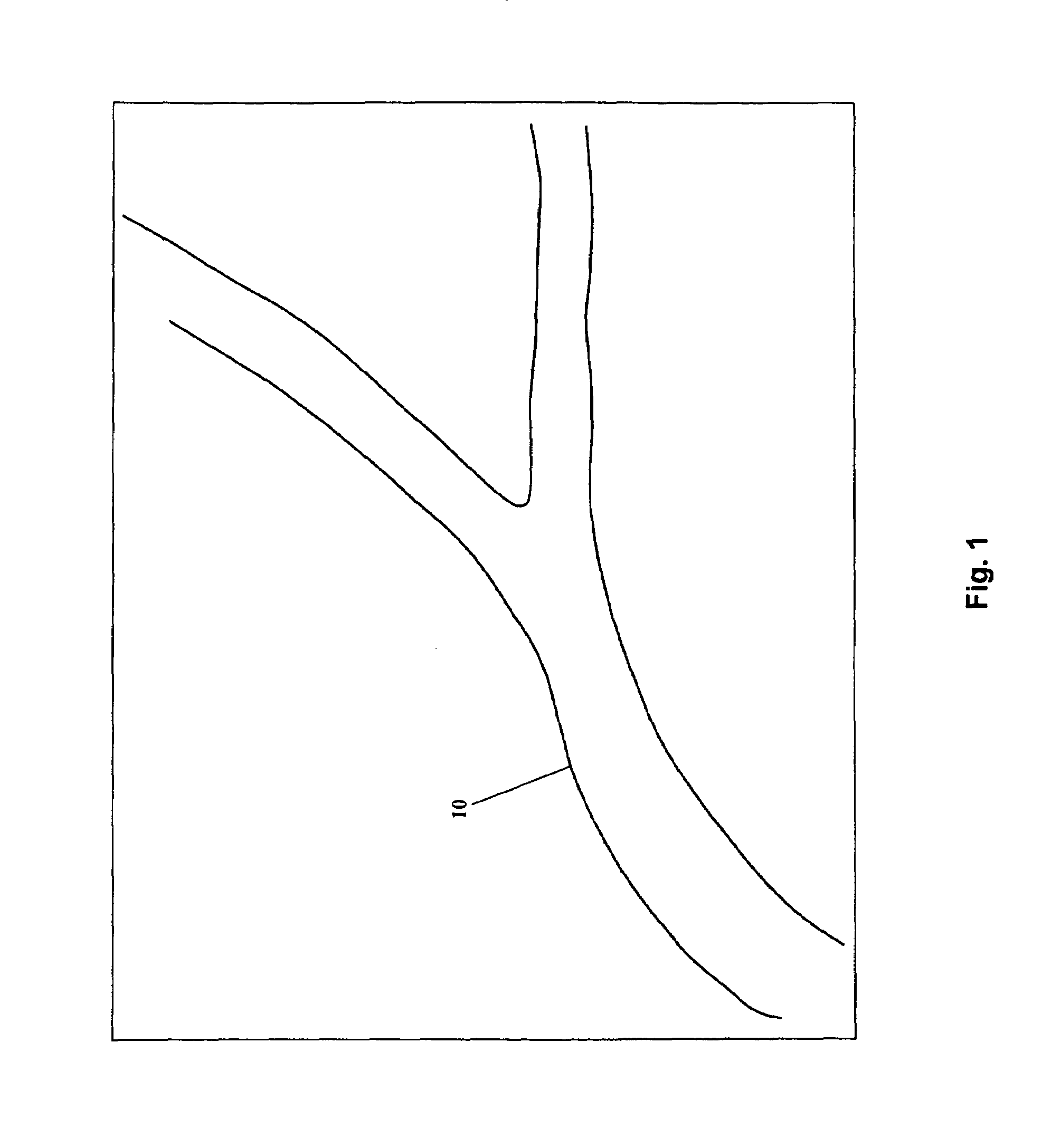
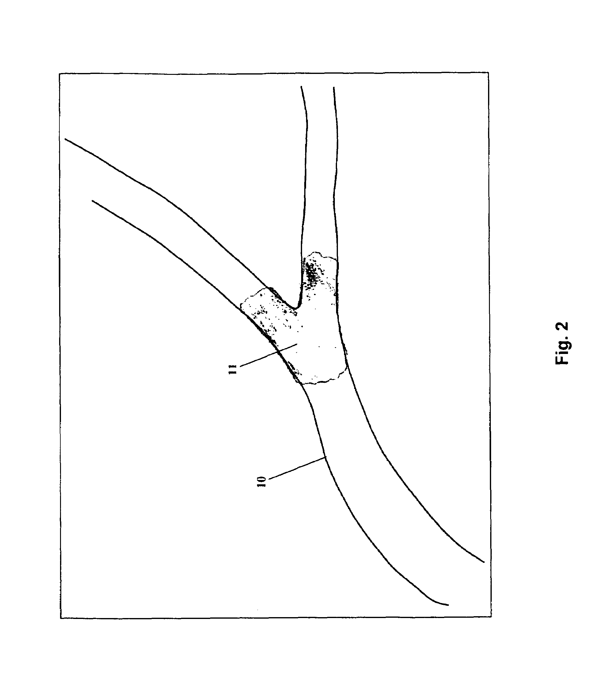
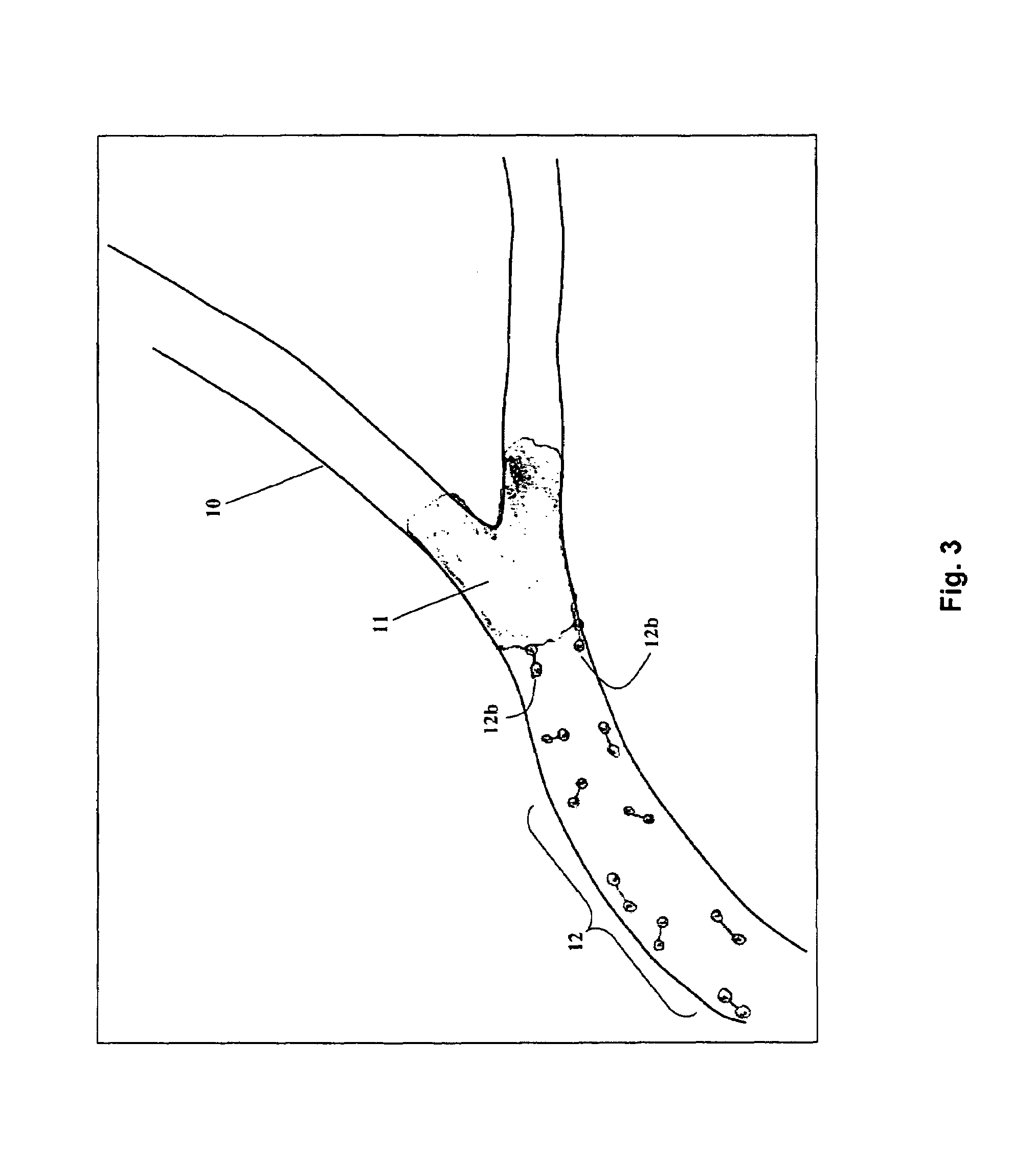
Detection of Blocked Blood Vessel

[0056]A 45-year-old male presents with substernal chest pain. He has a complex medical history, and possible etiologies for his pain include an acute heart attack, a pulmonary embolus, or his chronic pulmonary disease. His initial ECG in non-diagnostic. In addition to standard treatment, the fibrin-binding protein-contrast agent 50 mg / kg is given as a bolus intravenous injection. After 10 minutes a CT of his chest is preformed which shows a “blush” of contrast in the area of the heart, consistent with a blocked coronary blood vessel. The patient is then quickly seen by the cardiologist and definitive treatment (e.g., tPA) is given without delay.

example 2

Detection of Pulmonary Embolus

[0057]A 75-year-old female presents with shortness of breath. The patient has a long history of smoking, and has a slight fever on presentation. She has just returned from vacation, which included a 6-hour trip. The fibrin-binding protein-contrast agent compound is given as a bolus intravenous injection. 10 minutes later, a chest x-ray is taken that is clear except for a slight blush in the right middle lobe. She is then taken to the CT scan, which shows a contrast “blush” in the right middle lobe, consistent with a blood clot, or pulmonary embolus. The patient is admitted for definitive treatment.

example 3

Detection of Blood Clot in Leg Vessel

[0058]A 28-year-old male presents with increasing swelling and pain of his left leg. The patient broke the same leg three weeks ago, and it is now in a cast. The patient was doing well, with minimal pain until four days ago. His exam is normal except for mildly swollen toes at the far end of his cast. The fibrin-binding protein-contrast agent compound is administered by intravenous bolus to his descending artery. The patient then has an x-ray of his upper and lower leg 10 minutes later, which shows a contrast “blush” just above the knee. Treatment (e.g., coumidin) is started for the blood clot, and a potential pulmonary embolus is avoided.


PUM

PropertyMeasurementUnit
computed tomographyaaaaaaaaaa
nucleic acid sequenceaaaaaaaaaa
timeaaaaaaaaaa

Abstract

This invention describes how to modify a fibrin-binding protein, such as tPA, with a contrast agent, such as iodine. This substance could then be given to a patient suspected of having a blood clot by an intravenous route, and then detected by a radiographic study at a short pre-determined time later. Appropriate therapy is started immediately as determined by the radiographic study.

Description

CROSS-REFERENCE TO RELATED APPLICATIONS[0001]This application claims the benefit of U.S. Ser. No. 60 / 674,369, filed Apr. 25, 2005, which is incorporated herein by reference in its entirety.STATEMENT OF GOVERNMENTAL SUPPORT[0002]None.BACKGROUND OF THE INVENTION[0003]1. Field of the Invention[0004]This invention describes how to modify a fibrin-binding protein, such as tPA, to produce a fibrin-binding protein without fibrinolytic activating properties. This modified protein is then combined (and / or covalently bonded) with a contrast agent, such as iodine. This combination substance is then administered to a patient suspected of having a blood clot by a standard route. Next, any blockage or clot is detected by a radiographic study at a short pre-determined time later, and the patient is immediately treated with drugs and / or therapy based on the diagnosis / results of the radiographic study.[0005]2. Related ArtBackground[0006]Currently, there are numerous tests that may be used when tryin...

Claims


Application Information

Patent Timeline
Patent Type & Authority Patents(United States)
IPC IPC(8): A61K49/04
CPCC12N9/6424C12N9/6459G01N33/58G01N33/86C12Y304/21069
Inventor BENFORD, JACOB
Owner BENFORD JACOB
Why Eureka
Try Eureka