Method of quickly detecting and/or assaying antigen by fluorescence correlation spectrometry

a fluorescence correlation and antigen technology, applied in the direction of fluorescence/phosphorescence, material analysis through optical means, instruments, etc., can solve the problems of animal death, brain spongiform, abnormal prions at those particular sites, etc., and achieve the effect of quick and convenien

Inactive Publication Date: 2009-01-13
JAPAN SCI & TECH CORP
View PDF19 Cites 6 Cited by
  • Summary
  • Abstract
  • Description
  • Claims
  • Application Information

AI Technical Summary

Benefits of technology

[0029]An object of the present invention is to provide a quick and convenient method of detecting and / or assaying an antigen or an antigenic protein such as a pathogenic protein (e.g., an abnormal prion) or a harmful protein contained in a food material.
[0030]During diligent studies to attain the above object, the present inventors have focused their attention on fluorescence correlation spectroscopy (FCS). Recently, FCS has been known as a method that is frequently used in the analysis and the like of molecules derived from organisms and that can detect and assay, in almost real time, the physical parameters of protein molecules, such as number, size, or shape without undergoing a physical separation step. The present inventors learned that fluorescently-labeling the antigen molecule to be detected, and using a fluorescence-labeled antibody fragment and a non-fluorescence-labeled intact antibody that binds together via the antigen in the fluorescence labeling, can allow a significant difference in diffusion rate to arise between the fluorescence-labeled antibody fragment not bonded to the antigen and the complex formed by the fluorescence-labeled antibody fragment, the antigen, and the non-fluorescence-labeled intact antibody. The present inventors discovered that, according to this method, a wide scope of antigens can be detected and assayed using FCS, including antigens that have relatively low molecular weight such as an antigenic protein, independent from the shape or molecular weight of the antigen. Based on these findings, the present invention has been completed.
[0031]Specifically, the present invention may comprise a method of detecting and / or assaying an antigen, such as an antigenic protein, by using a fluorescence-labeled antibody fragment targeted to an epitope of the antigen and a non-fluorescence-labeled intact antibody targeted to another epitope of the antigen which together forms an antigen / antibody complex among the antigen, the fluorescence-labeled antibody fragment, and the non-fluorescence-labeled intact antibody. The method may further comprise detecting and analyzing the formed antigen / antibody complex by fluorescence correlation spectroscopy. The detection and / or assay method of the present invention can be applied widely to the detection and / or assay of an antigenic protein, for example, a pathogenic protein (e.g., an abnormal prion) or a harmful protein contained in a food material, whereby the antigenic protein can be detected and / or assayed quickly and accurately by a simple procedure.
[0034]The present invention may also relate to: (6) a method of quickly detecting and / or assaying an antigen by fluorescence correlation spectroscopy, comprising the addition of a fluorescence-labeled antibody fragment targeted to an epitope of an antigen and a non-fluorescence-labeled intact antibody targeted to another epitope of the antigen to a test sample, performing antigen / antibody reaction thereamong, and detecting and analyzing an antigen / antibody complex formed among the antigen, the fluorescence-labeled antibody fragment, and the non-fluorescence-labeled intact antibody by fluorescence correlation spectroscopy; (7) the method of quickly detecting and / or assaying an antigen by fluorescence correlation spectroscopy according to (6), wherein the antigen is an antigenic protein; (8) the method of quickly detecting and / or assaying an antigen by fluorescence correlation spectroscopy according to (6) or (7), wherein the detection and / or assay of an antigen by fluorescence correlation spectroscopy are performed without undergoing a step of physically separating the antigen contained in the test sample; (9) the method of quickly detecting and / or assaying an antigen by fluorescence correlation spectroscopy according to any one of (6) to (8), wherein the step of adding a fluorescence-labeled antibody fragment targeted to an epitope of the antigen and a non-fluorescence-labeled intact antibody targeted to another epitope of the antigen to a test sample, the step of performing antigen / antibody reaction among the test sample, the fluorescence-labeled antibody fragment, and the non-fluorescence-labeled intact antibody, and the step of detecting and analyzing the test sample that has undergone the antigen / antibody reaction by fluorescence correlation spectroscopy are performed automatically or semi-automatically; and (10) the method of quickly detecting and / or assaying an antigen by fluorescence correlation spectroscopy according to any one of (6) to (9), wherein the test sample is a biological protein ample, and the antigen is a pathogenic protein antigen.
[0035]The present invention may further relate to: (11) the method of quickly detecting and / or assaying an antigen by fluorescence correlation spectroscopy according to (10), wherein the pathogenic protein antigen is an abnormal prion; (12) the method of quickly detecting and / or assaying an antigen by fluorescence correlation spectroscopy according to any one of (6) to (9), wherein the test sample is a food material, and the antigen is a harmful protein antigen contained in the food material; (13) a detection reagent for quickly detecting and / or assaying an antigen by fluorescence correlation spectroscopy, comprising a fluorescence-labeled antibody fragment targeted to an epitope of an antigen to be detected and / or assayed and a non-fluorescence-labeled intact antibody targeted to another epitope of an antigen to be detected and / or assayed; and (14) a kit for quickly detecting and / or assaying an antigen by fluorescence correlation spectroscopy, comprising the detection reagent according to (12).

Problems solved by technology

This results in the accumulation of the abnormal prions at those particular sites.
The accumulation of the abnormal prions in the brain renders the brain spongiform, leading to animal death.
These methods involve complexities and takes much time.
This centrifugation procedure can take much time.
In all, these methods involve complicated treatments and therefore present a problem of being unsuitable for testing a large number of samples in a short period of time.
As a result, the method assays the abnormal prion without performing the centrifugation procedure or electrophoresis, and can test a large number of samples in a short time.
Thus, these methods are not necessarily sufficient for conveniently and quickly detecting and assaying an antigenic protein such as a prion.
Moreover, these methods are less-than-suitable for automatically or semi-automatically performing treatment steps for detection and assay, and for assaying large amounts of samples.
However, even if these methods were used, the applicability of FCS was limited regarding which objects could be detected.Patent Document 1: Japanese Patent Application No. 10-267928Patent Document 2: Japanese Laid-Open Patent Application No. 2001-272404Patent Document 3: Japanese Laid-Open Patent Application No. 2003-130880Patent Document 4: Japanese Laid-Open Patent Application No. 2003-215131Patent Document 5: Japanese Patent No. 3517241Non-Patent Document 1: J. Infect. Dis. 154: 518-521, 1986Non-Patent Document 2: Chem. Phys., 4, 390-401, 1974Non-Patent Document 3: Biopolymers, 13, 1-27, 1974Non-Patent Document 4: Physical Rev.

Method used


Image

  • Method of quickly detecting and/or assaying antigen by fluorescence correlation spectrometry
  • Method of quickly detecting and/or assaying antigen by fluorescence correlation spectrometry
  • Method of quickly detecting and/or assaying antigen by fluorescence correlation spectrometry

Examples


example 1

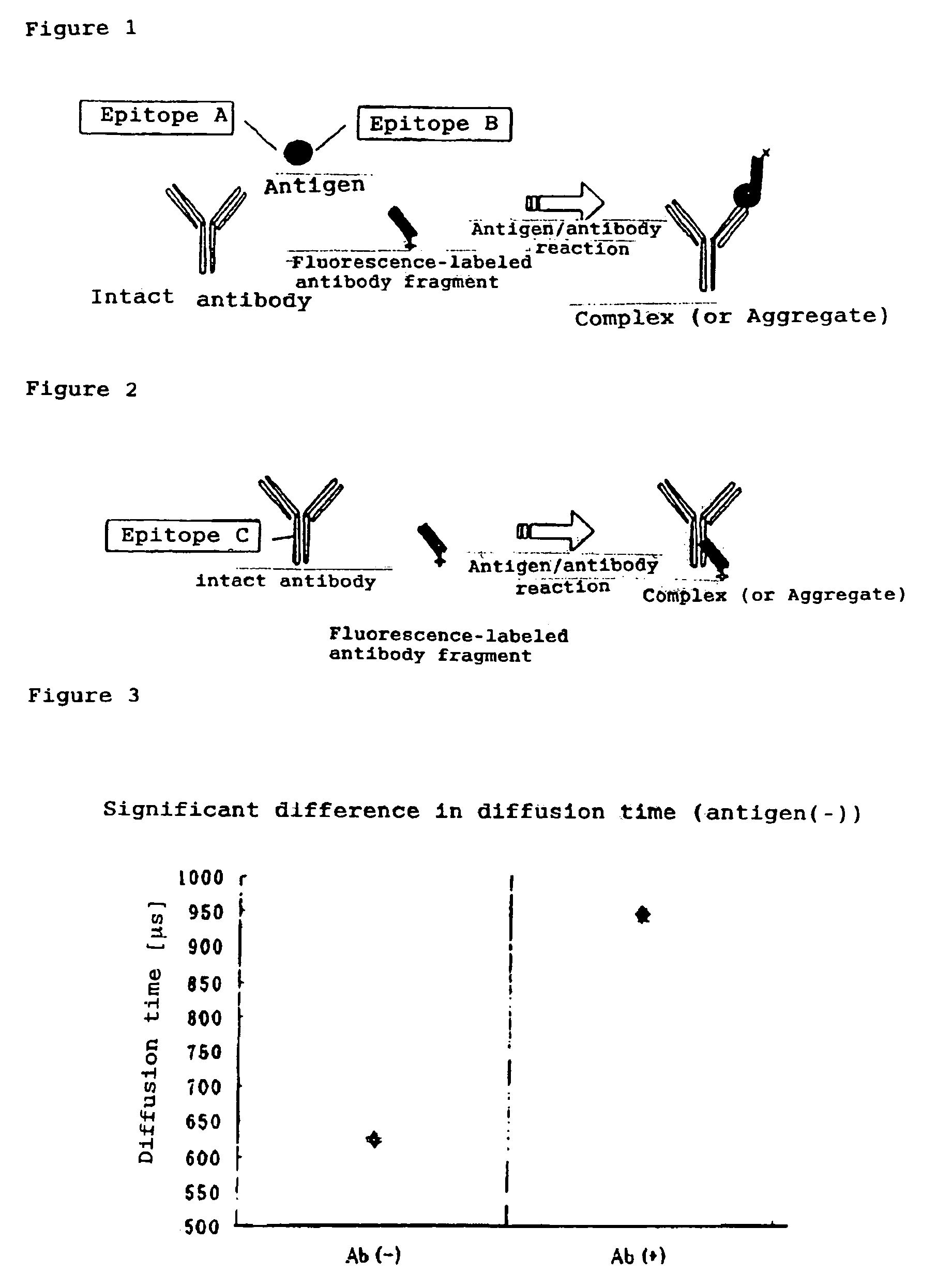
Influence of a Non-fluorescence-labeled Intact Antibody on Diffusion Rate

(1) Materials:

[0073]Alexa Fluor 647 (Zenon One Mouse IgG1 Labeling Kit)[0074]Fab 647 (Zenon One IgG1 Labeling Reagent)[0075]Antibody (indicated by Ab in FIG. 2) (Zenon One Blocking Reagent (mouse IgG))

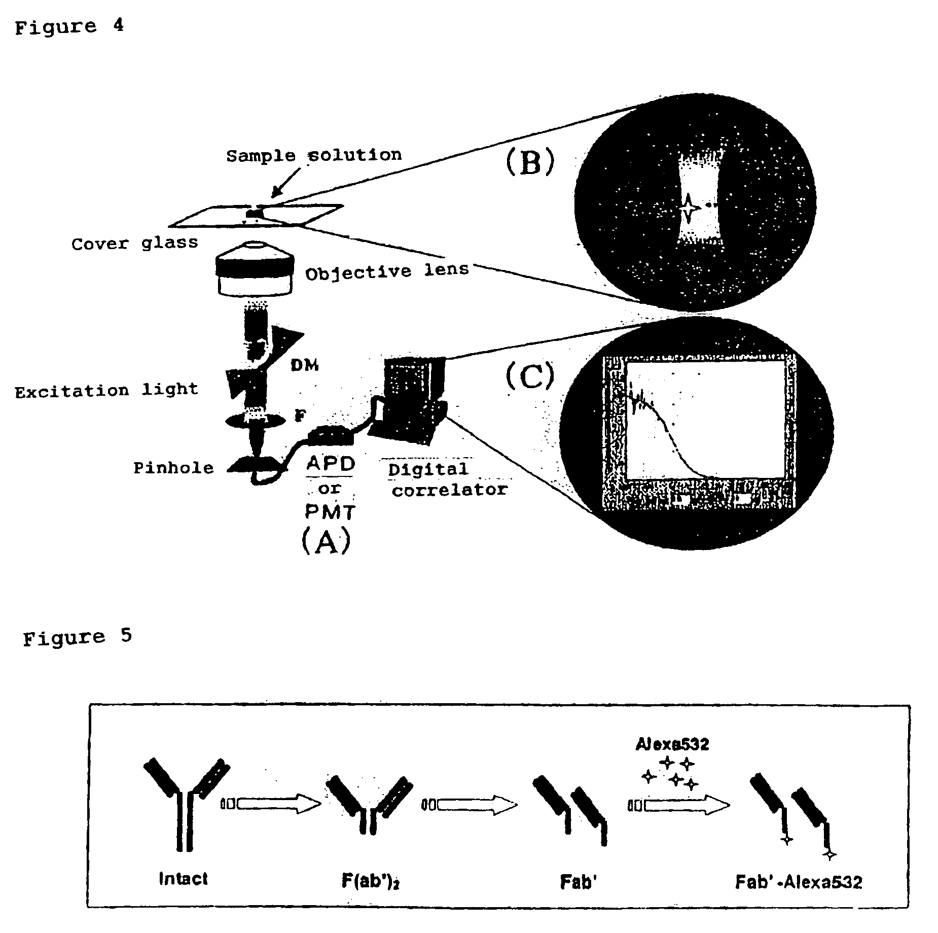
(2) Apparatus for FCS Assay:[0076]MF-20 (intermolecular interaction analysis system: Olympus Corp.)

(3) Procedures:

[0077]A solution of 10 nM Fab 647 (mouse IgG antibody) alone and a Fab 647 mixture obtained by mixing Fab 647 with 100 nM intact antibody were applied to a 384-well plate (Olympus) blocked with N101 (NOF Corp.), followed by assay with MF20 (Olympus). The assay was performed by 30 sec.×three measurements at a laser power set to 100 μW. The processing software in MF20 was used to derive each parameter including diffusion time.

[0078]To test how a non-fluorescence-labeled intact antibody can influence the diffusion rate in the Brownian motion of an antigen / antibody complex molecule, an intact antibody and ...

example 2

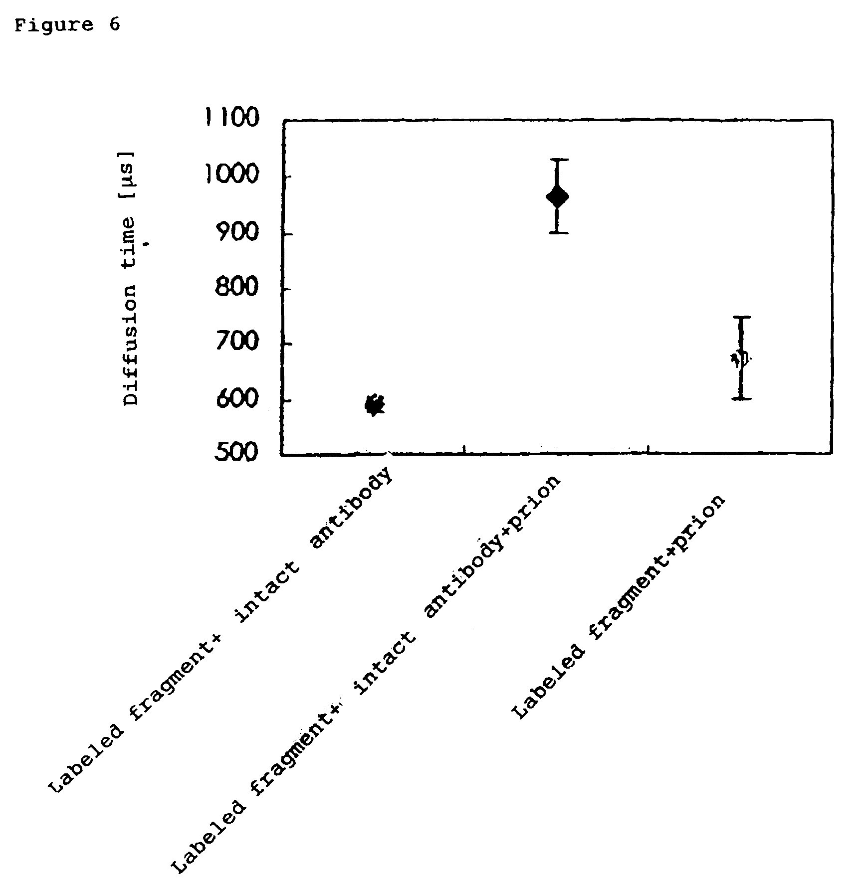
Detection and Assay of Antigen (Antigenic Protein) using FCS

(1) Apparatus

[0080]FCS apparatus[0081]Intermolecular interaction analysis system (MF-20, manufactured by Olympus Corp.)

(2) Materials[0082]Preparation of fluorescence-labeled antibody fragment

[0083]In this Example, the fluorescence-labeled antibody fragment (Fab′-Alexa 532) of an anti-prion antibody was taken as an example. The outline of preparation of Fab′-Alexa 532 is shown in FIG. 5. An anti-PrP antibody solution was equilibrated with a citric acid solution (pH 6.3) by use of a PD-10 column (Pharmacia) and then supplemented (37° C., approximately 30 minutes) with pepsin (1% (w / w)) to prepare F(ab′)2. The degree of the digestion was confirmed by HPLC (column: G300SWXL). Then, the fragment was purified by FPLC (column: Superdex 200 (16 / 60)) using 0.1 M phosphate buffer solution (pH6.3), then concentrated, and stored. It was further reduced (37° C., approximately 1.5 hours) by the addition of 2-mercaptomethylamine (0.01 M) ...


PUM

PropertyMeasurementUnit
molecular weightaaaaaaaaaa
volumeaaaaaaaaaa

Abstract

The present invention provides a method of quickly and accurately detecting and / or assaying an antigen using fluorescence correlation spectroscopy (FCS), which involves a fluorescence-labeled antibody fragment and a non-fluorescence-labeled intact antibody that form a complex with the antigen. There is a significant difference in diffusion rate between the fluorescence-labeled antibody fragment not bonded to the antigen and the complex formed by the the fluorescence-labeled antibody fragment, the antigen, and the non-fluorescence-labeled intact antibody, and this diffusion rate can be determined using FCS. The antigen can be an antigenic protein, such as an abnormal prion or a harmful protein contained in a food material. According to this method, antigens over a wide scope can be assayed regardless of the shape or molecular weight.

Description

INCORPORATION BY REFERENCE[0001]This application is a continuation-in-part application of international patent application Serial No. PCT / JP2005 / 010043 filed Jun. 1, 2005, which published as international publication No. WO 2005 / 119256 on Dec. 15, 2005, which claims priority to Japanese patent application Serial No. JP 2004-166440 filed Jun. 3, 2004.[0002]The foregoing applications, and all documents cited therein or during their prosecution (“appln cited documents”) and all documents cited or referenced in the appln cited documents, and all documents cited or referenced herein (“herein cited documents”), and all documents cited or referenced in herein cited documents, together with any manufacturer's instructions, descriptions, product specifications, and product sheets for any products mentioned herein or in any document incorporated by reference herein, are hereby incorporated herein by reference, and may be employed in the practice of the invention.FIELD OF THE INVENTION[0003]Th...

Claims


Application Information

Patent Timeline
Patent Type & Authority Patents(United States)
IPC IPC(8): G01N21/76G01N21/64G01N21/78G01N33/536G01N33/542G01N33/558G01N33/58G01N33/68
CPCG01N33/536G01N33/582G01N33/6857G01N33/6896G01N2800/2828
Inventor KINJO, MASATAKAHORIUCHI, MOTOHIROFUJII, FUMIHIKOSAKATA, HIROSHITAMURA, MAMORUUENO, MASAYOSHIYANAGIYA, TAKAYUKI
Owner JAPAN SCI & TECH CORP
Why Eureka
Try Eureka