Computerized tomography device using X rays and image processing method

a computerized tomography and image processing technology, applied in the field of computerized tomography (ct) devices using x rays and image processing methods, can solve the problems of large work, reduced objectivity or reliability of the quantification result of brown adipose, and inability to automatically identify and extract brown adipose images, etc., to achieve the improvement of the reliability of the quantification calculation result and the accuracy of the identification of brown adipos

Inactive Publication Date: 2009-04-28
HITACHI LTD
View PDF10 Cites 18 Cited by
  • Summary
  • Abstract
  • Description
  • Claims
  • Application Information

AI Technical Summary

Benefits of technology

[0008]The present invention also advantageously enables obtaining information for evaluating the brown adipose while precisely identifying the brown adipose based on the CT image.
[0011]The X-ray CT device according to the present invention has a brown adipose measuring function which is not provided in the X-ray CT devices of the related art. By executing this function, it is possible to automatically extract the brown adipose through analysis of the CT image, and, thus, various problems caused in the case of the manual extraction can be resolved. In other words, with the above-described structure, it is possible to extract the brown adipose with a high precision and the extraction process is automatically executed under an objective standard. Therefore, the device has a superior reproducibility and the reliability of the measurement result can be improved because of this characteristic. It is also possible to employ a configuration in which the brown adipose is automatically extracted within a two-dimensional region of interest or a three-dimensional region of interest designated by the user.
[0014]According to the above-described method, it is possible to identify candidate pixels based on the CT value on the CT image and then remove erroneous pixels included in the candidate pixels, to identify the brown adipose pixels. For example, in a region such as a boundary between the white adipose and muscle and a thin muscle layer, a pixel having a CT number similar to the CT number of the brown adipose pixel may be observed. By applying a process to remove these erroneous pixels, the identification precision of the brown adipose and reliability of quantification calculation result can be improved.
[0015]According to another aspect of the present invention, it is preferable that, in the image processing method, an evaluation value useful for health management and disease diagnosis is calculated based on the extracted brown adipose pixels. With the evaluation value, it is possible to objectively or comprehensively evaluate a sample.

Problems solved by technology

There is, however, no device or method for automatically identifying and extracting images of brown adipose.
With such manual determination, however, the objectivity or reliability of the quantification result of the brown adipose is reduced.
Moreover, with the manual operation, a large work is required and the process cannot be quickly performed.
In particular, when a volume of the brown adipose is to be determined based on a plurality of CT images, these problems become more significant.

Method used


Image

  • Computerized tomography device using X rays and image processing method
  • Computerized tomography device using X rays and image processing method
  • Computerized tomography device using X rays and image processing method

Examples


Embodiment Construction

[0035]A preferred embodiment of the present invention will now be described.

(1) Summary of X-ray CT Device in the Preferred Embodiment

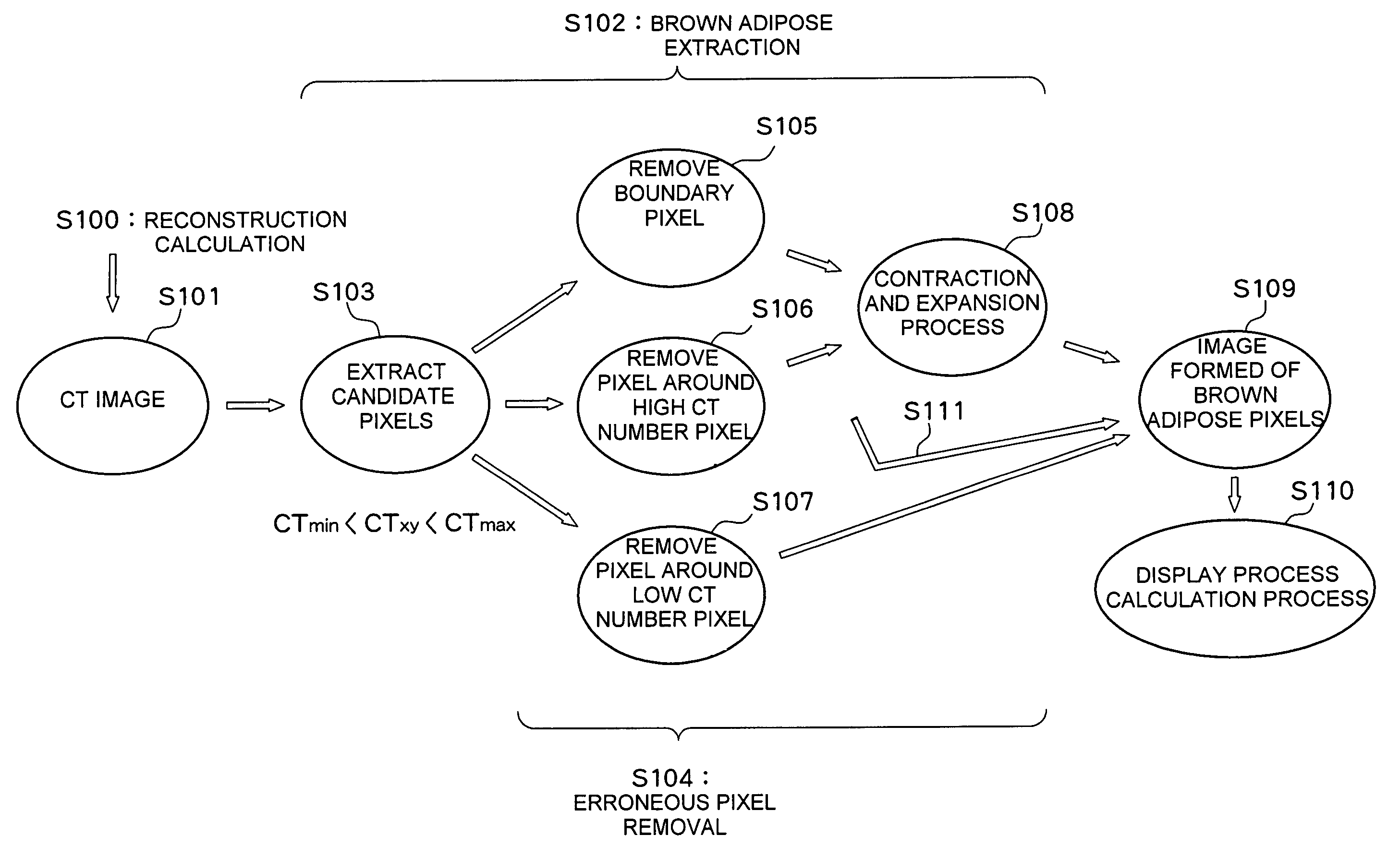
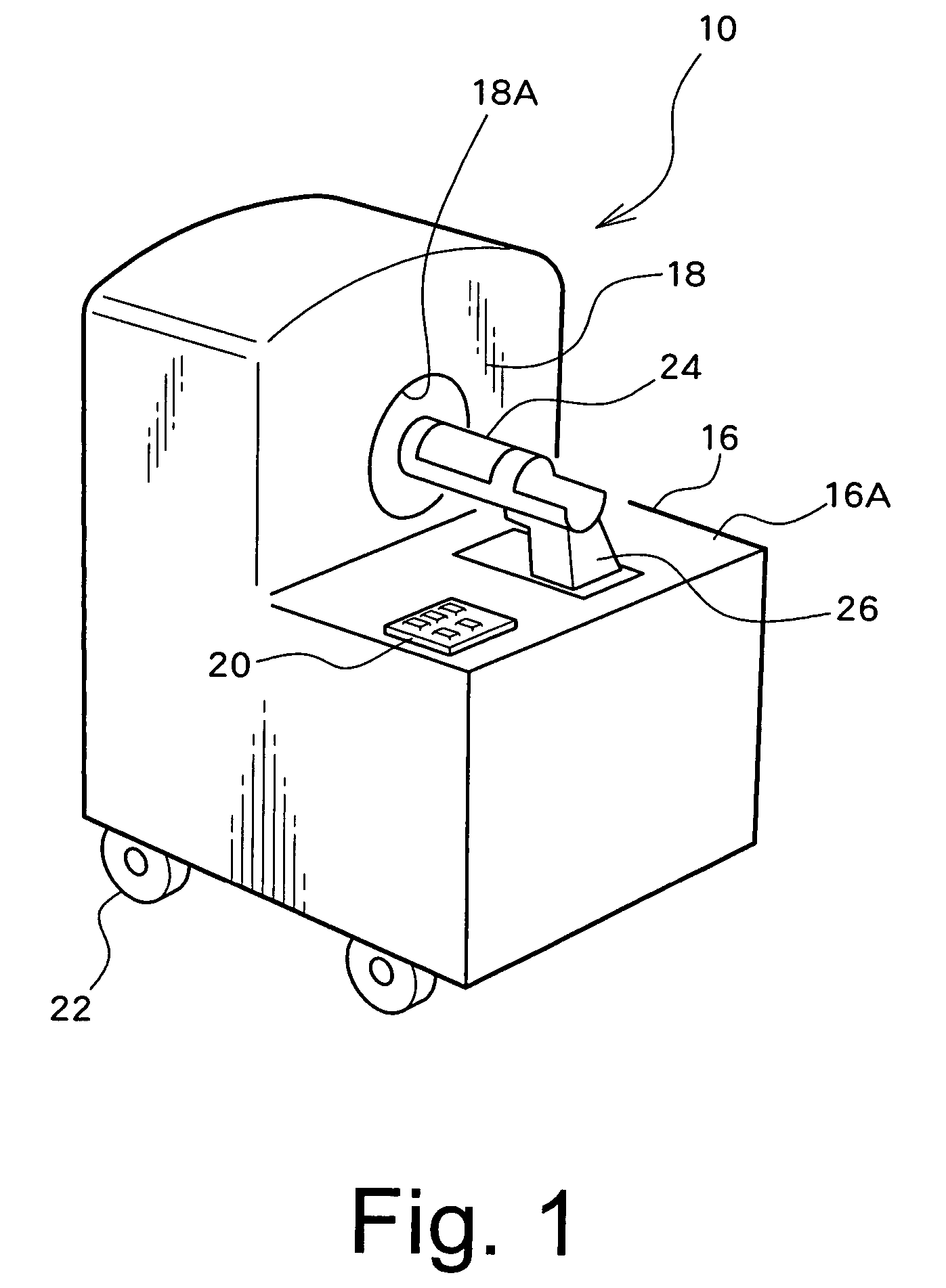
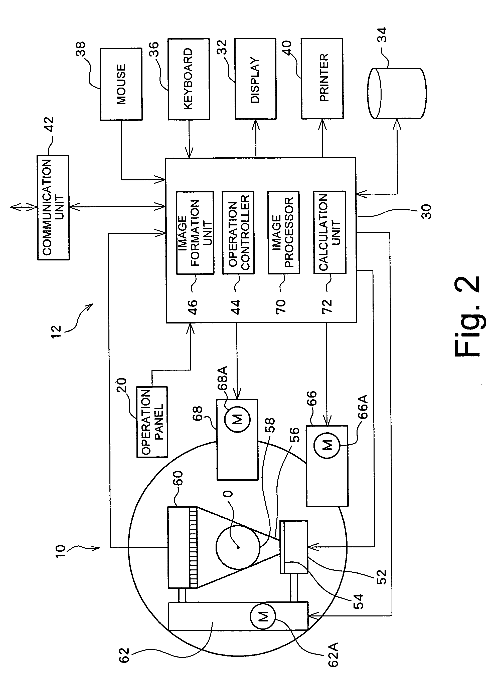
[0036]An X-ray computerized tomography (CT) device of the preferred embodiment of the present invention comprises an X-ray generator, an X-ray detector, a rotation mechanism, a scan mechanism, an image formation unit, an image processor, and a calculation unit, as will be described later in detail. The image formation unit comprises a first extraction unit (first extraction function) and a second extraction unit (second extraction function). The first extraction unit extracts candidate pixels from among a group of pixels reconstructing a CT image based on a CT number of each pixel. Each candidate pixel is a pixel which may depict brown adipose. The second extraction unit applies an erroneous pixel removal process to the candidate pixels, to extract brown adipose pixels. The extracted brown adipose pixels form a brown adipose image. When there are no e...


PUM


Abstract

A computerized tomography device using an X-ray reconstructs a CT image of a sample and processes the CT image. Brown adipose candidate pixels are extracted based on a CT number of each pixel reconstructing the CT image. An erroneous pixel removal process is applied to the candidate pixels. In the erroneous pixel removal process, a boundary pixel removal process, a contraction and expansion process, or the like are applied. With this process, only the brown adipose pixels are extracted. An amount of brown adipose is determined from the brown adipose pixels and an evaluation value such as a brown adipose percentage is calculated based on the amount of brown adipose and amounts of other tissues.

Description

BACKGROUND OF THE INVENTION[0001]1. Field of the Invention[0002]The present invention relates to a computerized tomography (CT) device using X rays and an image processing method, and, in particular, to a CT image processing technique for extracting a predetermined tissue.[0003]2. Description of the Related Art[0004]It is known that adipose tissues (fat cells) present in a body of a living body (humans and other animals) can be divided into two types, white adipose tissue and brown adipose tissue. White adipose tissue stores energy as a triglyceride and provides energy to the body as prompted. Brown adipose tissue provides a highly advanced thermogenesis or calorigenic action. An increase and an activation of the brown adipose cause reduction in the white adipose which causes adiposis or obesity. Because of this, brown adipose has recently attracted much attention in the medical field. White adipose is present in a large amount in the abdomen, the buttocks, the thighs, the back, the...

Claims


Application Information

Patent Timeline
Patent Type & Authority Patents(United States)
IPC IPC(8): A61B6/03
CPCA61B5/4872A61B6/032A61B6/04A61B6/0428A61B6/508G06T7/0012A61B6/583A61B2503/40Y10S378/901
Inventor KOBAYASHI, MASAKI
Owner HITACHI LTD
Why Eureka
Try Eureka