Method of deriving a quantitative measure of a degree of calcification of an aorta

a quantitative measure and aortic calcification technology, applied in the field of quantitative measure of the degree of calcification of the blood vessel, can solve the problems of not being able to describe, the method being used in more routine diagnoses, and the method not being able to give more than. to achieve the effect of accurate measuremen

Active Publication Date: 2009-07-14
BIOCLINICA
View PDF12 Cites 4 Cited by
  • Summary
  • Abstract
  • Description
  • Claims
  • Application Information

AI Technical Summary

Problems solved by technology

There are, however, no methods available at present that use inpainting to give more than a basic indication of the presence of a calcification.
However, the expense involved in CT scans prevents this method from being used in more routine diagnoses.
A manual scoring system as described has been successfully applied in epidemiological studies but this method does not allow for describing subtle changes in disease progression and the method is both labour-intensive and prone to inter- and intra-observer variations.

Method used


Image

  • Method of deriving a quantitative measure of a degree of calcification of an aorta
  • Method of deriving a quantitative measure of a degree of calcification of an aorta
  • Method of deriving a quantitative measure of a degree of calcification of an aorta

Examples


Embodiment Construction

[0039]To generate a quantitative measure of the degree of calcification in the aorta the following steps can be taken:

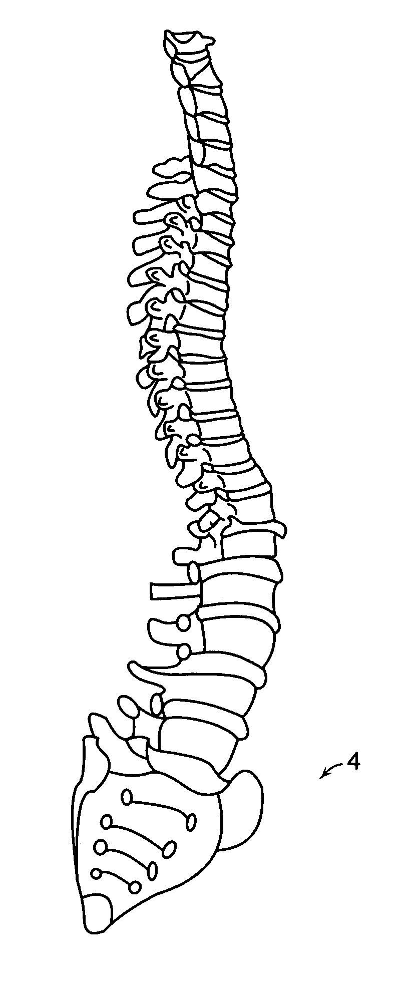
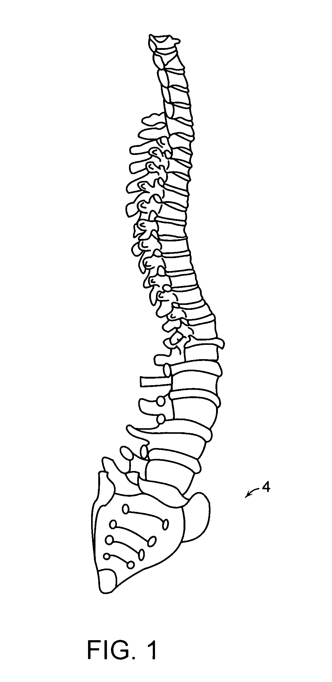
[0040]1. Obtain lateral 2-D x-ray images of the spine.

[0041]2. Estimate the location of the aorta.

[0042]3. Detect calcified areas.

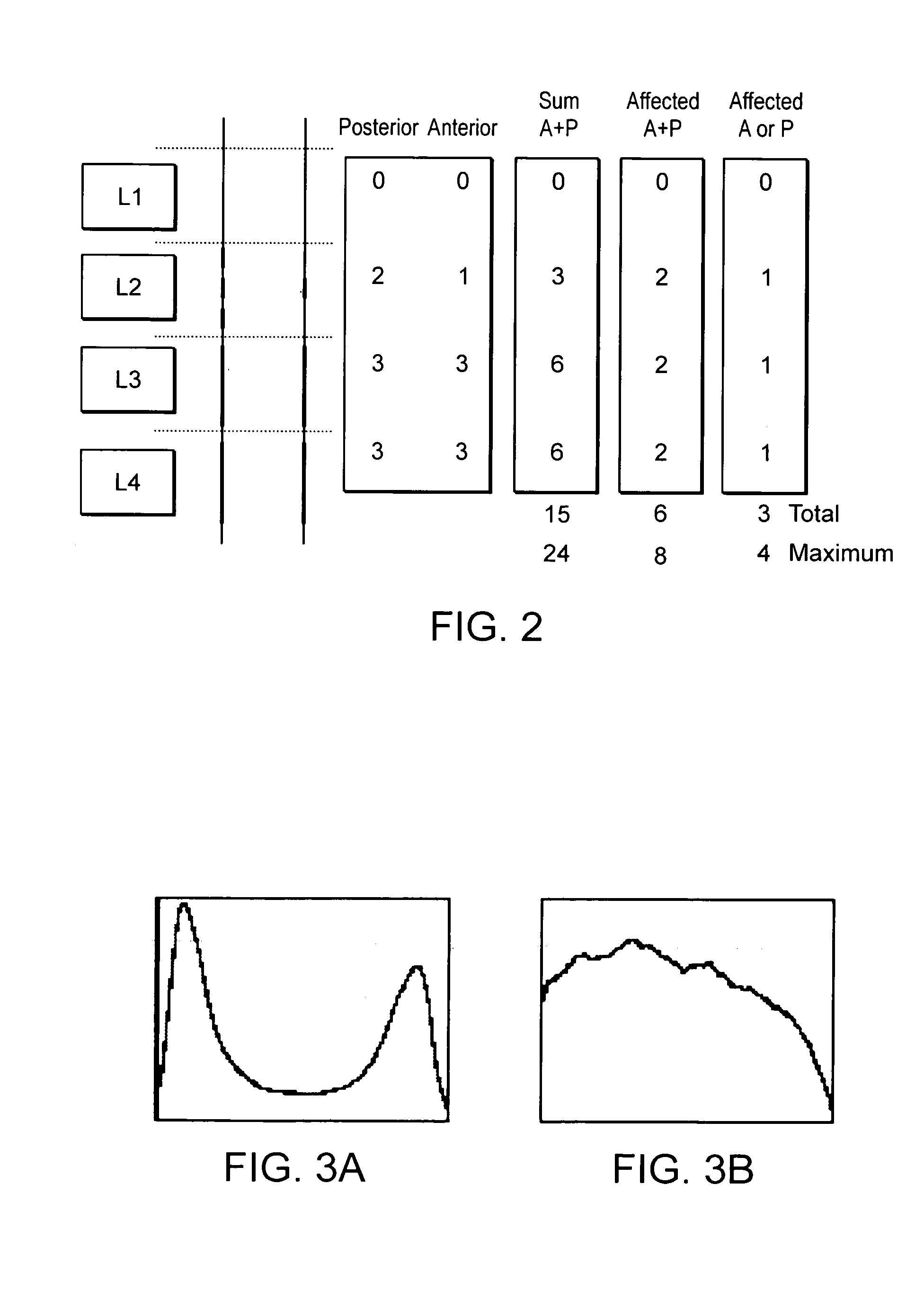
[0043]4. Obtain a quantitative measure of the degree of calcification.

[0044]5. Output the result.

[0045]The present invention is primarily concerned with Steps 3 and 4, the actual process involved in detecting calcified areas of an aorta and obtaining a quantitative measure of the degree of calcification.

[0046]Methods of obtaining x-ray images are well known. This step will therefore not be described herein.

[0047]In the described embodiment, lateral 2-D x-rays are used to provide a cost-efficient way of delivering reliable and easily reproducible data which may be used to extract information regarding the degree of calcification of the aorta.

[0048]Calcifications usually can be depicted in x-rays as small and usually elongated bright structu...


PUM

PropertyMeasurementUnit
areaaaaaaaaaaa
signal to noise ratioaaaaaaaaaa
noise ratioaaaaaaaaaa

Abstract

A method of deriving a quantitative measure of a degree of calcification of a blood vessel such as an aorta by processing an image such as an X-ray image of at least a part of the blood vessel containing said calcification comprises:taking a starting set of digital data representative of an image of at least part of a blood vessel containing a calcification set against a background;estimating the boundary of the calcification;using inpainting to replace digital data in said starting set representing the calcification with data extrapolating the boundary of the background to extend over the area of calcification, and so generating an inpainted set of digital data; andcomputing the difference between the starting set of digital data and the inpainted set of digital data to obtain a quantitative measure of the degree of calcification of the blood vessel.

Description

CROSS-REFERENCE TO RELATED APPLICATION[0001]The present application is a continuation-in-part of U.S. patent application Ser. No. 11 / 144,488 as filed on Jun. 2, 2005 now U.S. Pat. No. 7,463,758 the disclosure of which is incorporated herein by reference.BACKGROUND OF THE INVENTION[0002]The present invention relates to a method of deriving a quantitative measure of a degree of calcification of a blood vessel, e.g. an aorta.[0003]Inpainting is a technique that originates from retouching paintings where one wants to recreate lost or damaged structures in a legible way. Digital inpainting uses spatial or frequency information to restore partially damaged / removed images.[0004]Various inpainting techniques are known that enable image restoration, in particular for photographs, videos and films.[0005]It is known to detect and inpaint small regions in mammograms that possibly define a micro calcification to enable detection of calcified regions. Subsequently, features such as the average an...

Claims


Application Information

Patent Timeline
Patent Type & Authority Patents(United States)
IPC IPC(8): G06K9/00B41M5/00
CPCG06T7/0012G06T7/0083G06T2207/30101G06T2207/30012G06T2207/10116G06T7/12
Inventor CONRAD-HANSEN, LARS A.DE BRUIJNE, MARLEENLAUZE, FRANCOIS BERNARDCHRISTIANSEN, CLAUSNIELSEN, MADS
Owner BIOCLINICA
Why Eureka
Try Eureka