

Method for differentiating tissues in magnetic resonance imaging
a tissue and magnetic resonance imaging technology, applied in the field of tissue differentiation methods in magnetic resonance imaging, can solve the problem of not providing tissue differentiation methods
- Summary
- Abstract
- Description
- Claims
- Application Information
AI Technical Summary
Benefits of technology
Problems solved by technology
Method used
Image

Examples
example 1
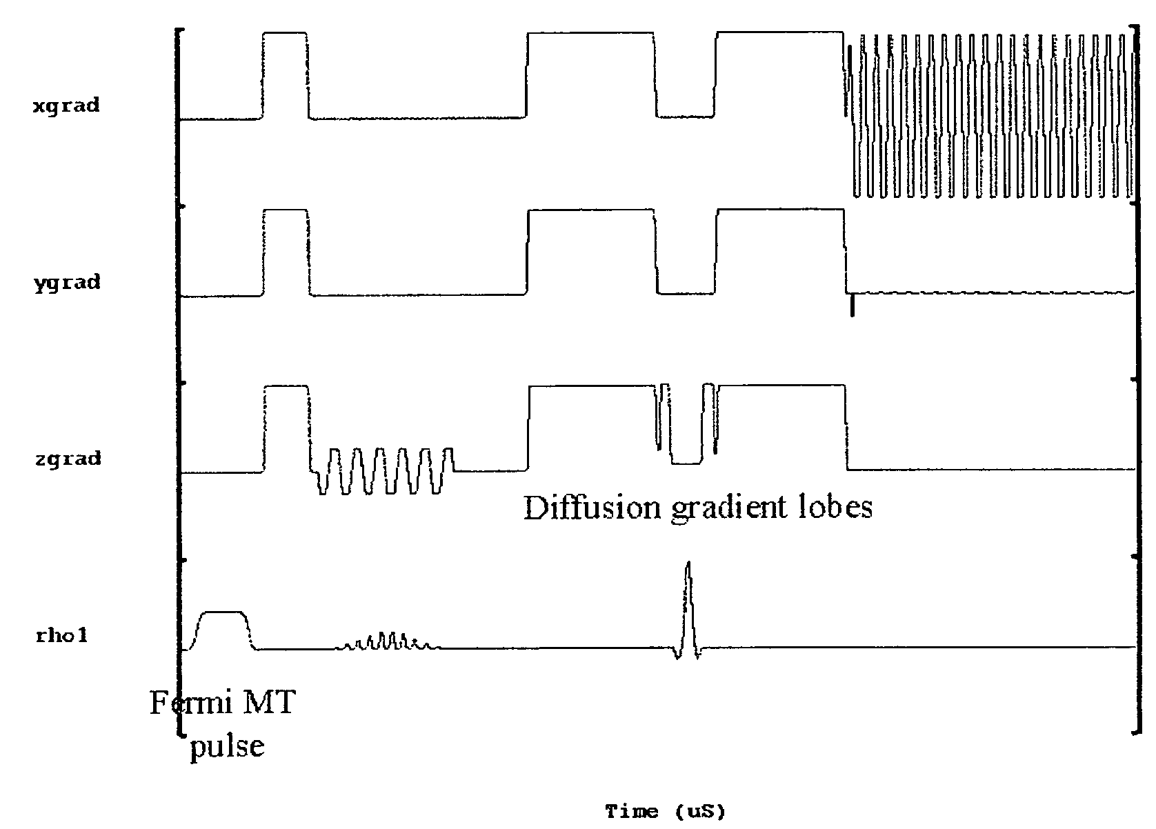
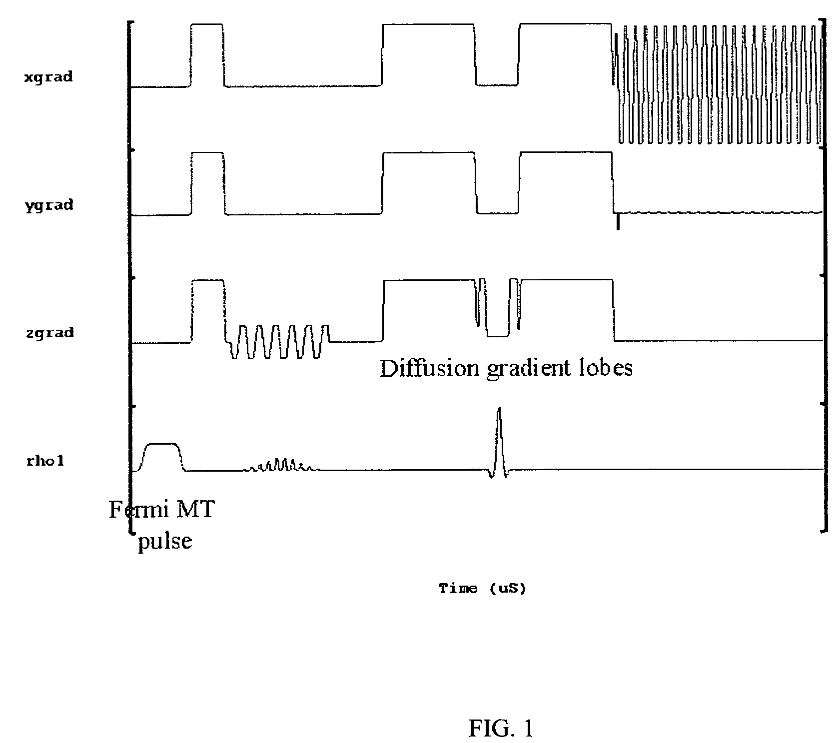
[0019]A total of 10 healthy age and sex-matched volunteers were scanned after taking their informed consent. All these volunteers were male between 27-35 years of age. Imaging experiments were performed on 1.5-T clinical MR scanner (Signa; GE Medical Systems, Milwaukee, Wis.), using a quadrature head coil available with the scanner. All experiments were carried out with a modified single shot SE-DWEPI pulse sequence, with and without MT preparation pulse.
[0020]The imaging parameters used were:[0021]TR / TE=10.5 s / 10 ms (minimum TE),[0022]FOV=40 cm, number of excitation=2,[0023]slice thickness 5 mm,[0024]interslice gap=0.1 mm with matrix size of 128×128.
[0025]Diffusion gradients were applied along all orthogonal directions (namely X, Y and Z) with a “b” value of 1000 s / mm2. The MT RF pulses used were Fermi pulses of 16 ms duration with a flip angle of 670 deg: and resonance offset of 1200 Hz. The MT pulses were applied in every TR.
[0026]It was found that a short (8 ms) Fermi-shaped off...
example 2
[0033]Preliminary results on in vivo MR experiments and further physical measurements on excised cystic tumors and aspirated abscesses on patients further supported and validated the postulates using the longer, sharper excitation profile off-resonance MT pre-pulse. The abscesses have large cellular component and are highly viscous, whereas fluid from the cystic tumors is less viscous (observed from physical measurements in Table 2).
[0034]
TABLE 2Summary of biological parameters and computed ADC with andwithout MT pre-pulse for abscess and tumorsTumor cavity(n = 6)Param-Abscess cavity (n = 12)Me-etersMedianRangedianRange*p-valueADC0.700.52-1.792.991.99-3.180.000*ADC_MT0.760.54-1.963.092.09-3.380.000*Protein40.31 13-13438.00 27-1140.892molecularweight(kD)Cell32000 45-2800000 0-250.000*densityViscosity16.06 2.60-10191.251-40.001**using Mann-Whitney U Test
[0035]Fluid from cystic lesions was collected at the time of surgery and various biological parameters that are supposed to influenc...
PUM
 Abstract
Description
Claims
Application Information