Medical training simulator including contact-less sensors

a technology of contactless sensors and simulators, which is applied in the field of medical simulators, can solve the problems of not being well suited for all types of training and not always readily available cadavers, and achieve the effect of facilitating proper positioning of simulated tissu

Inactive Publication Date: 2010-02-23
TOLY CHRISTOPHER C
View PDF87 Cites 51 Cited by
  • Summary
  • Abstract
  • Description
  • Claims
  • Application Information

AI Technical Summary

Benefits of technology

[0007]In the present invention, contact-less sensors and corresponding detection objects are used to enhance the performance of medical training systems. By using contact-less sensors and corresponding detection objects, each contact-less sensor and detection object can be incorporated into components of the medical training system in a way that hides the sensors and detection objects from view. This approach not only enhances a realistic appearance of the components of the medical training system, it also reduces a likelihood that any trainee will be able to infer a particular significance for any portion of a component of the medical training system by recognizing that a sensor or a detection object is disposed in a particular portion of the medical training system. Many different types of contact-less sensors can be employed, such as magnetic sensors, capacitance-based sensors, and inductive-based sensors.
[0009]Embodiments in which the detection object is incorporated into the medical model enable less-expensive medical models to be produced, because detection objects are generally less expensive than their corresponding sensors. A single sensor integrated into a simulated instrument can detect a plurality of different detection objects distributed throughout a medical model. Furthermore, if the sensors are included in the simulated medical instrument rather than incorporated into the medical model, the medical model should not require wiring (which would complicate the manufacturing process of the medical model) to logically couple each sensor to the processor.
[0011]In another embodiment, at least one detection object is incorporated into a disposable simulated physiological structure, such as a simulated tissue. The detection object is incorporated into a portion of the simulated tissue that must be dissected in a training exercise. Before the exercise, the simulated tissue is placed adjacent to a sensor bed, such that the sensor is triggered by the detection object in the simulated tissue structure. When the student successfully dissects the portion of the simulated tissue structure incorporating the detection object, the sensor is no longer triggered by the detection object, and the processor can provide feedback indicating that the student has successfully completed the exercise. Such a sensor bed can be beneficially incorporated into a box trainer. Further, it should be understood that the sensor bed can be used with the simulated tissue, and without a box trainer, if so desired. Additional detection objects can be incorporated into the simulated tissue, with additional sensors being incorporated into the sensor bed to facilitate proper positioning of the simulated tissue relative to the sensor bed.
[0012]The sensor data collected during a training exercise can be used to immediately provide feedback to a student during the training exercise, or can be stored for later review, or the sensor data can be manipulated to provide feedback only to a proctor monitoring the training exercise, so that the proctor knows whether the student has successfully completed the training exercise. Sensor data (or feedback generated by the processor after receiving the sensor data) can be communicated to remote locations over a network, so that the proctor and student do not need to be in the second location. Thus, the present invention is well-suited for remote learning environments.

Problems solved by technology

Although cadavers have traditionally been beneficially employed for this purpose, cadavers are not always readily available and are not well suited for all types of training.
Even if a simulated physiological structure having simulated tissue faithfully portrays finer details of an actual physiological structure and provides a realistic tactile sensation during a simulated procedure, the few prior art simulators that may both tactilely and visually resemble the anatomical structure being simulated do not include means for producing objective and measurable results that can be used to evaluate how well a simulated procedure is performed.

Method used


Image

  • Medical training simulator including contact-less sensors
  • Medical training simulator including contact-less sensors
  • Medical training simulator including contact-less sensors

Examples


Embodiment Construction

Overview of the Present Invention

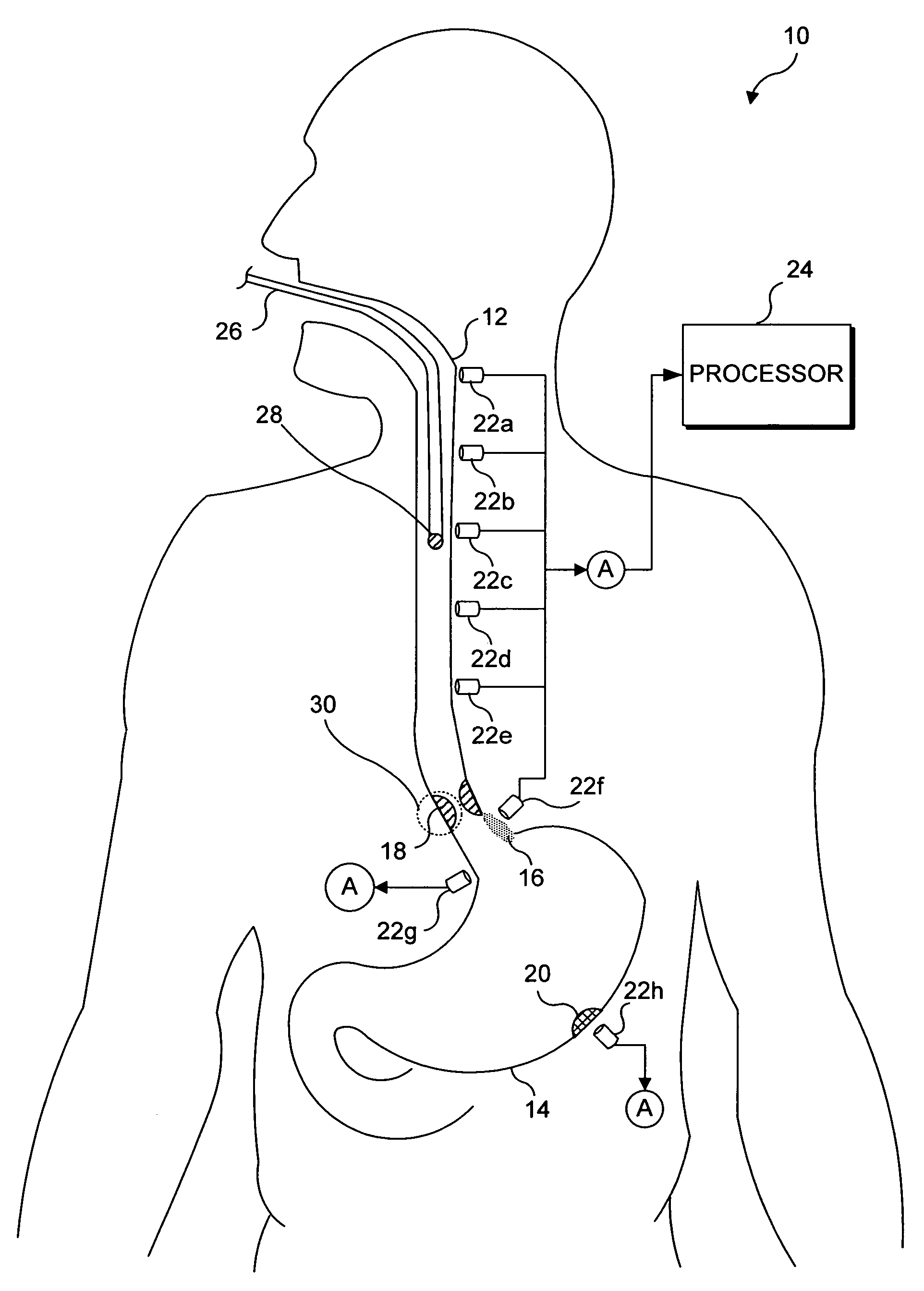
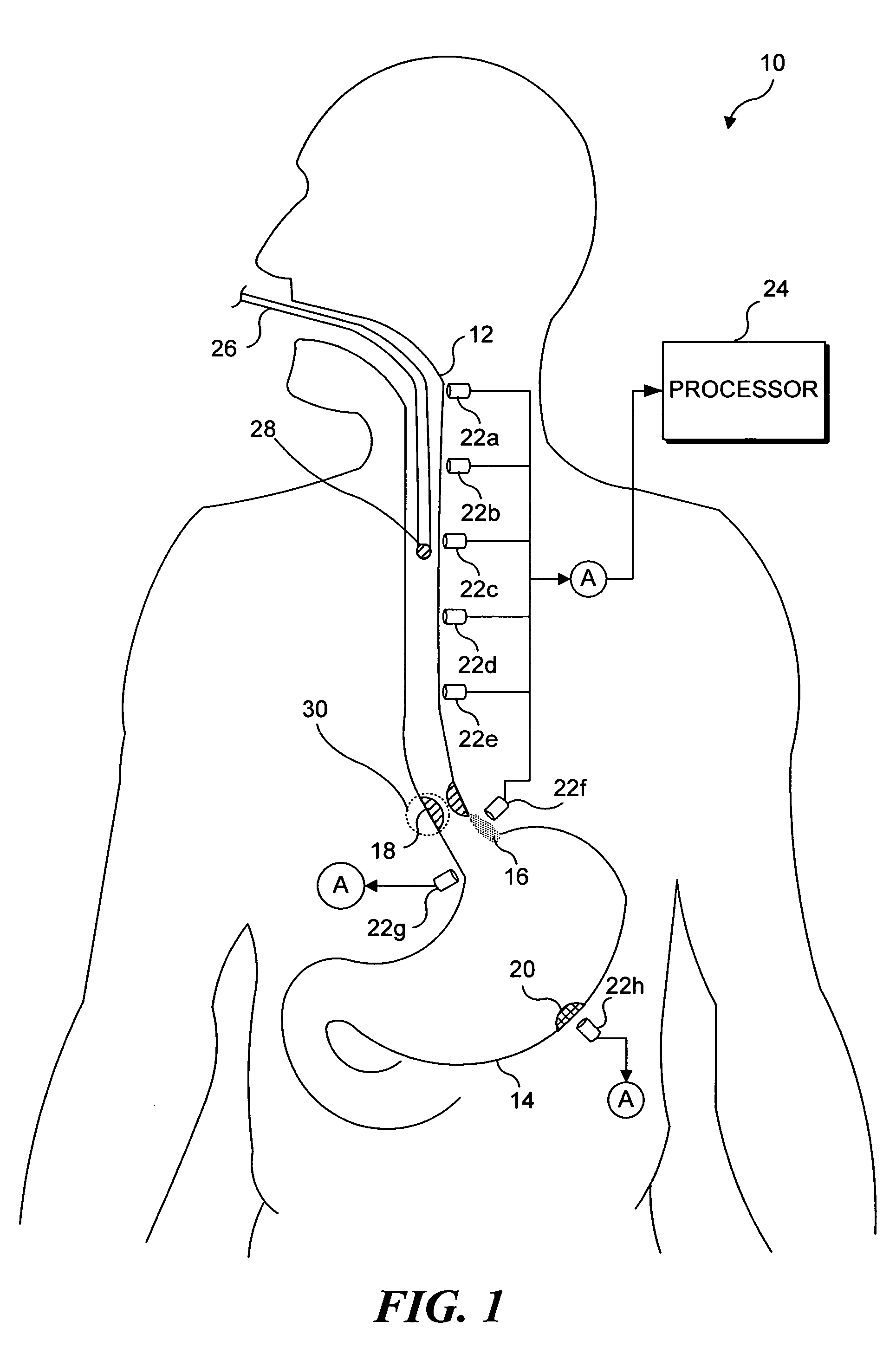
[0029]In the present invention, one or more sensors are incorporated into a medical training model that can be used for teaching and / or testing. Such sensors can be employed to provide feedback indicating how well a simulated procedure was performed using the medical training model. Each medical model preferably includes one or more simulated physiological structures. Preferably, much of the model, and in particular the simulated physiological structures, are formed out of elastomeric materials to enhance a realism of the medical training model.

[0030]The sensors used in the present invention do not require physical contact between the sensor and an object that is to be detected and are therefore referred to as “contact-less sensors.” In the context of the present invention, the object to be detected is generally a tool used during a simulated medical procedure (although in some embodiments, a sensor is incorporated into such a tool, to detect objects...


PUM


Abstract

A medical training simulator includes contact-less sensors and corresponding detection objects, configured to enable sensor data collected during a training exercise to be used to evaluate the performance of the training exercise. The simulator includes a simulated anatomical structure, at least one contact-less sensor, and at least one detection object. During a training exercise, a spatial relationship between the contact-less sensor and the detection object produces data for evaluating performance of the training exercise. Either the contact-less sensor or the detection object is embedded in the simulated physiological structure, while the other is included in either a support for the simulated physiological structure, or as part of a tool used during the training exercise. Many types of contact-less sensors can be employed, including capacitance sensors, impedance sensors, inductive sensors, and magnetic sensors.

Description

RELATED APPLICATIONS[0001]This application is a continuation-in-part of a copending patent application, Ser. No. 10 / 718,492, filed on Nov. 20, 2003, which itself is a continuation-in-part of a copending patent application Ser. No. 09 / 695,380, filed on Oct. 23, 2000, the benefit of the filing dates of which is hereby claimed under 35 U.S.C. § 120.FIELD OF THE INVENTION[0002]The present invention generally pertains to medical simulators for use as medical training aids, and more specifically, to medical simulators that include contact-less sensors and corresponding detection objects, which facilitate the evaluation of a student's performance during a training exercise implemented with the medical simulator.BACKGROUND OF THE INVENTION[0003]The use of simulated physiological structures for training medical students and for providing skill training to practicing physicians is widespread. Although cadavers have traditionally been beneficially employed for this purpose, cadavers are not al...

Claims


Application Information

Patent Timeline
Patent Type & Authority Patents(United States)
IPC IPC(8): G09B23/28G09B23/30
CPCG09B23/285G09B23/30
Inventor TOLY, CHRISTOPHER C.
Owner TOLY CHRISTOPHER C
Why Eureka
Try Eureka