Method and composition for diagnosis of melanocytic lesions

a technology of melanocytic lesions and compositions, applied in the field of melanocytic antigens and antibodies against melanomaassociated antigens, can solve the problems of limiting the usefulness of nki/c3 antibodies in the diagnosis of melanocytic lesions, anti-s100 antibodies are notoriously unspecific,

Inactive Publication Date: 2010-02-23
ONCOMAX ACQUISITION CORP +1
View PDF18 Cites 8 Cited by
  • Summary
  • Abstract
  • Description
  • Claims
  • Application Information

AI Technical Summary

Problems solved by technology

Early diagnosis is required for surgery to be effective, which is unfortunately hampered by the lack of markers that are sensitive and specific for melanoma.
However, anti-S100 antibodies are notoriously unspecific.
These unspecific activities limit the usefulness of NKI / C3 antibody in the diagnosis of melanocytic lesions.

Method used


Image

  • Method and composition for diagnosis of melanocytic lesions

Examples


Embodiment Construction

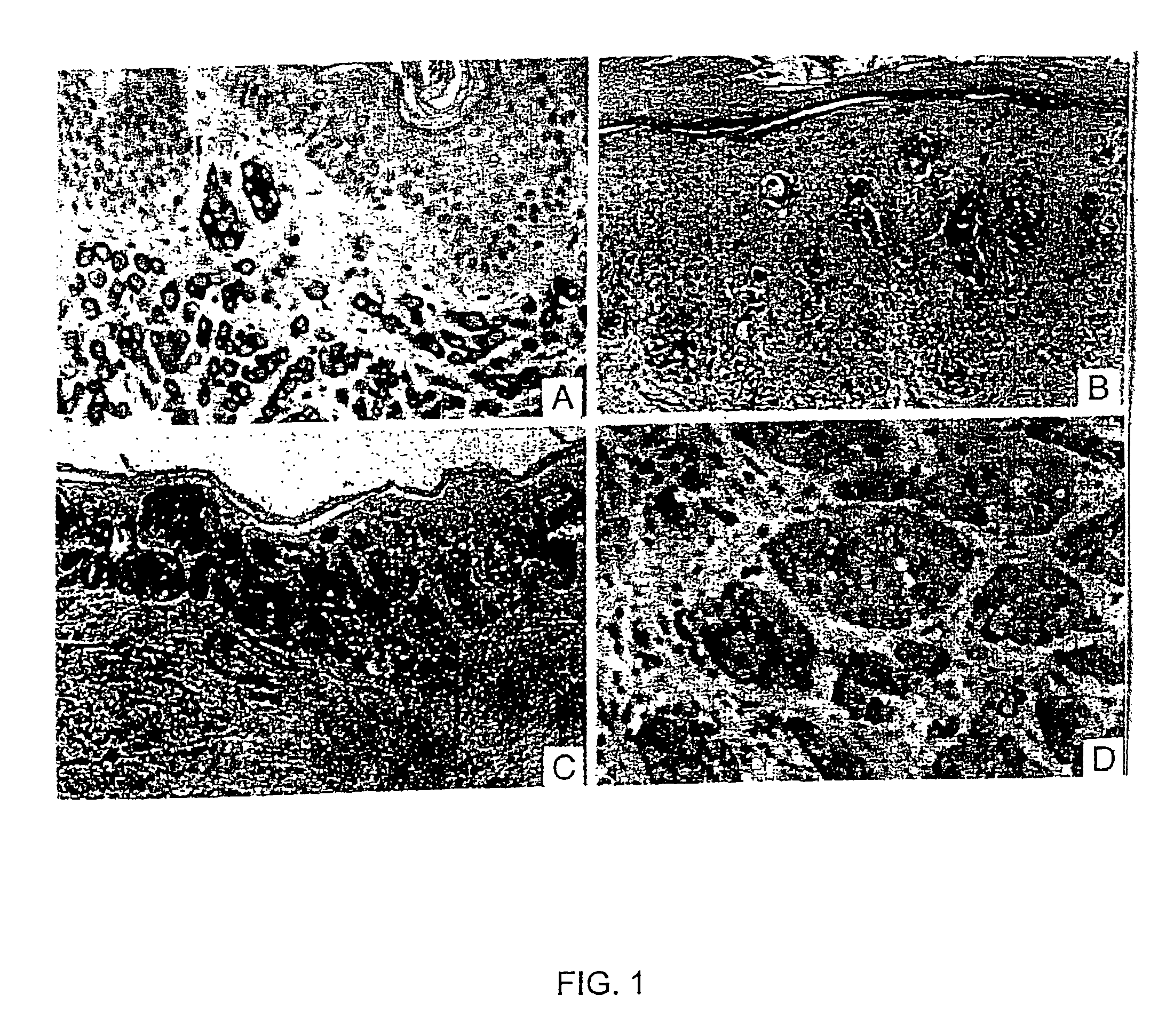
[0078]A. Preparation of SM5-1 Antibody

[0079]SM5-1 antibody is selected from a panel of monoclonal antibodies generated by using a subtractive immunization protocol as previously described in Williams et al., (1992) Biotechniques 12:842–8477, and Brooks et al., (1993) J. Cell Biol. 22:1351–1359, both of which are incorporated by reference herein.

[0080]Briefly, mice were immunized with human melanoma cell line SMMU-1, which was obtained from the primary melanoma of a patient (Guo et al., (1994) Cancer Res. 54:561–1565, incorporated by reference herein). Next, the mice were treated with cyclophosphamide to abrogate activated B cells that produce antibody against epitopes expressed by the primary melanoma. The mice were then immunized with human melanoma cell line SMMU-2, which was obtained from the metastatic lesion of the same patient. The splenocytes from the mice were used for making hybridomas using standard techniques. Köhler G, Milstein C: Derivation of specific antibody producin...


PUM

PropertyMeasurementUnit
apparent molecular weightaaaaaaaaaa
spectrumaaaaaaaaaa
antibody titersaaaaaaaaaa

Abstract

This invention relates to monoclonal antibodies that recognize an antigen specific to melanocytic lesions. These antibodies are useful in methods of isolating melanoma cells and diagnosing melanocytic lesions. These antibodies are also useful for immunotherapy against melanoma.

Description

[0001]This application is a continuation of application Ser. No. 09 / 451,353, filed Dec. 1, 1999, entitled: METHODS AND COMPOSITION FOR DIAGNOSIS OF MELANOCYTIC LESIONS, now abandoned.I. FIELD OF THE INVENTION[0002]This invention relates to melanoma-associated antigens, antibodies against melanoma-associated antigens, and diagnostic and therapeutic methods concerning melanoma.II. BACKGROUND OF THE INVENTION[0003]Human malignant melanoma usually starts as harmless moles that undergo radial to invasive growth and end in the destructive stage of metastatic melanoma. Melanoma usually resists chemotherapy and radiotherapy. Surgery is the most effective treatment. Early diagnosis is required for surgery to be effective, which is unfortunately hampered by the lack of markers that are sensitive and specific for melanoma.[0004]The commonly used antibodies for immuno-histochemical diagnosis of melanoma are HMB-45, anti-S-100 and NKI / C3 (for a review, please see Smoller B R, “Immunohistochemist...

Claims


Application Information

Patent Timeline
Patent Type & Authority Patents(United States)
IPC IPC(8): G01N33/53G01N33/574C07H21/04C07K16/30C12N15/09C12Q1/68
CPCC07K16/3053
Inventor GUO, YAJUNMA, JING
Owner ONCOMAX ACQUISITION CORP
Why Eureka
Try Eureka