Subcutaneous cardioverter-defibrillator

a cardioverter and subcutaneous technology, applied in the field of implantable cardioverter defibrillators, can solve the problems of prohibitively expensive implanting of currently available imds in all such patients, difficult to implant in some patients, and high cost of imds and implant procedures

Active Publication Date: 2010-03-23
MEDTRONIC INC
View PDF25 Cites 97 Cited by
  • Summary
  • Abstract
  • Description
  • Claims
  • Application Information

AI Technical Summary

Problems solved by technology

Such devices are often difficult to implant in some patients (particularly children and thin, elderly patients) and typically require the sacrifice of 1 or 2 veins to implant the lead system because leads associated with the implantation of an IMD utilize a transvenous approach for cardiac electrodes and lead wires.
Although IMDs and implant procedures are very expensive, most patients who are implanted have experienced and survived a sudden cardiac death episode because of interventional therapies delivered by the IMDs.
However, implanting currently available IMDs in all such patients would be prohibitively expensive.
Further, even if the cost factor is eliminated there is shortage of trained personnel and implanting resources.
Further, endocardial lead placement is not possible with patients who have a mechanical heart valve implant and is not generally recommended for pediatric cardiac patients.

Method used


Image

  • Subcutaneous cardioverter-defibrillator
  • Subcutaneous cardioverter-defibrillator
  • Subcutaneous cardioverter-defibrillator

Examples


first embodiment

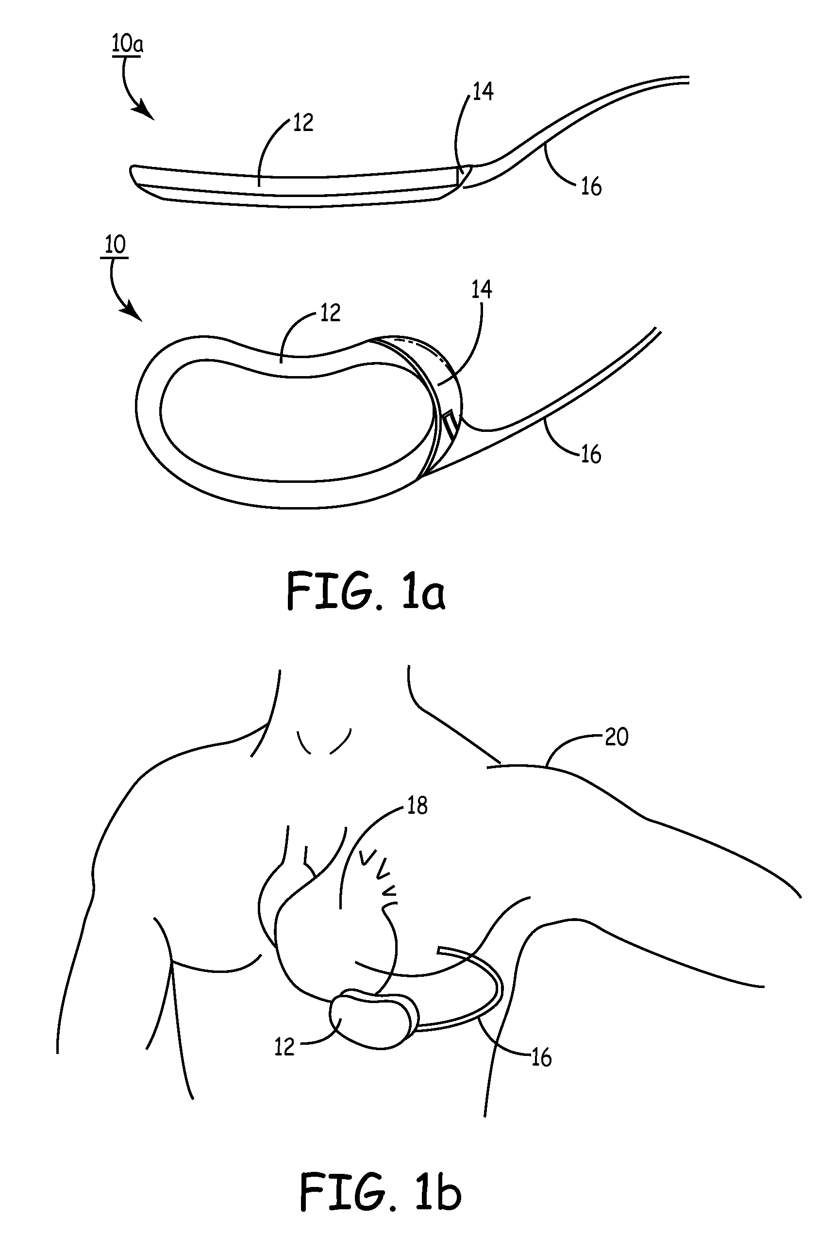
[0061]FIG. 1A depicts a multi-planar view of the present invention. SubQ ICD 12 is an ovoid, substantially, kidney-shaped housing with connector 14 for attaching a subcutaneous sensing and cardioversion / defibrillation therapy delivery lead 16. SubQ ICD 12 may be constructed of stainless steel, titanium or ceramic as described in U.S. Pat. Nos. 4,180,078 “Lead Connector for a Body Implantable Stimulator” to Anderson and 5,470,345 “Implantable Medical Device with Multi-layered Ceramic Enclosure” to Hassler, et al. The electronics circuitry of SubQ ICD 10 (described herein pertaining to FIG. 21) may be incorporated on a polyamide flex circuit, printed circuit board (PCB) or ceramic substrate with integrated circuits packaged in leadless chip carriers and / or chip scale packaging (CSP). In one of the views, the concave construction of SubQ ICD 12 is illustrated. The minor concavity of the housing of SubQ ICD 12 follows the natural curve of the patient's median ribcage at about the cardia...

second embodiment

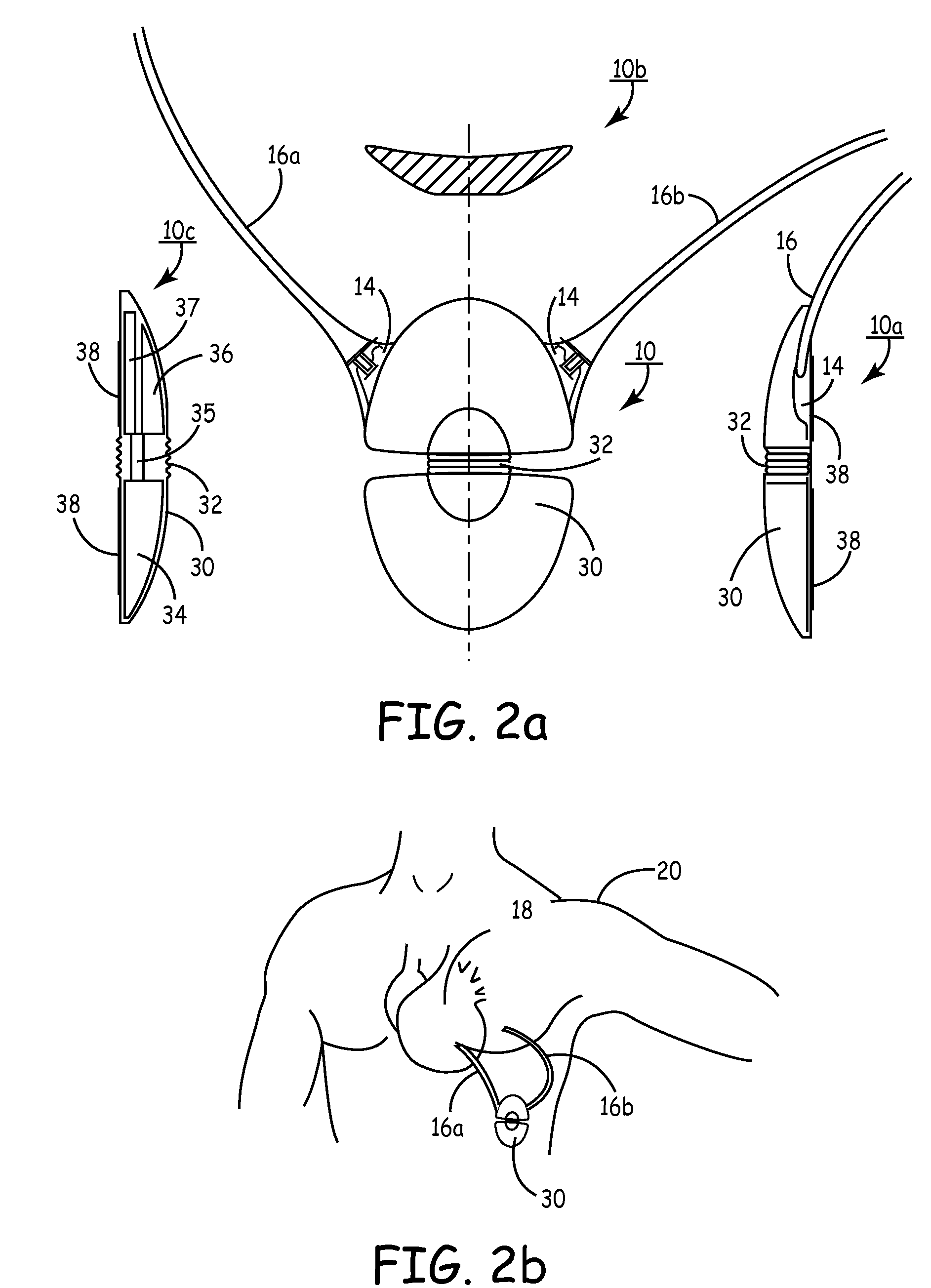
[0064]FIG. 2A is a multi-planar view of SubQ ICD 30, the present invention. SubQ ICD 30 is a convex, flexible ovaloid-shaped housing with connectors 14 (2 shown) for attaching 2 subcutaneous sensing and cardioversion / defibrillation therapy delivery leads 16A and 16b. SubQ ICD 30 may be constructed of stainless steel, titanium or ceramic. View 10A is a side view of SubQ ICD 30 showing the tapered housing 30, a mid-line flexible joint 32, connector 14, lead 16 and active can electrode 38. The active can electrode 38 allows sensing and cardioversion, defibrillation and / or pacing therapy delivery between the SubQ ICD 30 and one or both leads 16A or 16b. The jointed housing 30 allows physician flexibility in selecting implant locations and accommodates variances in size and weight of patients for implant. Additionally, the flexible housing provides less patient discomfort in sitting, bending over and / or during normal torso movement because the configurations allows dynamic adjustment to ...

third embodiment

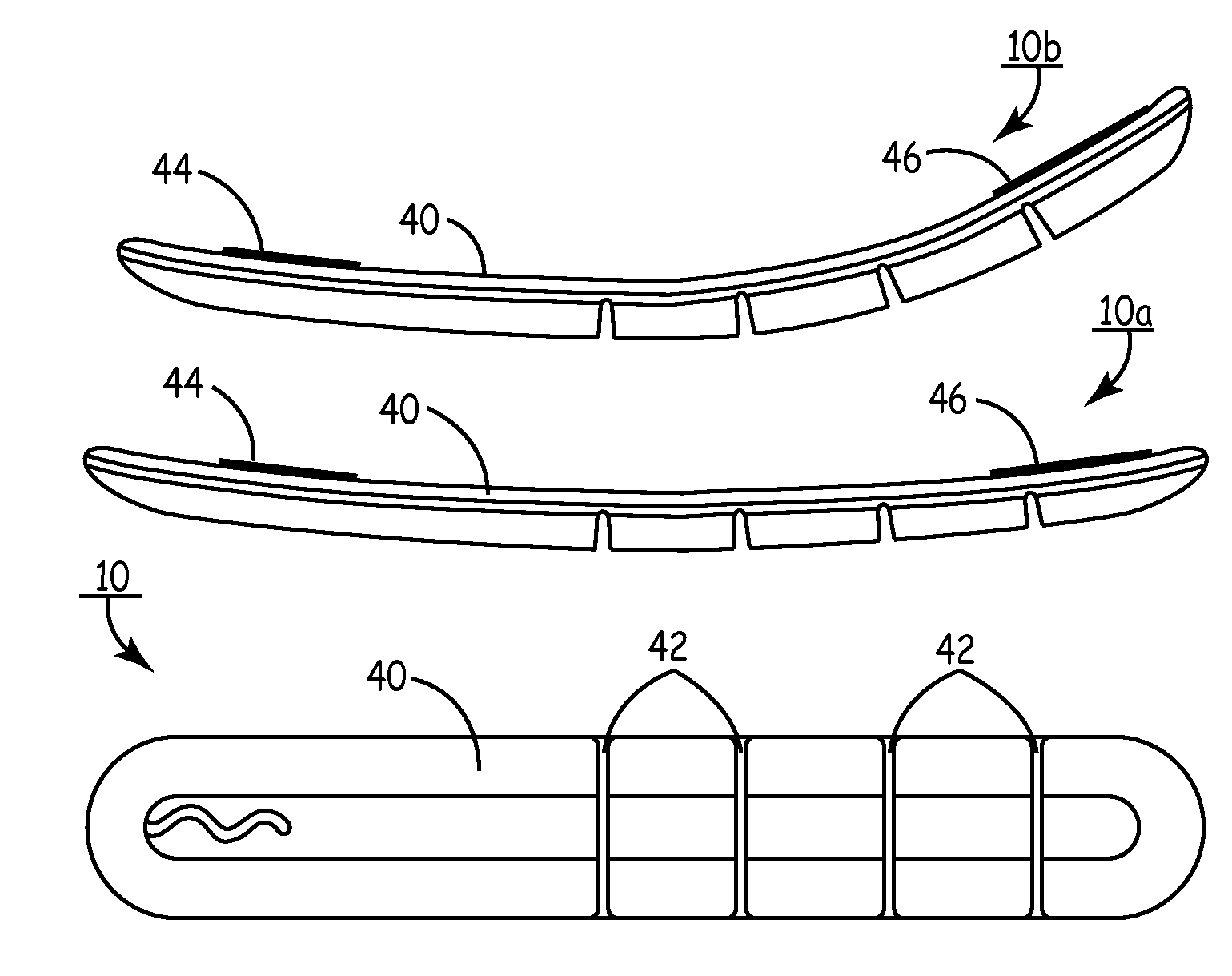
[0066]FIG. 3A is a multi-planar view of SubQ ICD 40. SubQ ICD 40 is an elongated slender ellipsoid with sections of partially articulating dynamic segments having surface mounted subcutaneous sensing and cardioversion / defibrillation therapy delivery electrodes 44 and 46. SubQ ICD 40 may be constructed of stainless steel, titanium or ceramic or equivalent. View 10A is a top view of SubQ ICD 40 showing the segmented construction. Electrodes 44 and 46 located at opposite ends of SubQ ICD 40 are typically 100 mm2 to 1000 mm2. View 10b is a further top view showing the dynamic flexibility of SubQ ICD 40 in which it assumes a dynamically adjustable, compressive and tensile opposing surfaces when implanted outside the thoracic cavity over the ribs. Specifically, in its normal position, SubQ ICD 40 is substantially flat both at the top and bottom surfaces. However, when implanted, SubQ ICD 40 dynamically forms a concave and convex surface at the flat top and segmented bottom surfaces when t...


PUM


Abstract

SubQ ICDs are disclosed that are entirely implantable subcutaneously with minimal surgical intrusion into the body of the patient and provide distributed cardioversion-defibrillation sense and stimulation electrodes for delivery of cardioversion-defibrillation shock and pacing therapies across the heart when necessary. Configurations include one hermetically sealed housing with 1 or, optionally, 2 subcutaneous sensing and cardioversion-defibrillation therapy delivery leads or alternatively, 2 hermetically sealed housings interconnected by a power / signal cable. The housings are generally dynamically configurable to adjust to varying rib structure and associated articulation of the thoracic cavity and muscles. Further the housings may optionally be flexibly adjusted for ease of implant and patient comfort.

Description

FIELD OF THE INVENTION[0001]The present invention generally relates to implantable medical devices, particularly implantable cardioverter-defibrillators that are entirely implanted subcutaneously (SubQ ICD) and, more particularly, have no leads or electrodes contacting the heart or extending into the thoracic cavity.BACKGROUND OF THE INVENTION[0002]Many types of implantable medical devices (IMDs) have been clinically implanted over the last twenty years that deliver relatively high-energy cardioversion and / or defibrillation shocks to a patient's heart when a malignant tachyarrhythmia, e.g., atrial or ventricular fibrillation, is detected. Cardioversion shocks are typically delivered in synchrony with a detected R-wave when fibrillation detection criteria are met, whereas defibrillation shocks are typically delivered when fibrillation criteria are met and an R-wave cannot be discerned from the EGM.[0003]The current state of the art of ICDs or implantable pacemaker / cardioverter / defibr...

Claims


Application Information

Patent Timeline
Patent Type & Authority Patents(United States)
IPC IPC(8): A61N1/02
CPCA61N1/375A61N1/3968A61N1/0504A61N1/3756A61N1/37518
Inventor OLSON, WALTER H.MOY, KING Y.NAKAYAMA, JOEY G.PROKOP, GARY F.STILWELL, GARY J.
Owner MEDTRONIC INC
Why Eureka
Try Eureka