Methods and devices for endoscopic imaging

a technology of endometrial imaging and endometrium, applied in the field of endometrial imaging, can solve the problems of no current screening test for endometrial cancer, no endometrial imaging is used in the above techniques, and no live or stored 2-d visual image inspection may yield adequate information for detailed evaluation

Inactive Publication Date: 2010-06-29
ZICKFELD ASSOC LLC
View PDF28 Cites 86 Cited by
  • Summary
  • Abstract
  • Description
  • Claims
  • Application Information

AI Technical Summary

Problems solved by technology

This live or stored 2-D visual image inspection may not yield adequate information for detailed evaluation.
On the other hand, there is no current screening test for endometrial cancer, the most prevalent form of gynecological cancer.
None of the above techniques use imaging of the endometrium.
However, the majority of gynecologists do not perform hysteroscopy.
Beyond the issues of pain, invasiveness, and morbidity, there is a steep learning curve.
In addition, the use of a distending media, for example, saline or a gas (e.g., CO2) to create open space in the uterus may lead to problems.
In addition, because the hysteroscope can only image the tissue in front of it, experience and manual dexterity are required in order to examine the whole endometrium.

Method used


Image

  • Methods and devices for endoscopic imaging
  • Methods and devices for endoscopic imaging
  • Methods and devices for endoscopic imaging

Examples


Embodiment Construction

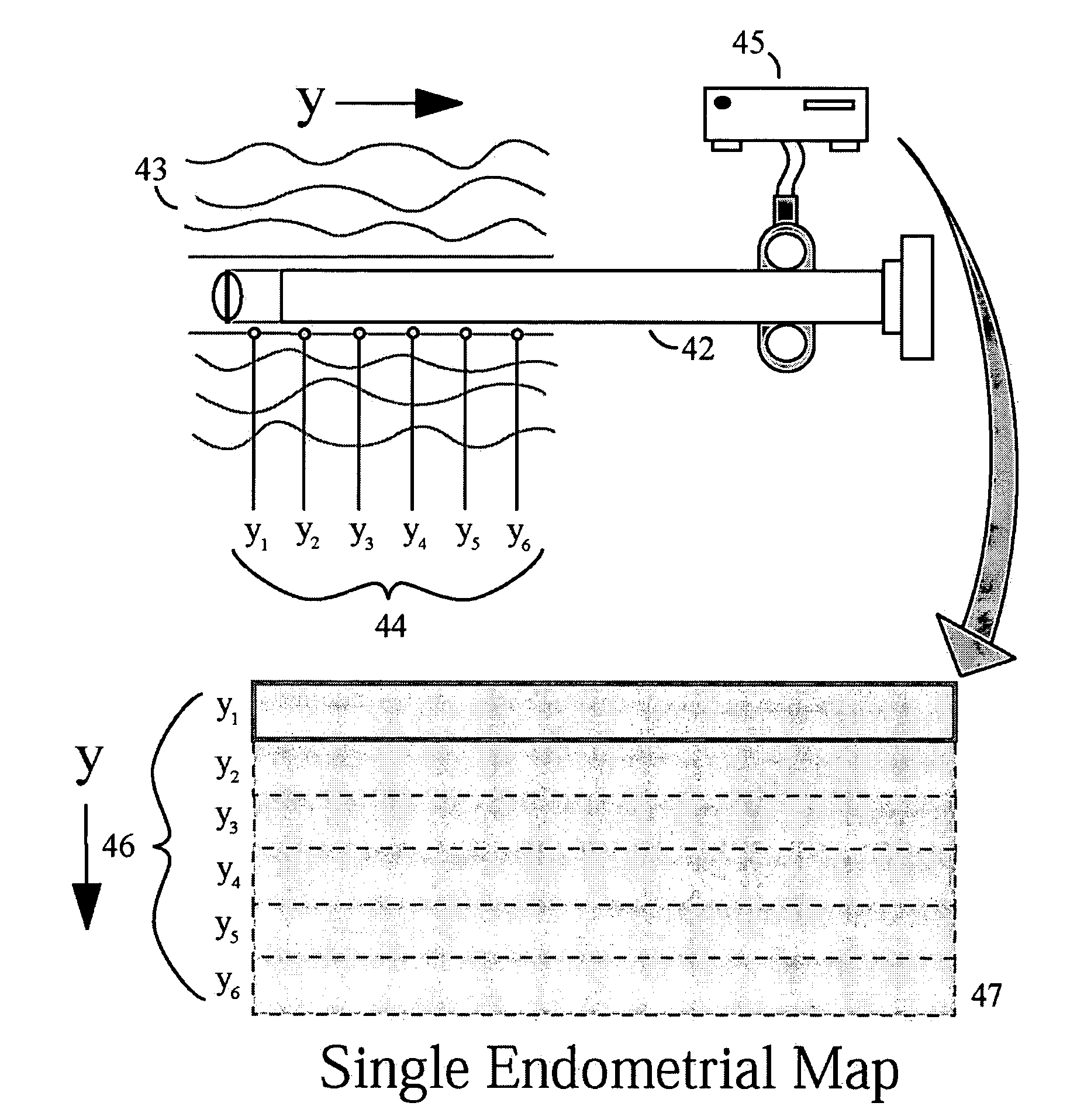
[0027]As described above, the visual inspection of a live or stored 2-D visual image may not provide sufficient information for a detailed evaluation. Such visual inspection does not involve the incorporation of additionally data from other dimensions, such as images acquired at other instances in time, images which use alternate modalities, or images at other depths (3-D images above and below the surface of the tissue). It does not incorporate physiological data such as blood flow or evidence of pathology.

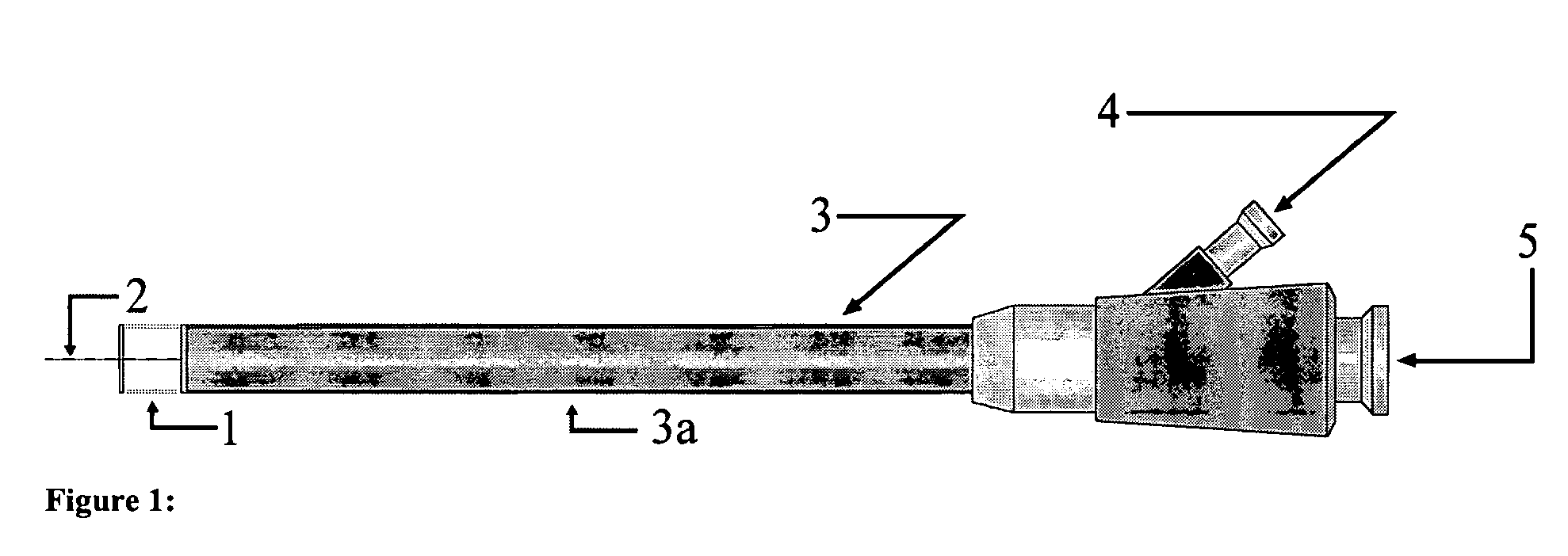
[0028]Certain embodiments of the present invention may pertain to minimally invasive imaging systems and methods used to identify or diagnose pathology of an organ system cavity and / or provide guidance for imaging guided procedures, including but not limited to biopsy and therapy delivery. Such organ system cavities may include, but are not limited to, an endometrial cavity, a gastrointestinal lumen, an orthopedic cavity, an orthopedic joint cavity, a sinus cavity, a nasal passag...


PUM


Abstract

Embodiments include devices and methods. One embodiment includes a method for imaging an endometrial cavity, including acquiring a plurality of images using an imaging system. A first part of the imaging system is positioned within the endometrial cavity. At least portions of two or more of the images are combined into a representation of at least a portion of the endometrial cavity. The combining at least portions of two of the images may include determining any motion of the first part of the imaging system, between the two or more of the images. Other embodiments are described and claimed.

Description

[0001]This application is a continuation-in-part of U.S. application Ser. No. 10 / 785,802, filed Feb. 24, 2004, now U.S. Pat No. 7,559,890 which claims priority in U.S. Provisional Application No. 60 / 450,224, filed Feb. 26, 2003; and this continuation-in-part application claims priority in U.S. Provisional Application No. 60 / 619,736, filed on Oct. 18, 2004. The content of each application number listed above is hereby incorporated by reference in its entirety.RELATED ART[0002]Many types of imaging, include endoscopy, are based on the visual inspection of a live or stored 2-D visual image. This live or stored 2-D visual image inspection may not yield adequate information for detailed evaluation. This shortcoming is present in a number of different fields of medicine, including, but not limited to, gynecology.[0003]A common practice in gynecology is for a woman to have an annual examination including speculum and bimanual examination and a Papanicolau smear (which primarily screens for...

Claims


Application Information

Patent Timeline
Patent Type & Authority Patents(United States)
IPC IPC(8): A61B1/00A61B1/04A61B1/06A61B1/303A61B10/00A61B10/02A61B17/42
CPCA61B1/00096A61B1/00177A61B1/06A61B1/303A61B10/0291A61B17/42A61B1/0607A61B10/0275A61B1/0646A61B1/0625
Inventor WALLACE, JEFFREY M.NATARAJAN, ANANTHVENKATESHA, SANTOSHPEACOCK, KEITHTHAKOR, NITISH V.
Owner ZICKFELD ASSOC LLC
Why Eureka
Try Eureka