
Graft core for seal and suture anastomoses with devices and methods for percutaneous intraluminal excisional surgery (PIES)

a technology of percutaneous intraluminal excision and graft core, which is applied in the field of graft core for seal and suture anastomosis with devices and methods for percutaneous intraluminal excisional surgery, can solve the problems of physiological damage, premature death in industrialized societies, and severe strain on patients

Inactive Publication Date: 2010-08-10
SHRIVER EDGAR LOUIS
View PDF6 Cites 22 Cited by
  • Summary
  • Abstract
  • Description
  • Claims
  • Application Information

AI Technical Summary

Benefits of technology

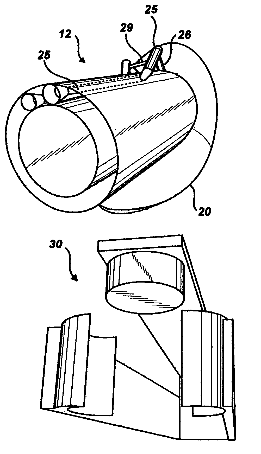
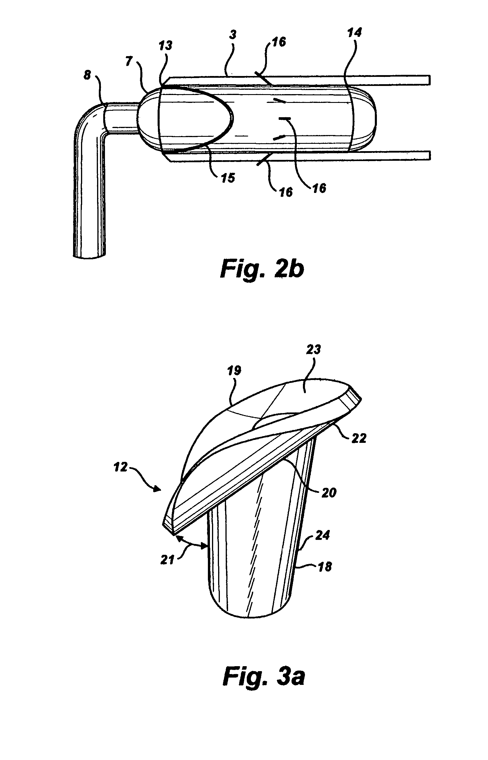
[0017]A delivery catheter slidingly fitted inside the clamping catheter is advanced on the explorer guidewire to the anchored target site. It contains a previously prepared length of graft attached to graft cores at each end by hollow sutures, posts and a circular suture. The brims of the distal and proximal graft cores are slightly compressed to enter the delivery tube. The delivery catheter also contains a catheter with a rodding balloon on its end fitted inside the graft core. The delivery catheter is capped by a holding balloon on which is mounted a cutting device. This makes a slit on the longitudinal axis of the second tube. The delivery tube is immediately pushed through the opening far enough that the brim of the graft core and holding balloon are in the lumen and the stem is in the opening. To remove the graft core from the delivery tube, the rodding balloon is partially inflated to engage the base of the graft core stem. The holding balloon is pulled back and the rodding balloon advanced sufficiently to grip the graft core while the delivery catheter is withdrawn. This allows the brim to expand in the lumen of the second tube and the wall of the second tube to close around the stem of the graft core. This forms a seal as pressure inside the lumen presses the graft core brim against the wall of the lumen which has the same shape as the brim. The key on the distal end of the rodding balloon is in a keyway inside the stem which aligns each rod with the proper stiff suture. The rodding balloon is fully inflated to bring the rods in line with the stiff sutures each is assigned to drive. The stiff suture that must travel the longest distance to enter the brim is in contact with the rod that will drive it first and therefore farthest and the stiff suture with the shortest distance to travel is placed in contact last. This may be accomplished by rods of different length or by stiff sutures at different distances from the base of the graft core. Either way, the rods are advanced through the hollow cones of the hollow sutures in which the stiff sutures are lodged. With the holding balloon pulling the brim of the graft core against the lumen wall, the rodding balloon is advanced, driving the sharp ends of the stiff sutures through the wall of the second tube and into the brim. Barbs prevent the stiff sutures from backing up from this position.
[0021]The devices and methods revealed here utilize the two transected ends of a graft, and thus do not produce a side-to-side graft. If adjacent tubes are to be joined with the devices and methods revealed here, the graft or grafts can be cut so short that the end-to-end graft functions like a side-to-side anastomosis. It may be noted that commonly, and particularly herein, the third tube is called the graft. So the terms used here for the body parts being joined are first tube, second tube and graft. These terms are generic and exact as well as simple and direct, an advantage in a discussion as abstract as a patent application.

Problems solved by technology

This is because there are a large number of tubes carrying essential body fluids for circulation and excretion and they often suffer physiological damage.
Disease in coronary arteries is the leading cause of premature death in industrialized societies.
MIDCABG procedures are endoscopic and involve less collateral damage but still represent a severe strain on patients and have replaced CABG procedures in only a small percentage of cases.
Anastomoses of coronary arteries are challenging because they are tiny, friable, intolerant of injury and moving with each beat of the heart.
The manual skills for sewing extremely small sutures in tiny frail tubes are well perfected by those who perform CABG surgeries but each anastomosis represents a chance for error or stress beyond what the patient can tolerate.
This represents further collateral damage.
The risk in heart stoppage is always high, but goes up sharply if time on bypass machine lasts longer than an hour.
Sometimes the suturing is inadvertently loose, allowing fluid to leak which can cause acute or chronic loss of blood pressure and possible scarring which results in another blockage.
In addition to the threat of leakage, fluid mechanics are such that introducing a stream into a tube at a substantially different angle or velocity of flow than is normal for the tube can cause damage.
Though they appear to have certain advantages over manual suturing, they are limited to CABG and MIDCABG operations.

Method used


Image

  • Graft core for seal and suture anastomoses with devices and methods for percutaneous intraluminal excisional surgery (PIES)
  • Graft core for seal and suture anastomoses with devices and methods for percutaneous intraluminal excisional surgery (PIES)
  • Graft core for seal and suture anastomoses with devices and methods for percutaneous intraluminal excisional surgery (PIES)

Examples


Embodiment Construction

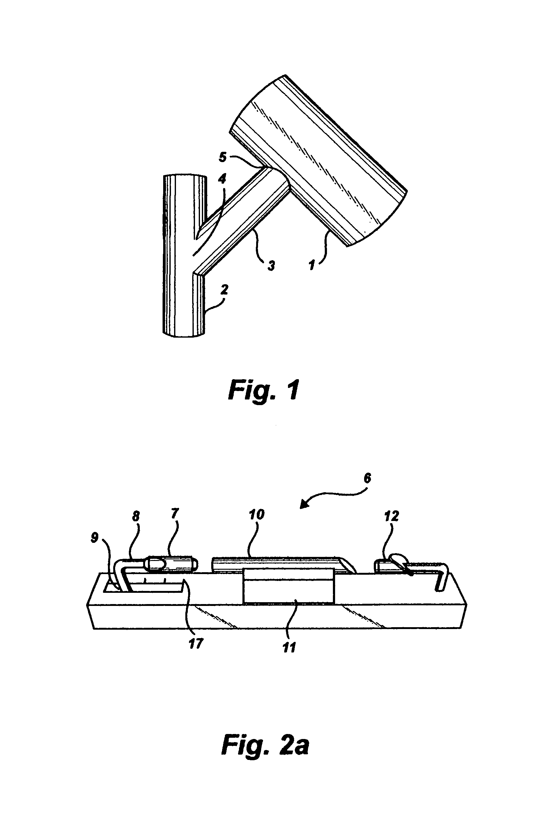
[0065]FIG. 1 shows the product of the present invention that is visually the same as that of prior art; the sides of a mammalian first tube 1 and second tube 2 connected by the transected ends of a graft 3. The transected ends of said graft are also identified 4, 5. The present invention has additional features that are not part of prior art, including a doubly sealed graft, no more collateral damage to the body than percutaneous entry, suturing accomplished in as many seconds as manual suturing requires minutes, no need to stop the heart more than for a few seconds in coronary bypass applications and potential for a larger population who can tolerate a procedure that may be virtually out-patient.

[0066]The innermost endothelial linings of two tubes, called intima, should be in contact all along their circumference for a graft to grow properly. The prior art leaves this to the surgeon's eye-hand coordination skill and a scissors. Many patent applications ignore the need or even thwar...


PUM


Abstract

The present invention is a combination anastomosis device that both sutures and seals connections between two native body tubes and a graft—better proof against leaks than prior art of suturing alone or as some propose, by sealing. The invention is also a combination of supporting devices and methods that allow the anastomoses to be performed in seconds rather than the minutes required by present art, causing no more collateral bodily damage than percutaneous entry, requiring no time on a cardiac bypass system, either no heart stoppage or less than a minute thus potentially increasing the population who can tolerate coronary bypass as an out-patient procedure. The tract of application is not limited to coronary but includes vascular, urinary, pulmonary, alimentary, cerebral-spinal or other mammalian tract. May be manufactured of biodegradeable or biocompatible material and graft may be harvested or synthetic.

Description

BACKGROUND OF THE INVENTION[0001]Wounds of battle created a need for surgeons. The point of body entry was the choice of battle, not the surgeon. The devices were the knife, saw, clamp, needle and thread. The methods and skills for using the tools were not much different than those of the ship's carpenter or sailmaker. The term “surgery” made no distinction between collateral damage and damage necessary to effect the desired bodily change and that remains essentially true today. Skill with new devices, methods and pharmaceuticals have improved, making elective surgery feasible.[0002]A substantial percentage of elective surgical procedures involve joining or anastomizing tubes that are not joined naturally. This is because there are a large number of tubes carrying essential body fluids for circulation and excretion and they often suffer physiological damage. Such sites in tubes are frequently by-passed with another tube extracted from the body or made of artificial material. A few e...

Claims


Application Information

Patent Timeline
IPC IPC(8): A61B17/08
CPCA61F2/06A61F2/064A61B2017/1107A61B2017/1135
Inventor SHRIVER, EDGAR LOUIS
Owner SHRIVER EDGAR LOUIS
Learn More