
Method of analyzing lymphocytes

a lymphocyte and lymphocyte technology, applied in the field of lymphocyte analysis, to achieve the effect of convenient and effective methods

Active Publication Date: 2010-10-19
BECTON DICKINSON & CO
View PDF6 Cites 16 Cited by
  • Summary
  • Abstract
  • Description
  • Claims
  • Application Information

AI Technical Summary

Benefits of technology

The invention provides methods and compositions for analyzing white blood cells, specifically lymphocytes, in biological samples. The methods involve using probes that bind to specific surface markers on lymphocytes, such as CD2, CD3, and CD45RA molecules, to identify and measure the presence of lymphocytes in a sample. The probes can be labeled with different optical labels to distinguish between different types of lymphocytes, such as CD4+ cells. The invention allows for the accurate and efficient identification of lymphocytes in blood samples without the need for light scatter-based measurements. The invention is particularly useful for image-based cell identification devices.

Problems solved by technology

These are challenging measurements by immunophenotyping because the characteristic cell surface markers of such cells are shared to varying degrees with non-lymphocyte white blood cells.

Method used


Image

  • Method of analyzing lymphocytes
  • Method of analyzing lymphocytes
  • Method of analyzing lymphocytes

Examples


examples

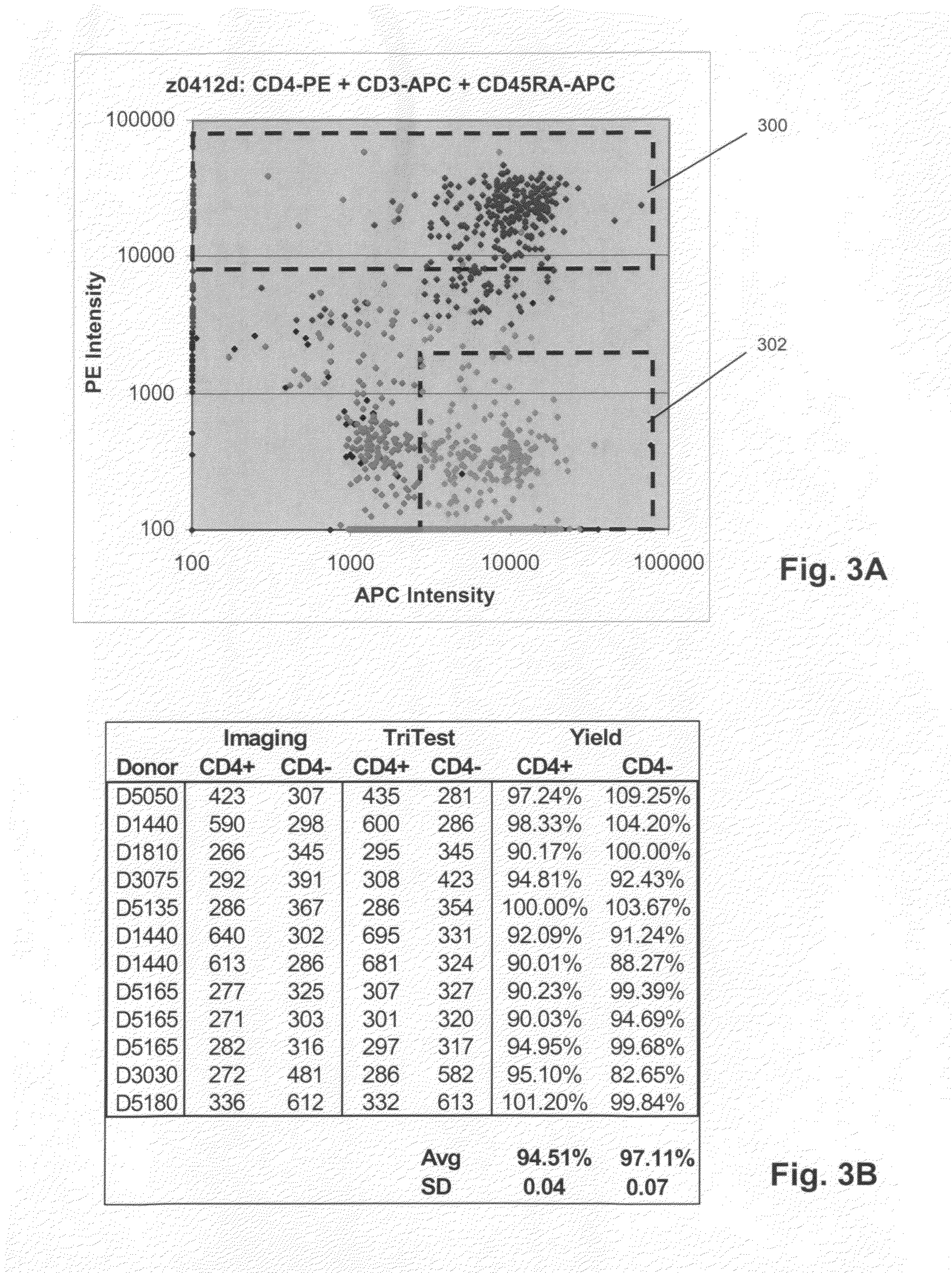
[0032]Several examples of the probes of the invention were prepared and tested on whole blood samples from normal individuals. Commercially available reagents (e.g. BD Biosciences, San Jose, Calif.) were employed and used in accordance with manufacturer's instructions. Unless otherwise noted, scatter and fluorescent intensity measurements were made on a FACSCanto flow cytometer (BD Biosciences, San Jose, Calif.).

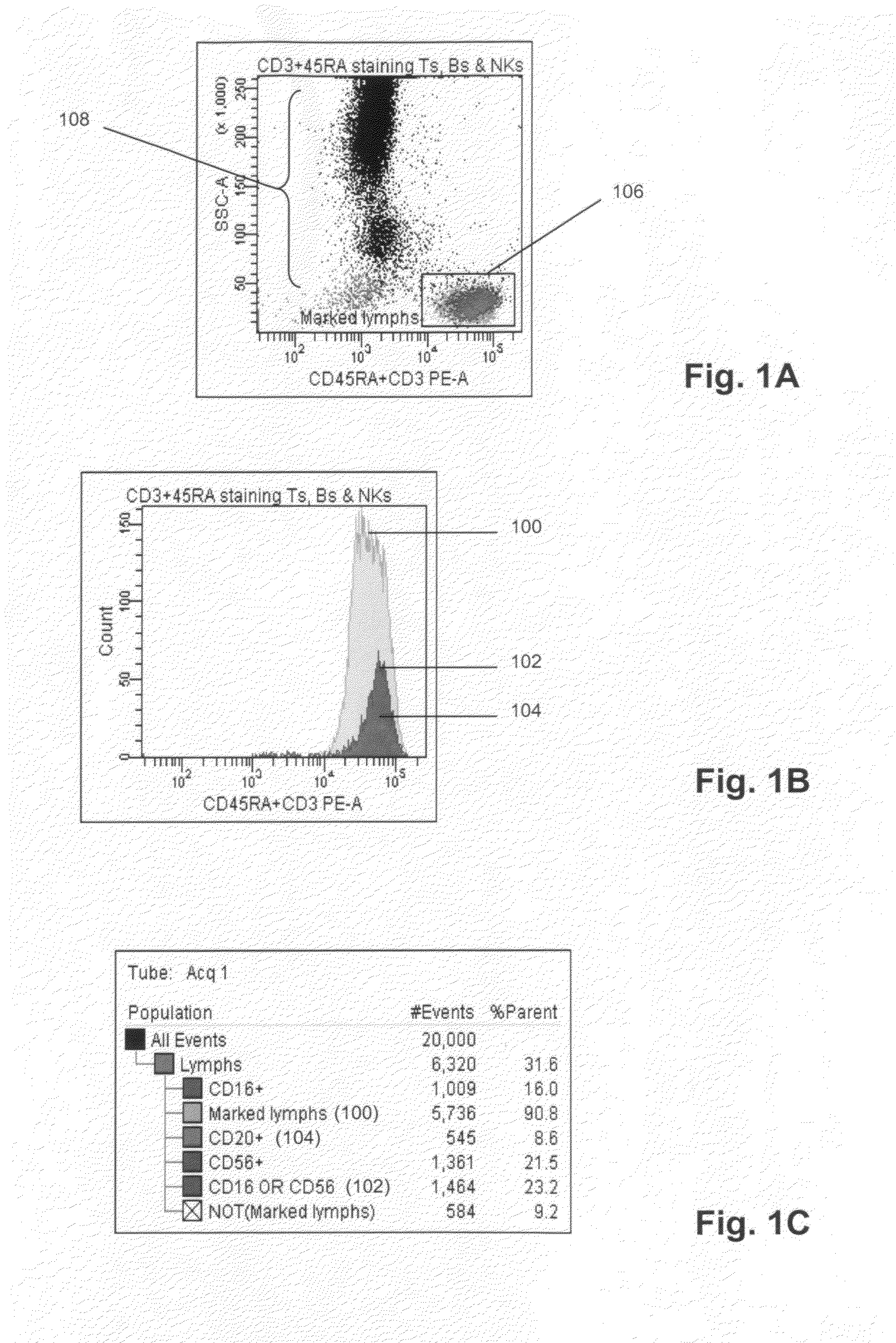
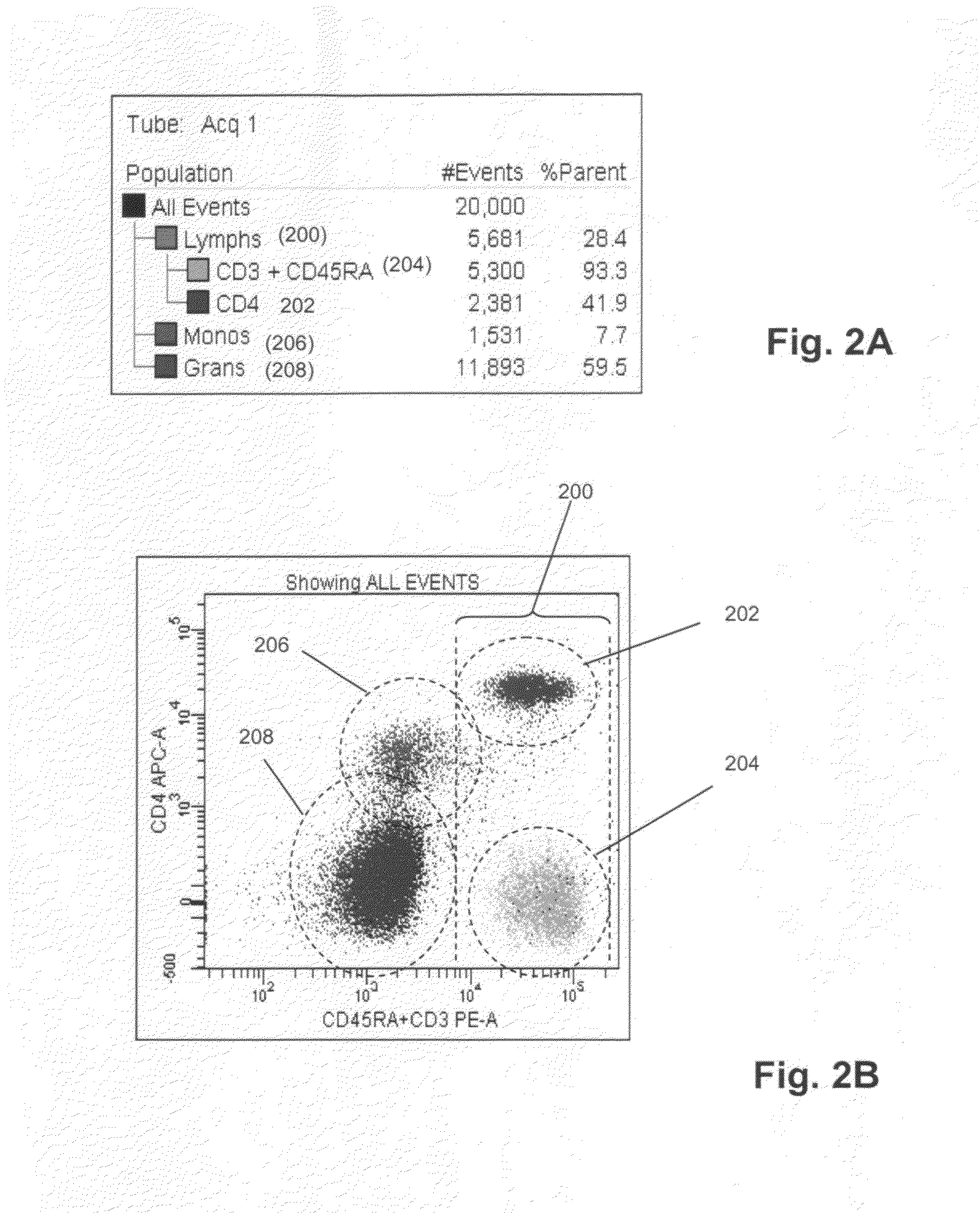
[0033]50 μL whole blood was combined with 20 μL anti-CD45RA antibody labeled with phycoerythrin (PE) (BD Biosciences, San Jose, Calif.), 20 μL anti-CD3 antibody labeled with phycoerythrin (PE) (BD Biosciences, San Jose, Calif.), 5 μL anti-CD16 antibody labeled with PE-Cy7 (BD Biosciences, San Jose, Calif.), 5 μL anti-CD56 antibody labeled with APC (BD Biosciences, San Jose, Calif.), and 20 μL anti-CD20 antibody labeled with PerCP-Cy5.5 (BD Biosciences, San Jose, Calif.), after which side scatter and PE fluorescence measurements were made with a FACSCanto flow cytometer. Port...


PUM

PropertyMeasurementUnit
fluorescentaaaaaaaaaa
fluorescenceaaaaaaaaaa
imagingaaaaaaaaaa

Abstract

The invention provides methods and compositions for identifying and counting lymphocytes in a biological sample, such as whole blood, by means of a probe comprising at least one binding compound specific for a T lymphocyte-specific marker, e.g. a CD2 or CD3, and at least one binding compound specific for CD45RA. Lymphocytes within the sample combine with such a probe to form a distinguishable subpopulation based on the amount of probe that specifically binds to their surfaces, thereby permitting such lymphocytes to be detected and enumerated on the basis of the intensity of the signal generated by the probe, and without the need of a separate physical measurement, such as light scatter. With additional probes specific for additional blood cell markers, percentages of lymphocytes, monocytes and granulocytes in a sample may be determined.

Description

[0001]This application claims priority from U.S. provisional applications 60 / 958,382 filed Jul. 5, 2007, which is hereby incorporated by reference in its entirety.BACKGROUND[0002]Cell surface markers permit the identification of cellular phenotypes characteristic of both healthy and disease states, e.g. Maecker et al, J. Clin. Immunol., 20: 391-399 (2000); Rothe et al, Leukemia, 10: 877-895 (1996); Reilly et al, J. Clin. Pathol., 54: 508-511 (2001). Such markers are typically measured by staining a cell sample with a selection of labeled antibodies specific for different markers, followed by multi-parameter analysis by imaging or by flow cytometry, e.g. Stewart, Immunophenotyping (Wiley-Liss, 2000). Multi-parameter analysis using labeled antibodies, or immunophenotyping, has been particularly useful for identifying distinct functional and developmental classes of white blood cells, which has important applications in the classification of blood-related diseases, such as leukemias an...

Claims


Application Information

Patent Timeline
Patent Type & Authority Patents(United States)
IPC IPC(8): C12N5/071
CPCG01N33/5047G01N33/505G01N33/5052G01N33/56972G01N2333/705G01N2333/70596G01N2333/7051G01N2333/70514G01N2333/70535G01N2333/70553G01N2333/70589G01N2333/70507
Inventor GOLDBERG, EDWARD MICHAEL
Owner BECTON DICKINSON & CO
Learn More