Method for detecting cytosine methylation in DNA samples

a dna sample and cytosine technology, applied in the field of detecting 5methylcytosine in genomic dna samples, can solve the problems of incomplete loss of epigenetic information carried by the 5-methylcytosine during pcr amplification, and the inability to identify the 5-methylcytosine position by sequencing, and achieve the effect of improving detectability

Inactive Publication Date: 2010-11-02
EPIGENOMICS AG
View PDF9 Cites 1 Cited by
  • Summary
  • Abstract
  • Description
  • Claims
  • Application Information

AI Technical Summary

Benefits of technology

[0041]According to the invention it is furthermore preferred that the produced fragments have a single positive or negative net charge for better detectability in the mass spectrometer.

Problems solved by technology

However, 5-methylcytosine positions cannot be identified by sequencing since 5-methylcytosine has the same base pairing behavior as cytosine.
Moreover, the epigenetic information carried by the 5-methylcytosines is completely lost during a PCR amplification.
However, 5-methylcytosine remains unmodified under these conditions.
Up to now, however, only individual regions of a length of up to approximately 3000 base pairs are analyzed; a global analysis of cells for thousands of possible methylation analyses is not possible.
However, this method cannot reliably analyze very small fragments from small sample quantities either.
These are lost in spite of the diffusion protection by the matrix.
DNA itself does not possess any functionalization which is suitable.
A difficulty consists in introducing such a functionalization into a DNA.
For nucleic acids having a multiply negatively charged backbone, the ionization process via the matrix is considerably less efficient.
For DNA, there are currently several matrixes in use, however, this has not reduced the difference in sensitivity.

Method used


Image

  • Method for detecting cytosine methylation in DNA samples

Examples


example 1

[0078]The following example relates to a fragment of exon 23 of the factor VIII gene in which a specific CG-position is to be analyzed for methylation.

[0079]In the first step, the fragment is amplified by primers of type A, namely by ATTATGTTGGAGTAGTAGAGTTTAAATGGTT (SEQ.-ID No.: 1) and ACTTAACACTTACTATTTAAATCACAACCCAT (SEQ.-ID No.: 2). The amplified DNA is hybridized to an oligonucleotide of type B (for example, GTTGGATGTTGTTGAGAAACG (SEQ.-ID No.: 3)) and elongated in a polymerase reaction with a labeled 2′,3′-didesoxythymidine triphosphat (type C 2). This elongation can only take place if a CG, that is, in the original genomic DNA sample, a methylated cytosine was present since otherwise, a mispairing at the 3′-end of the primer prevents the polymerase reaction. Thus, the methylation status of the specific cytosine to be analyzed decides on the elongation of the primer.

[0080]For control purposes, the reaction can be carried out with the primer GTTGGATGTTGTTGAGAAATG (SEQ.-ID No.: 4)...

example 2

[0081]The following example relates to a fragment of exon 23 of the factor VIII gene in which a specific CG-position is to be analyzed for methylation.

[0082]In the first step, the fragment is amplified by primers of type A, namely by ATTATGTTGGAGTAGTAGAGTTTAAATGGTT (SEQ.-ID No.: 1) and ACTTAACACTTACTATTTAAATCACAACCCAT (SEQ.-ID No.: 2). The amplified DNA is hybridized to an oligonucleotide of type B (for example, GTTGGATGTTGTTGAGAAACG (SEQ.-ID No.: 3)), which is immobilized to a solid phase surface with its 5′-end, and elongated in a polymerase reaction with a labeled 2′,3′-didesoxythymidine triphosphat (type C2). This elongation can only take place if a CG, that is, in the original genomic DNA sample, a methylated cytosine was present since otherwise, a mispairing at the 3′-end of the primer prevents the polymerase reaction. Thus, the methylation status of the specific cytosine to be analyzed decides on the elongation of the primer.

[0083]For control purposes, the reaction can be car...

example 3

[0084]The following example relates to a fragment of exon 23 of the factor VIII gene in which a specific CG-position is to be analyzed for methylation.

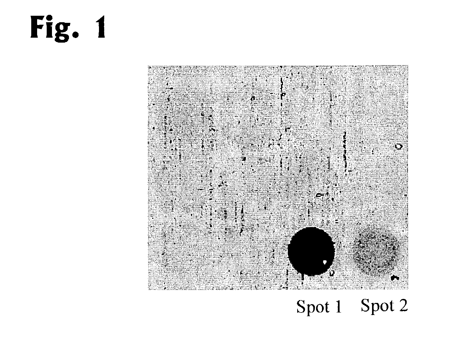
[0085]In the first step, the fragment is amplified by primers of type A, namely by ATTATGTTGGAGTAGTAGAGTTTAAATGGTT (SEQ.-ID No.: 1) and ACTTAACACTTACTATTTAAATCACAACCCAT (SEQ.-ID No.: 2). For this amplification, DNA treated with bisulphite was incubated for 5 min at 96° C., then denaturated by 40 cycles carried out for 55 sec at 96° C. each, incubated for 75 sec at 61.7° C. (annealing) and incubated for 100 sec at 72° C. (elongation). In a subsequent reaction (final extension) the reaction mixture is incubated for 15 min at 72° C. The amplified DNA is hybridized to the solid phase immobilized oligonucleotides AAAAACTACAAAAACTCT (SEQ-ID No.: 5) (spot 1 in FIG. 1) and to AAAACTACGAAAACTCT (SEQ-ID No.: 6 (spot 2 in FIG. 1). The elongation reaction affords 2′-desoxythymidine triphosphate (dTTP, as type C 1), 2′-desoxyguanosine triphosphate...


PUM

PropertyMeasurementUnit
elongationaaaaaaaaaa
fluorescenceaaaaaaaaaa
massaaaaaaaaaa

Abstract

Described is a method for detecting 5-methylcytosine in genomic DNA samples. First, a genomic DNA from a DNA sample is chemically converted with a reagent, 5-methylcytosine and cytosine reacting differently, and the pretreated DNA is subsequently amplified using a polymerase and at least one primer. In the next step, the amplified genomic DNA is hybridized to at least one oligonucleotide, forming a duplex, and said oligonucleotide is elongated by at least one nucleotide, the nucleotide carrying a detectable label, and the elongation depending on the methylation status of the specific cytosine in the genomic DNA sample. In the next step, the elongated oligonucleotides are analyzed for the presence of the label.

Description

BACKGROUND OF THE INVENTION[0001]The present invention relates to a method for detecting 5-methylcytosine in genomic DNA samples.[0002]The levels of observation that have been well studied by the methodological developments of recent years in molecular biology are the genes themselves, the translation of genes into RNA, and the resulting proteins. The question of which gene is switched on at which point in the course of the development of an individual, and how the activation and inhibition of specific genes in specific cells and tissues are controlled is correlatable to the degree and character of the methylation of the genes or of the genome.[0003]The present invention describes a method for detecting the methylation state of genomic DNA samples. The method can, at the same time, also be used for detecting point mutations and single nucleotide polymorphisms (SNPs).[0004]5-methylcytosine is the most frequent covalently modifiable base in the DNA of eukaryotic cells. It plays a role...

Claims


Application Information

Patent Timeline
Patent Type & Authority Patents(United States)
IPC IPC(8): C07H21/04C12Q1/68G01N27/62C12N15/09C12Q1/34C12Q1/48C12Q1/6816C12Q1/6827C12Q1/6837C12Q1/6872G01N33/483G01N33/566G01N33/58
CPCC12Q1/6816C12Q1/6837C12Q1/6872C12Q1/686C12Q1/6827C12Q2523/125C12Q2565/519C12Q2565/627
Inventor OLEK, ALEXANDERBERLIN, KURT
Owner EPIGENOMICS AG
Why Eureka
Try Eureka