Methods and apparatuses for analyzing images

a technology of image analysis and image, applied in image analysis, instruments, computing, etc., can solve the problems of affecting the detection of tissue pathology

Inactive Publication Date: 2011-02-08
UNIV OF IOWA RES FOUND
View PDF23 Cites 29 Cited by
  • Summary
  • Abstract
  • Description
  • Claims
  • Application Information

AI Technical Summary

Benefits of technology

[0014]One of the present apparatuses is an apparatus that includes an image input adapted to receive a diagnostic medical image. The image includes a plurality of pixels, and each pixel has a particular gray level. The apparatus also includes a display for displaying a graphical user interface and the received image; and a processor adapted to perform texture measures on one or more groups of pixels within the image and classify each group of pixels to a tissue patholo...

Problems solved by technology

The disorder is caused by destruction of the alveolar walls of the lung parenchyma (i.e., lung tissue), which results in an abnormal enlargement of air spaces distal to the terminal bronchiole.
Enlargement of these air spaces in the lungs impedes the exchange of oxygen in the air for carbon dioxide in the bloodstream.
As a result of this impeded process, an individual experiences breathlessness, making ordinary tasks, once thought simple, labor intensive.
That is, IPF and sarcoid also impede the carriage of oxygen from the lungs to the bloodstream like emphysema.
In addition to those pulmonary parenchymal diseases discussed above, peripheral small airways diseases, such as cystic fibrosis and asthma (along with over one-hundred other pathologies) also exist, which can adversely affect the lungs of an individual as well.
However, these tests are not always able to properly distinguish between the abnormalities of the lung that result from one particular disorder from another.
However, because of problems resulting from structural superposit...

Method used


Image

  • Methods and apparatuses for analyzing images
  • Methods and apparatuses for analyzing images
  • Methods and apparatuses for analyzing images

Examples


Embodiment Construction

[0034]Illustrative embodiments of the invention are described below. In the interest of clarity, not all features of an actual implementation are described in this specification. It will of course be appreciated that in the development of any such actual embodiment, numerous implementation-specific decisions must be made to achieve the developers' specific goals, such as compliance with system-related and business-related constraints, which will vary from one implementation to another. Moreover, it will be appreciated that such a development effort might be complex and time-consuming, but would nonetheless be a routine undertaking for those of ordinary skill in the art having the benefit of this disclosure.

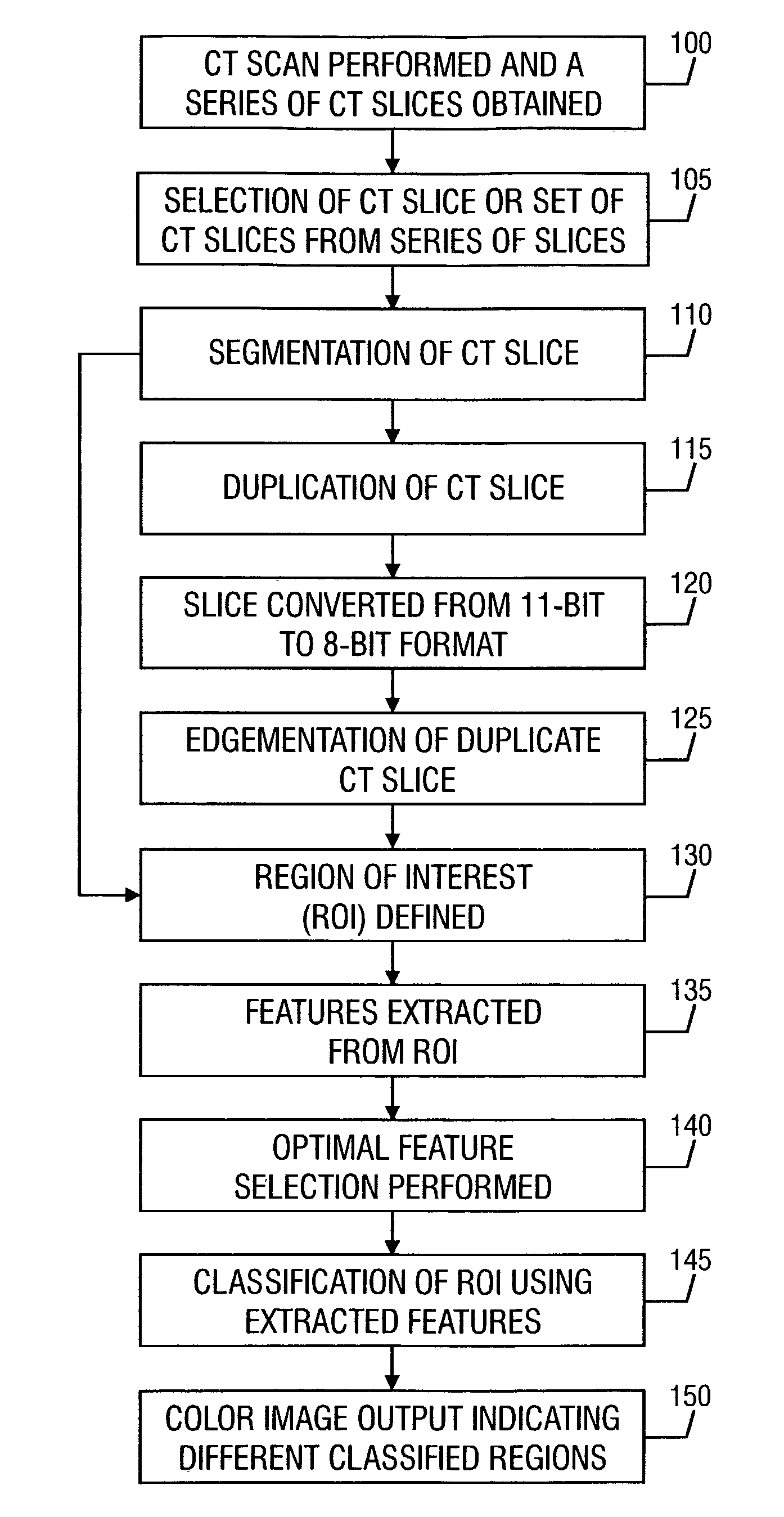
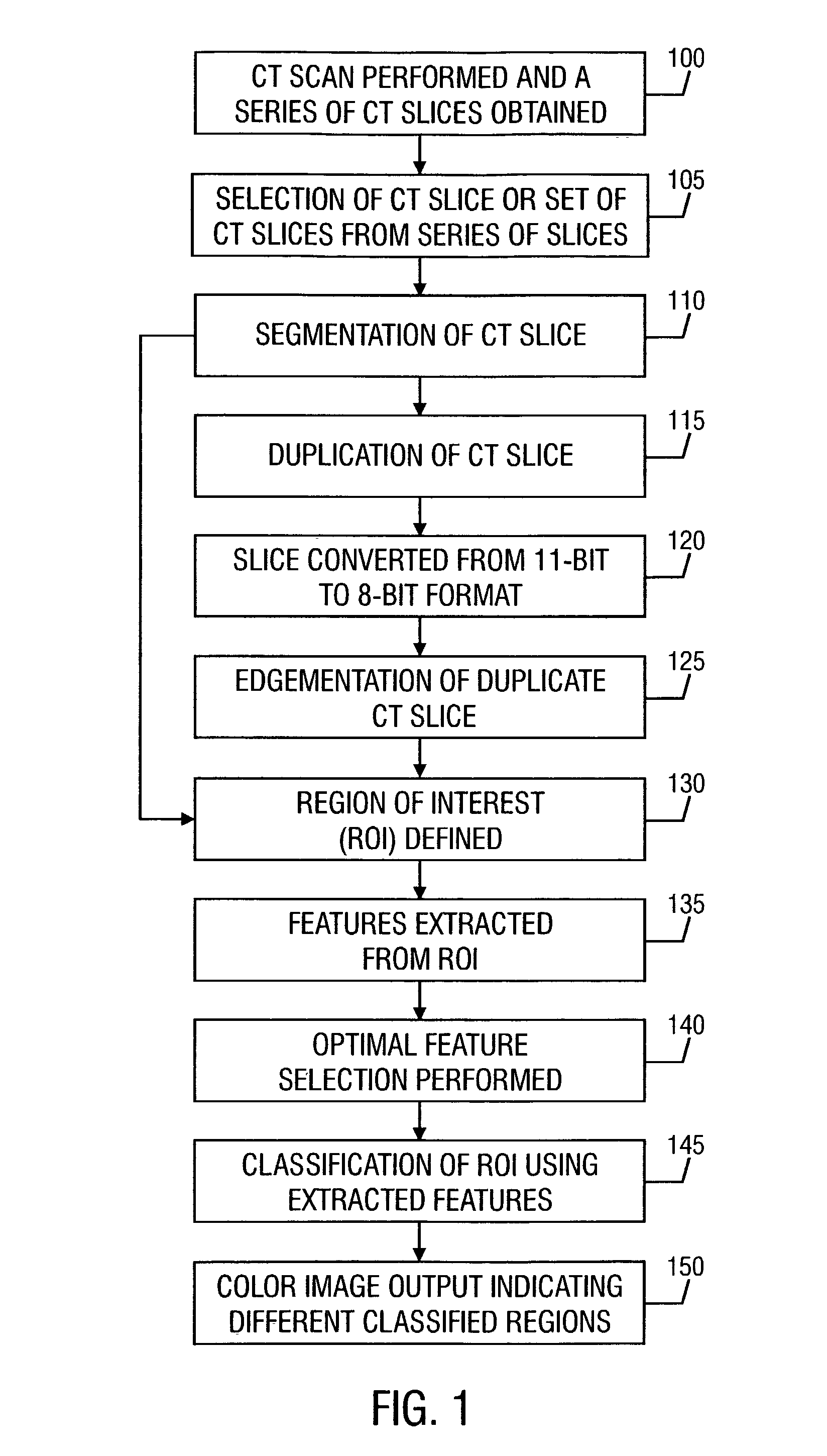
[0035]Turning now to the drawings and specifically referring to FIG. 1, a flowchart is shown illustrating the process used to perform an objective analysis of a diagnostic medical image, which in one embodiment is a computed tomography (CT) image. The process commences at step 100...


PUM


Abstract

A method for automated analysis of textural differences present on an image volume. The image volume includes a plurality of volume elements, and each volume element has a gray level. The method includes defining a volume of interest (VOI); performing texture measures within the VOI; and classifying the VOI as belonging to a tissue pathology class based upon the texture measures. Computer readable media encoded with computer readable instructions for carrying out these functions. An apparatus that includes an image input adapted to receive a diagnostic medical image. The image includes a plurality of pixels, and each pixel has a particular gray level. The apparatus also includes a display for displaying a graphical user interface and the received image; and a processor adapted to perform texture measures on one or more groups of pixels within the image and classify each group of pixels to a tissue pathology class based upon the textures measures. The processor is further adapted to (1) associate a color to each group of pixels indicative of the group's tissue pathology class, (2) cause the display to display one or more of the colors on the image at the location of the associated group or groups of pixels, (3) permit a user to manually associate a tissue pathology class to a group of pixels, and (4) cause the display to display the manually-associated tissue pathology class.

Description

PRIORITY CLAIM[0001]This is a continuation of co-pending application Ser. No. 09 / 022,093, filed Feb. 11, 1998 now U.S. Pat. No. 6,466,687, which claims priority to U.S. Provisional Patent Application Ser. No. 60 / 037,067 filed Feb. 12, 1997. The entire text of this provisional patent application is specifically incorporated by reference without disclaimer.BACKGROUND OF THE INVENTION[0002]A portion of the disclosure of this patent document contains material that is subject to copyright protection. The copyright owner has no objection to the facsimile reproduction by anyone of the patent document or the patent disclosure, as it appears in the Patent and Trademark Office patent files or records, but otherwise reserves all copyright rights whatsoever.1. FIELD OF THE INVENTION[0003]The present invention relates generally to the detection and diagnosis of tissue pathology using an image.2. DESCRIPTION OF THE RELATED ART[0004]Pulmonary emphysema is a common, debilitating, and progressive di...

Claims


Application Information

Patent Timeline
IPC IPC(8): G06K9/00G06T7/00
CPCG06T7/0012
Inventor UPPALURI, RENUKAMITSA, THEOPHANOHOFFMAN, ERIC A.MCLENNAN, GEOFFREYSONKA, MILAN
Owner UNIV OF IOWA RES FOUND
Why Eureka
Try Eureka