System and method for segmenting chambers of a heart in a three dimensional image

a three-dimensional image and chamber technology, applied in image enhancement, instruments, applications, etc., can solve problems such as poor contrast, difficult segmentation, and cardiac anatomy

Inactive Publication Date: 2011-03-29
SIEMENS HEALTHCARE GMBH +1
View PDF5 Cites 107 Cited by
  • Summary
  • Abstract
  • Description
  • Claims
  • Application Information

AI Technical Summary

Problems solved by technology

The complexity of cardiac anatomy, poor contrast, noise or motion artifacts makes this segmentation a challenging task.
There are a few limitations inherent in these techniques: 1) Most of them are semiautomatic and manual labeling of a rough position and pose of the heart chambers is needed.
2) They are likely to get stuck in local strong image evidence.
However, such techniques cannot fully exploit the benefit of 3D imaging in a natural way.
Exhaustive searching makes the system robust under local minima, however there are two challenges to extend the learning based approaches to 3D.
The computational demands are beyond the capabilities of current desktop computers.
Due to this limitation, previous approaches often constrain the search to a lower dimensional space.
The second challenge is that efficient features are needed to search the orientation and scale spaces.
However, when searching for rotation parameters, one either has to rotate the feature templates or rotate the volume which is very time consuming.

Method used


Image

  • System and method for segmenting chambers of a heart in a three dimensional image
  • System and method for segmenting chambers of a heart in a three dimensional image
  • System and method for segmenting chambers of a heart in a three dimensional image

Examples


Embodiment Construction

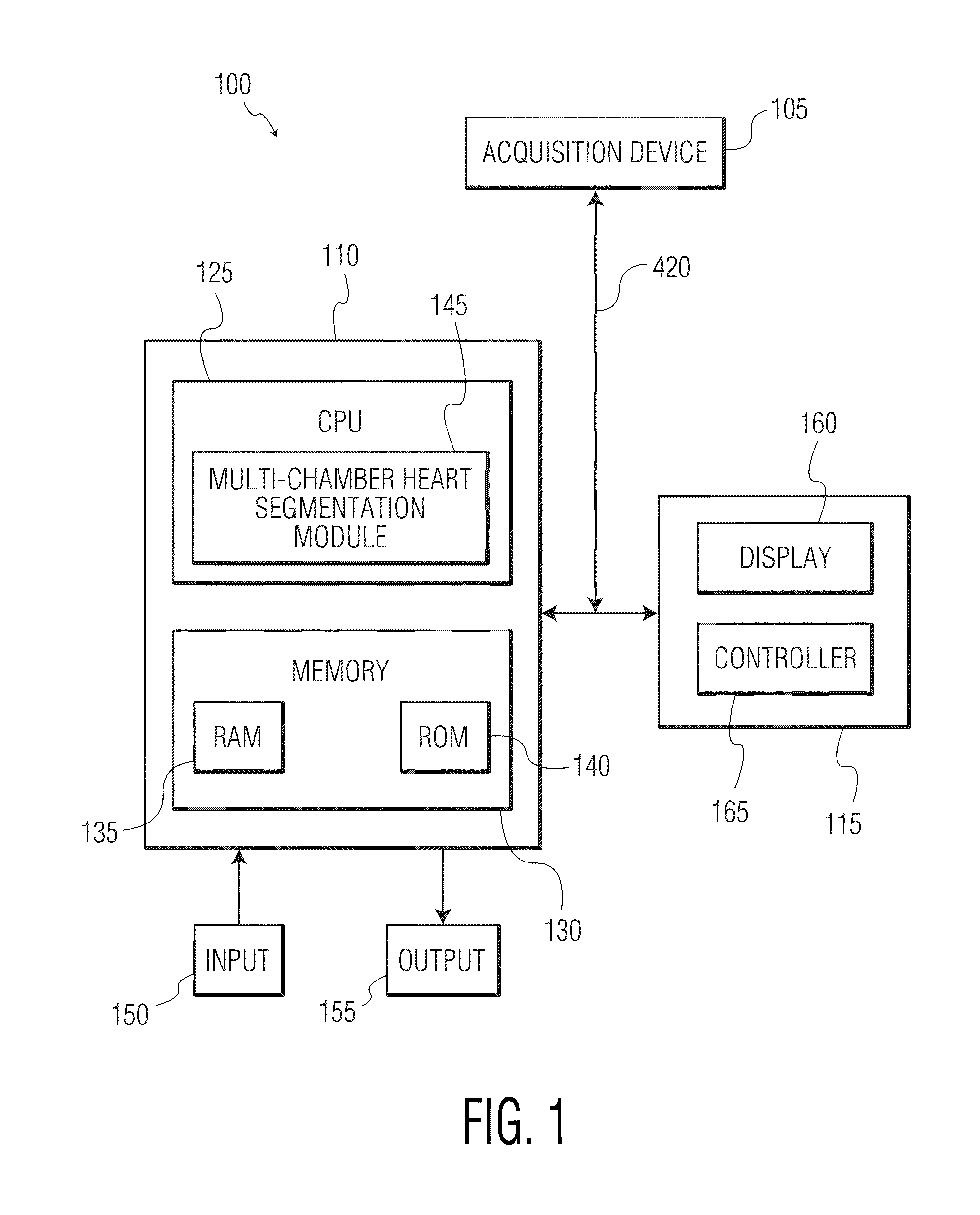
[0017]The present invention is directed to a system and method for multi-chamber heart segmentation. FIG. 1 illustrates a system 100 for segmenting a multi-chamber heart in three dimensional medical images according to an exemplary embodiment of the present invention. As shown in FIG. 1, the system 100 includes an acquisition device 105, a personal computer (PC) 110 and an operator's console 115 connected over a wired or wireless network 120.

[0018]The acquisition device 105 may be a computed tomography (CT) imaging device or any other three-dimensional (3D) high-resolution imaging device such as a magnetic resonance (MR) scanner or ultrasound scanner.

[0019]The PC 110, which may be a portable or laptop computer, a medical diagnostic imaging system or a picture archiving communications system (PACS) data management station, includes a central processing unit (CPU) 125 and a memory 130 connected to an input device 150 and an output device 155. The CPU 125 includes a multi-chamber heart...


PUM


Abstract

A system and method for segmenting chambers of a heart in three dimensional images is disclosed. A set of three dimensional images of a heart is received. The shape of the heart in the three dimensional images is localized. Boundaries of the chambers of the heart in the localized shape are identified using steerable features.

Description

CROSS REFERENCE TO RELATED APPLICATION[0001]This application claims the benefit of U.S. Provisional Application Ser. No. 60 / 827,235, filed Sep. 28, 2006 and Ser. No. 60 / 913,353, filed Apr. 23, 2007 which is are incorporated by reference in its their entirety.FIELD OF THE INVENTION[0002]The present invention is directed to a system and method for segmenting chambers of a heart in a three dimensional image, and more particularly, to a system and method for segmenting chambers of a heart in a three dimensional image using anatomical structure localization and boundary delineation.BACKGROUND OF THE INVENTION[0003]Cardiac computed tomography (CT) is an important imaging modality for diagnosing cardiovascular disease and it can provide detailed anatomic information about the cardiac chambers, large vessels or coronary arteries. Multi-chamber heart segmentation is a prerequisite for global quantification of the cardiac function. The complexity of cardiac anatomy, poor contrast, noise or mo...

Claims


Application Information

Patent Timeline
Patent Type & Authority Patents(United States)
IPC IPC(8): G06K9/00
CPCA61B6/503G06T7/0089G06K9/6247G06T7/0083A61B8/0883G06T2207/30048G06T2207/10081G06T7/12G06T7/149G06V10/7715G06V2201/031G06V2201/12G06F18/2135
Inventor ZHENG, YEFENGBARBU, ADRIANGEORGESCU, BOGDANSCHEUERING, MICHAELCOMANICIU, DORIN
Owner SIEMENS HEALTHCARE GMBH
Why Eureka
Try Eureka