Method, apparatus and computer program for contour detection of vessels using x-ray densitometry

a technology of densitometry and contour detection, applied in the field of method, apparatus and computer program for contour detection of vessels using densitometry, to achieve the effect of simplifying processing

Active Publication Date: 2011-06-28
PIE MEDICAL IMAGING
View PDF11 Cites 41 Cited by
  • Summary
  • Abstract
  • Description
  • Claims
  • Application Information

AI Technical Summary

Benefits of technology

[0007]According to a further aspect, under the assumption that the vessel has a known cross-sectional geometric shape (e.g., a circular shape), the densitometric information can be used to estimate the width of the vessel in question directly. This measure will simplify the processing and is in general accurate.
[0008]Advantageously, the invention for a predetermined vessel segment automatically finds a conversion factor between a densitometrically derived vessel diameter and a vessel diameter based on contour detection. Such conversion factor is found by fitting a first cumulative histogram of diameters found by contour detection with a second cumulative histogram of diameters found by the densitometry. This statistical processing is a mature art and will lead to a robust result of the operation.

Problems solved by technology

Generally, these overestimations are caused by noise and blur in the X-ray image.
Such apparent enlargement of a vessel may compromise the diagnostics of inter alia coronary stenosis or may influence the results of studies on the efficiency of new clinical treatments.

Method used


Image

  • Method, apparatus and computer program for contour detection of vessels using x-ray densitometry
  • Method, apparatus and computer program for contour detection of vessels using x-ray densitometry
  • Method, apparatus and computer program for contour detection of vessels using x-ray densitometry

Examples


Embodiment Construction

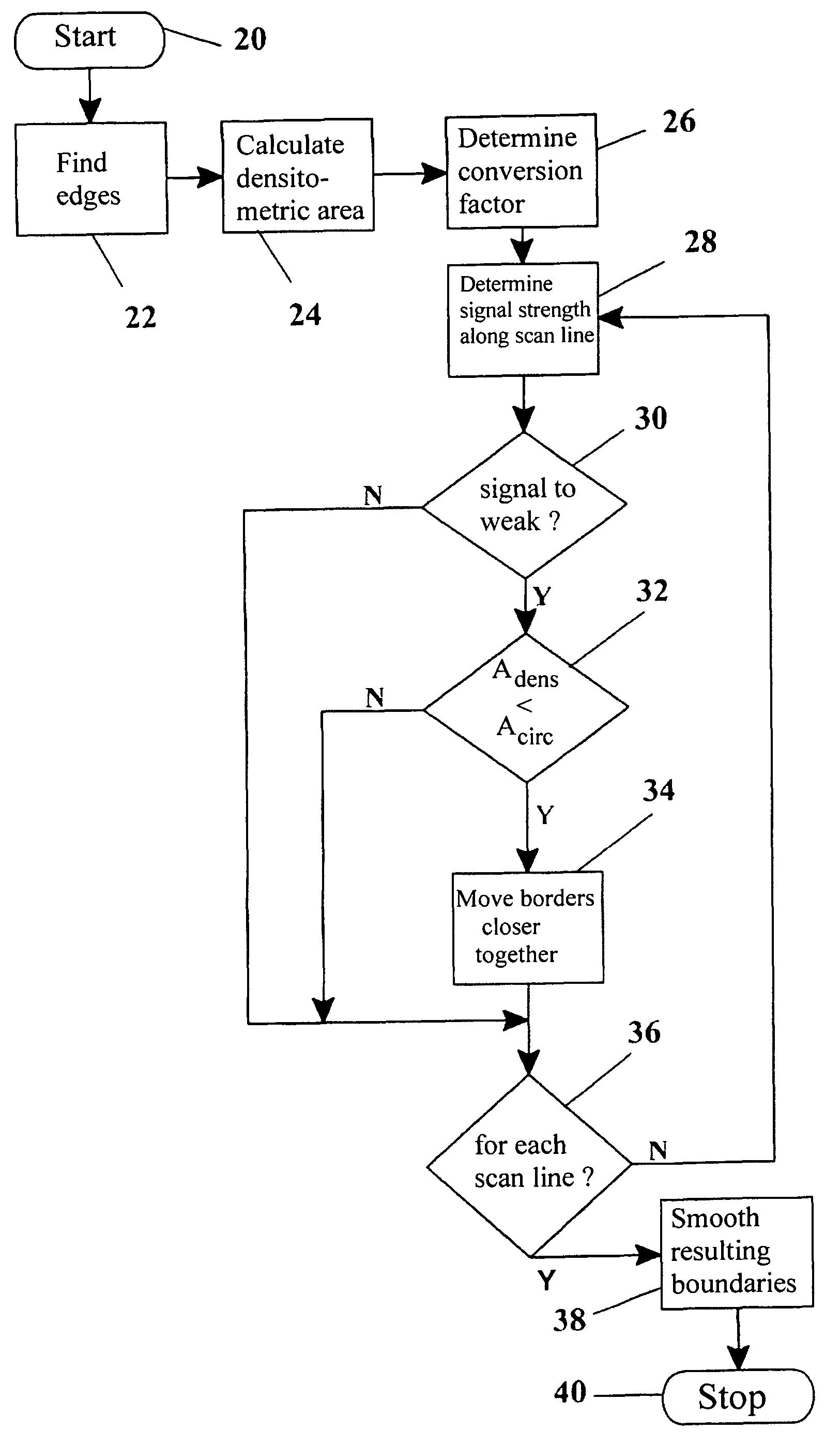
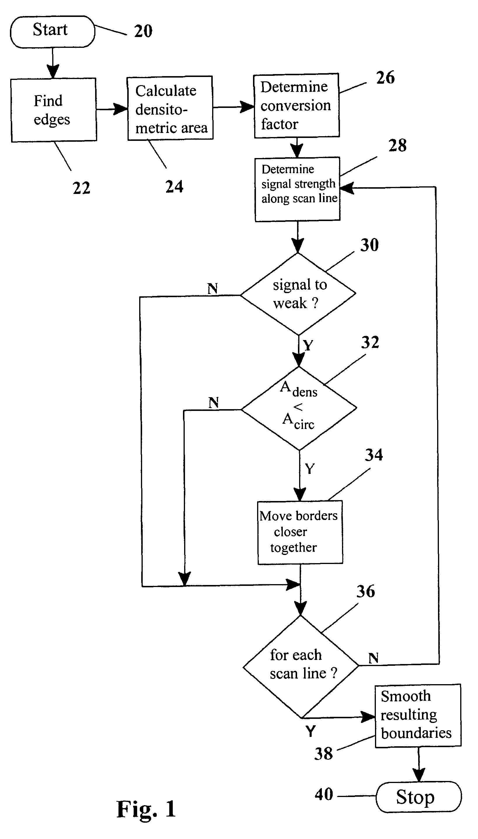
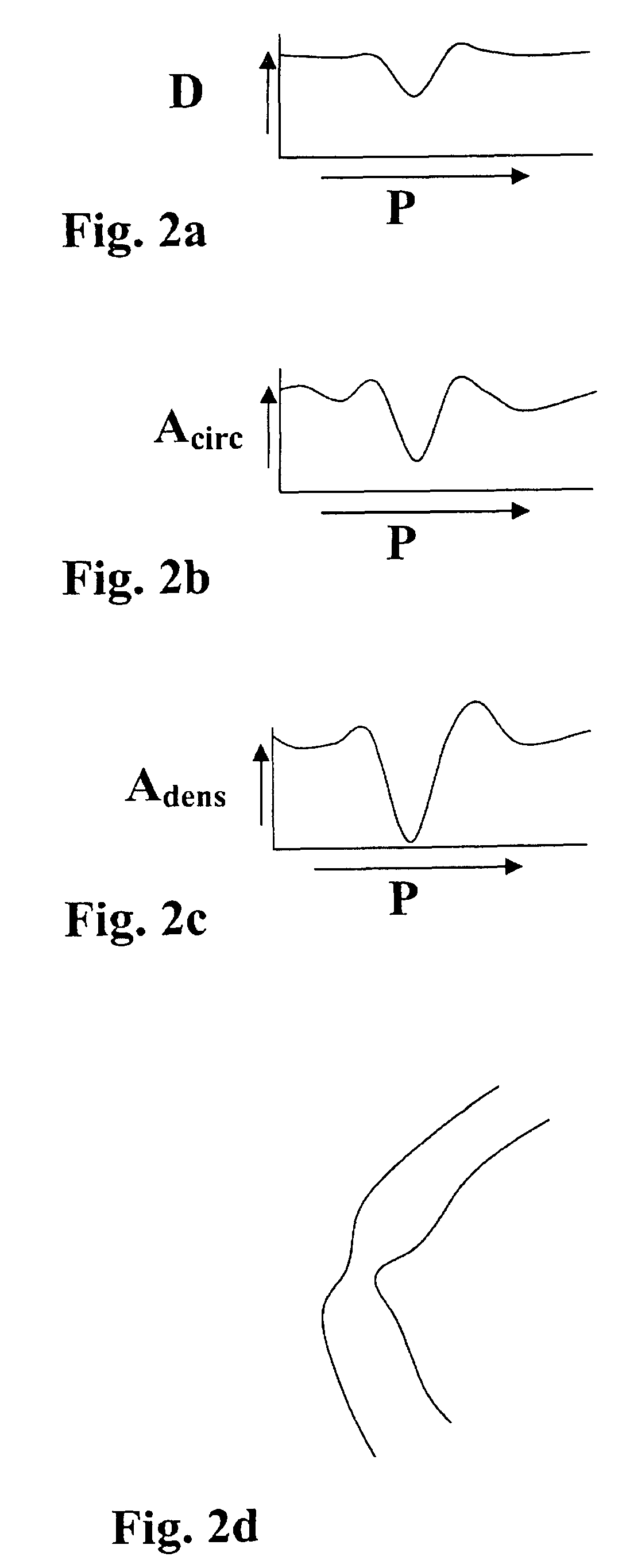
[0020]Hereinafter, a preferred embodiment of the invention will be discussed in detail. The reason why the contour detection goes wrong is that the contours may in certain areas have too little contrast. Although many other routes can be followed to determine if enough contrast is present, the inventors have by way of example used a Student's t-test for independent groups. An advantage of such statistical test is that it will also take the numbers of pixels into account. The Student's t-test is a statistical test that determines if the pixel values inside the found vessel differ statistically significantly from pixel values outside the vessel. A scan line is defined through the vessel that is approximately perpendicular to the local centerline of the vessel. Along this scan line the average pixel values inside the found vessel (μin) and outside the found vessel (μout) as well as the standard deviations inside (σin) and outside the found vessel (σout) are determined on the basis of n...


PUM


Abstract

A method has been described for deriving contour data in X-Ray images for vessels with differential absorption through applying a contour-finding algorithm on a shadow image and finding the vessel borders through segmentation based on image intensities. In particular, the method uses the following steps: finding a densitometric area of an above mentioned vessel, and displacing one or both of the borders inward until the densitometric measurement result between the borders after such displacing will start to change significantly. Furthermore, a specific procedure is introduced to automatically determine the conversion factor to equate the densitometrically based diameter to the contour based diameter of the vessel and to discriminate bifurcating or parallel vessels.

Description

BACKGROUND OF THE INVENTION[0001]The invention relates generally to computer-based methods for deriving contour data for vessels with differential absorption utilizing x-ray densitometry, and more particularly to such computer-based methodologies that detect vessel contours by the application of a contour-finding algorithm on a shadow image and segmentation based on image intensities. A prime field of application for such computer-based methodologies is in small, in particular sub-millimeter blood vessels. However, such computer-based methodologies may also be used for other vessels that may have differential X-Ray attenuation or for implanted objects such as stents. Such finding of contours or borders is standard technology in medical diagnostics and related arts.[0002]Prior art, in particular, S. Molloi et al, Quantification of Coronary Artery Lumen Volume by Digital Angiography, © 2001 to the American Heart Association, Reprints from symolloi@uci.edu, have described that contour ...

Claims


Application Information

Patent Timeline
Patent Type & Authority Patents(United States)
IPC IPC(8): G06K9/00G06K9/36G06V10/40
CPCA61B5/1075A61B6/504G06K9/46G06T7/0083G06T7/602A61B6/481G06T2207/30101G06K2209/05G06T2207/10116G06K2009/00932G06T7/12G06T7/62G06V40/14G06V10/40G06V2201/03
Inventor PUTS, MARCUS JOHANNES HUBERTUSABEN, JEAN-PAUL MARIA MICHELVERSTRAELEN, BOUDEWIJN JOSEPH ANGELUS
Owner PIE MEDICAL IMAGING
Why Eureka
Try Eureka