Methods and devices for quantitative analysis of x-ray images

a quantitative analysis and x-ray technology, applied in the field of radiographic imaging and analysis, can solve the problems of not always accurate current-available calibration phantoms, and cannot allow for the accurate determination of quantitative information contained in x-rays, and achieve the effect of improving the accuracy of quantitative information

Inactive Publication Date: 2011-10-04
IMATX INC
View PDF192 Cites 25 Cited by
  • Summary
  • Abstract
  • Description
  • Claims
  • Application Information

AI Technical Summary

Benefits of technology

In other embodiments, the quantitative information derived from the x-ray image includes one or more parameters relating to the acquisition of the x-ray image (e.g., x-ray tube voltage, x-ray energy, x-ray tube current, film-focus distance, object-film distance, collimation, focal spot size, spatial resolution of the x-ray system, filter technique, film focus distance, correction factor(s) or combinations thereof), for instance to improve the accuracy of the quantitative information. The x-ray acquisition parameters can be transferred over the network prior to, simultaneously or after transmission of the x-ray image. Furthermore, one or more of the x-ray acquisition parameters can be entered either manually or, alternatively, automatically into a computer.

Problems solved by technology

Current technology, however, does not allow for the accurate determination of quantitative information contained in the x-ray such as the density of an anatomic structure when x-ray images are transmitted in a network environment.
However, currently-available calibration phantoms are not always accurate, due to both the effect of structures or materials that project on or with the calibration phantom and, additionally, to the fact that one or more regions of the calibration phantom do not always appear on the x-ray image.

Method used


Image

  • Methods and devices for quantitative analysis of x-ray images
  • Methods and devices for quantitative analysis of x-ray images
  • Methods and devices for quantitative analysis of x-ray images

Examples


example 1

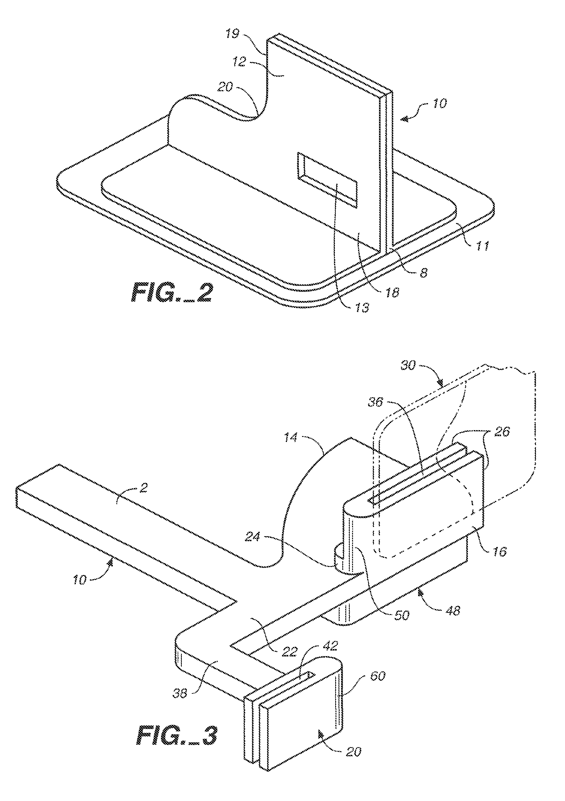
Calibration Phantom Integrated into Film Cover

The workflow presented herein constitutes one example for the use of a calibration phantom with the image acquisition. One skilled in the art will readily recognize other ways to include a calibration phantom in the acquisition process in order to normalize or standardize any form of measurement made from the x-ray image.

In this example, the calibration phantom is integrated into the cover of a dental x-ray film that protects it from exposure to light. The film is placed into the film holder that is used to hold the film in the patient's mouth. The film holder with the film is positioned inside the patient's mouth in such a way that the calibration phantom is not obstructed from the x-ray beam by any structures such as teeth or the lips etc.

After acquisition of the image, the film is taken to the darkroom and the cover with the calibration phantom is removed. The film is then processed in the same way as a conventional dental x-ray film....

example 2

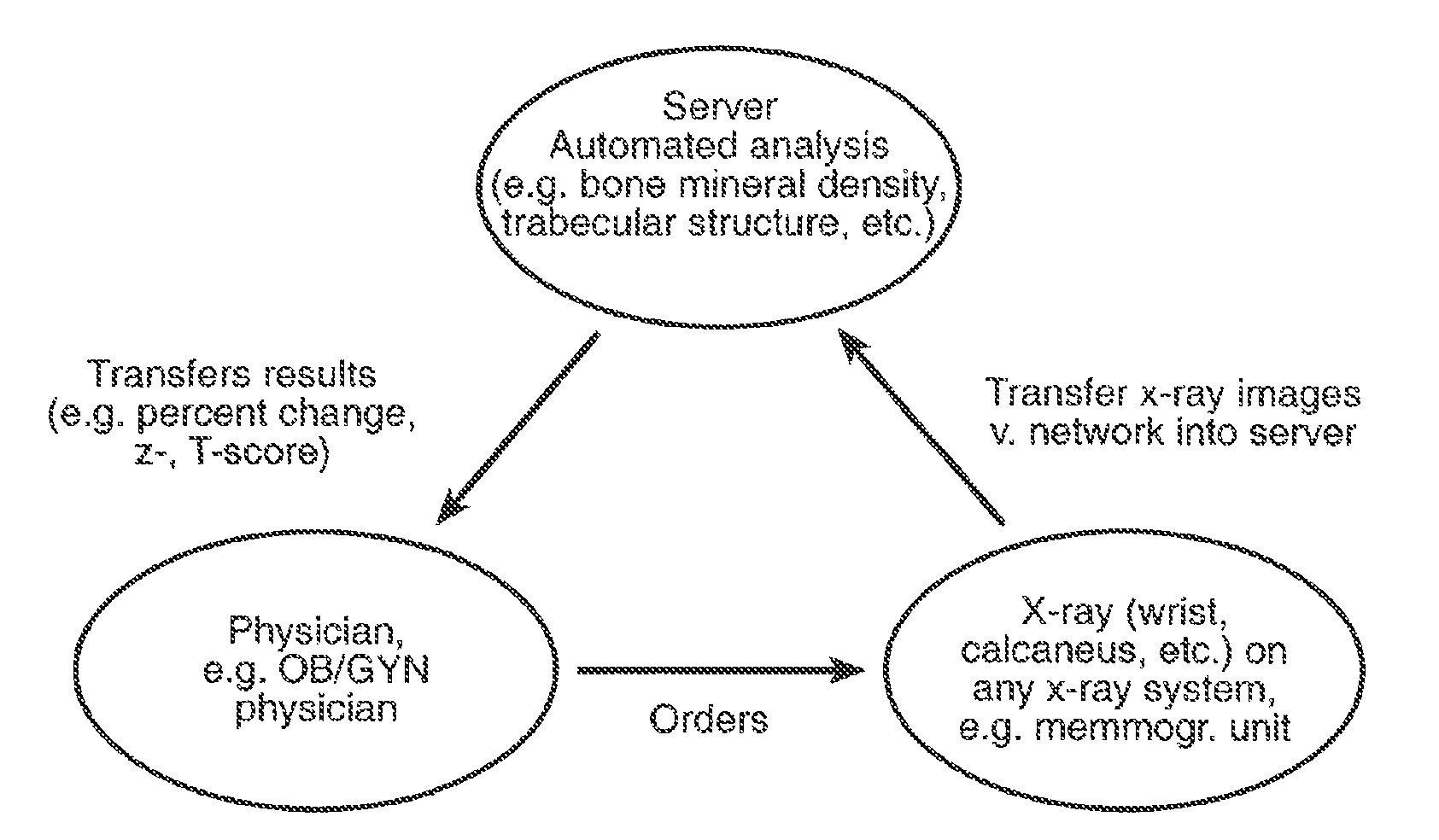
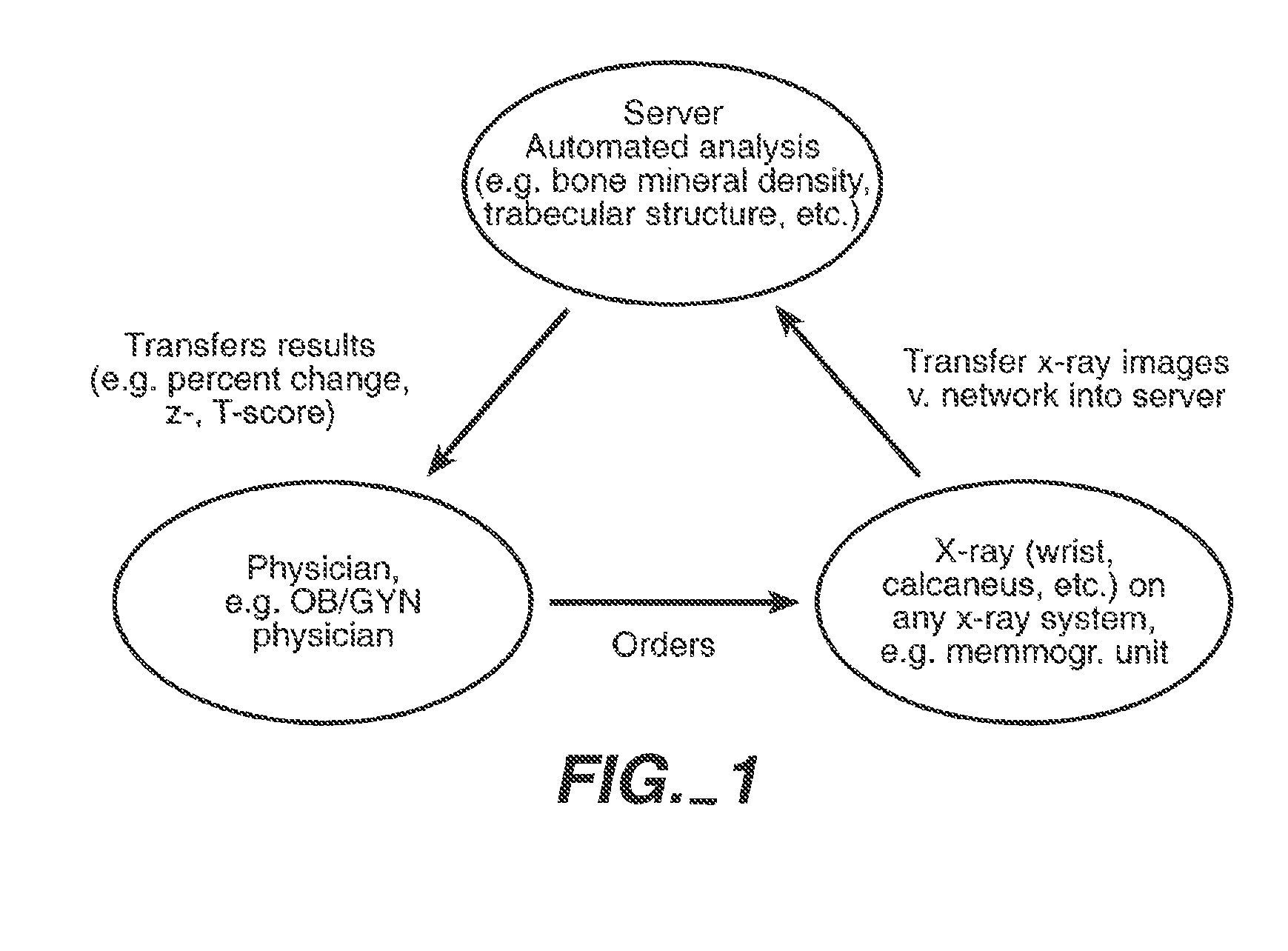
Transmission of X-Ray Image Including Image of Calibration Phantom over a Network

This example describes one possible typical application of the invention, in which a digitized x-ray image that includes the image of a calibration phantom is transmitted over a network. Similar applications of the invention can easily be recognized.

The x-ray image is acquired in such a way that the calibration phantom is projected onto the film. The film with the x-ray image and the image of the calibration phantom is developed. Subsequently, the film is digitized, for example using a flat-bed or slide film scanner, resulting in a digital image. The digital image, which includes the x-ray image and the image of the calibration phantom, is then transmitted over a network to a remote computer. The remote computer performs one or more measurements using information from the x-ray image and / or the image of the calibration phantom.

example 3

Transmission of X-Ray Image Including Image Acquisition Parameters

The transmission of image data over a network can also include data describing the image acquisition parameters. After acquisition and digitization of the x-ray image, the acquisition parameters are entered into the local computer system. These parameters can include, but are not limited to, voltage settings, tube current, or film-focus distance. The image and acquisition parameter data are then transmitted over the network to a remote computer.

At the remote computer, the image is analyzed. The acquisition parameters can be used in this evaluation to improve the accuracy of the measurements. The results can be sent back to the original location via a digital network or by fax transmission. The results can also be transmitted to third parties.


PUM


Abstract

The present invention relates to network enabled analysis of x-ray images. Also described are devices comprising calibration phantoms; methods of using these devices; methods of formulating databases containing information regarding x-ray images; the databases themselves; and methods of manipulating the information and databases.

Description

TECHNICAL FIELDThe present invention is in the field of radiographic imaging and analysis thereof. In particular, network enabled analyses and analysis techniques are described. Also described are devices comprising calibration phantoms and methods of using these devices.BACKGROUNDX-rays and other radiographic analysis are important diagnostic tools. Furthermore, it is common practice to transmit x-ray images via local and long-distance networks. Current technology, however, does not allow for the accurate determination of quantitative information contained in the x-ray such as the density of an anatomic structure when x-ray images are transmitted in a network environment.Calibration references (also known as calibration phantoms) for use in imaging technologies have also described. See, e.g., U.S. Pat. No. 5,493,601 and U.S. Pat. No. 5,235,628. U.S. Pat. No. 5,335,260 discloses a calibration phantom representative of human tissue containing variable concentrations of calcium that s...

Claims


Application Information

Patent Timeline
Patent Type & Authority Patents(United States)
IPC IPC(8): G01B15/02A61B5/00A61B6/00G01D18/00G06T7/00
CPCA61B6/508A61B6/14A61B6/505A61B6/563A61B6/583G01N23/04G06T7/0012G06T7/0018A61B5/4504A61B5/4547G06K2009/00946G06T2207/10116G06T2207/30036A61B5/7264A61B6/4423A61B5/002A61B5/0022A61B5/4509G06T7/80G06V40/155
Inventor LANG, PHILIPPSTEINES, DANIEL
Owner IMATX INC
Why Eureka
Try Eureka