Apparatus for ligating/suturing living tissues and system for resecting/suturing living tissues

a technology applied in the field of sutures for living tissues, can solve the problems of significant patient invasion, significant burden on the patient on the aspect of cost such as hospitalization cost, and significant patient pain, and achieves easy suture of a region, easy and reliable check, and easy observation

Active Publication Date: 2012-01-31
APOLLO ENDOSURGERY INC +1
View PDF21 Cites 75 Cited by
  • Summary
  • Abstract
  • Description
  • Claims
  • Application Information

AI Technical Summary

Benefits of technology

[0043]It is a second object of the present invention to provide a resecting / suturing system capable of reliably suturing a resection site by resecting tissues in the patient's body relevant to the patient in a lower invasive manner.
[0056]In the case where a chamber for suctioning the living tissues is mounted on the distal end of the endoscope, an element other than such chamber is composed of a sheath of the endoscope itself. Thus, the small outer diameter and excellent flexibility are achieved, thus making it easy to insert the entire system into the patient's body.

Problems solved by technology

However, in the case of surgical operation, of course, it is required to dissect the patient's body, and patient invasion is significant.
In addition, postoperative hospitalization is required, and a burden on the patient on the aspect of cost such as hospitalization cost is significant.
Although it is a benign disease, the patient's pain is significant.
However, a repair effect with medication is small in a case where the function of the LES is significantly degraded, or alternatively, in a case of severe diseases with anatomic problems such as Hiatal Hernia.
In addition, there is a problem that higher cost is indispensable because continuous administration is required.
There is a possibility that bleeding occurs at a part at which a mucous membrane or sub-mucous layer is resected immediately after resection, or alternatively, after an elapse of time after operation.
In addition, if the resected portion is kept as is, an ulcer or the like occurs.
Because of this, there is a problem that the operation technique is very complicated, and the treatment time is extended.
In this respect as well, the operation technique is complicated, and the treatment time is extended.
However, the following problem occur in the case of the device “a” having its constitution disclosed in U.S. Pat. No. 5,792,153.
As a result, the operation technique of suturing the living tissues in the patient's body is complicated, and the treatment time is extended as well.
In addition, in the case of carrying out a plurality of stitch manipulations as well, a distance between stitches cannot be controlled reliably.
Thus, it is difficult to reliably carry out suturing in a small number of stitch manipulations.
Thus, manipulation of the device “a” is very complicated, and the treatment time is extended.
However, considering a burden on the patient when the device “a” is inserted into the patient's body, the external diameter of the entire device “a” is dimensionally limited itself, and the external diameter of the entire device “a” is not increased dimensionally.
Thus, there is a problem that the size of the cavity “c” is reduced.
In the case where the suture range of the living tissue “m” is wider than that when absorbed into the cavity “c” by suction, its suturing is impossible, and its application is limited.
In this operation technique for repair of gastroesophageal reflux disease as well, there is a danger that a similar problem occurs with a large rise “x”.
However, in the device “n” of the above International Patent Application Publication WO99 / 22649, the size of the rise “x” is uniquely determined, thus making it difficult to have flexibility in the course of repairing the gastroesophageal reflux disease.
In addition, the female fastener “q” is exposed to the external surface of the device, and thus, there is a possibility that the female fastener “q” comes into contact with the body wall when this device is inserted into the body cavity.

Method used


Image

  • Apparatus for ligating/suturing living tissues and system for resecting/suturing living tissues
  • Apparatus for ligating/suturing living tissues and system for resecting/suturing living tissues
  • Apparatus for ligating/suturing living tissues and system for resecting/suturing living tissues

Examples


first embodiment

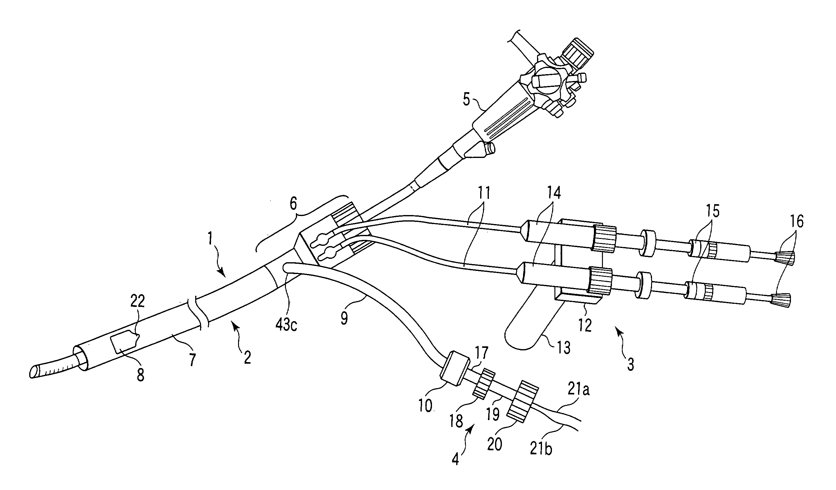
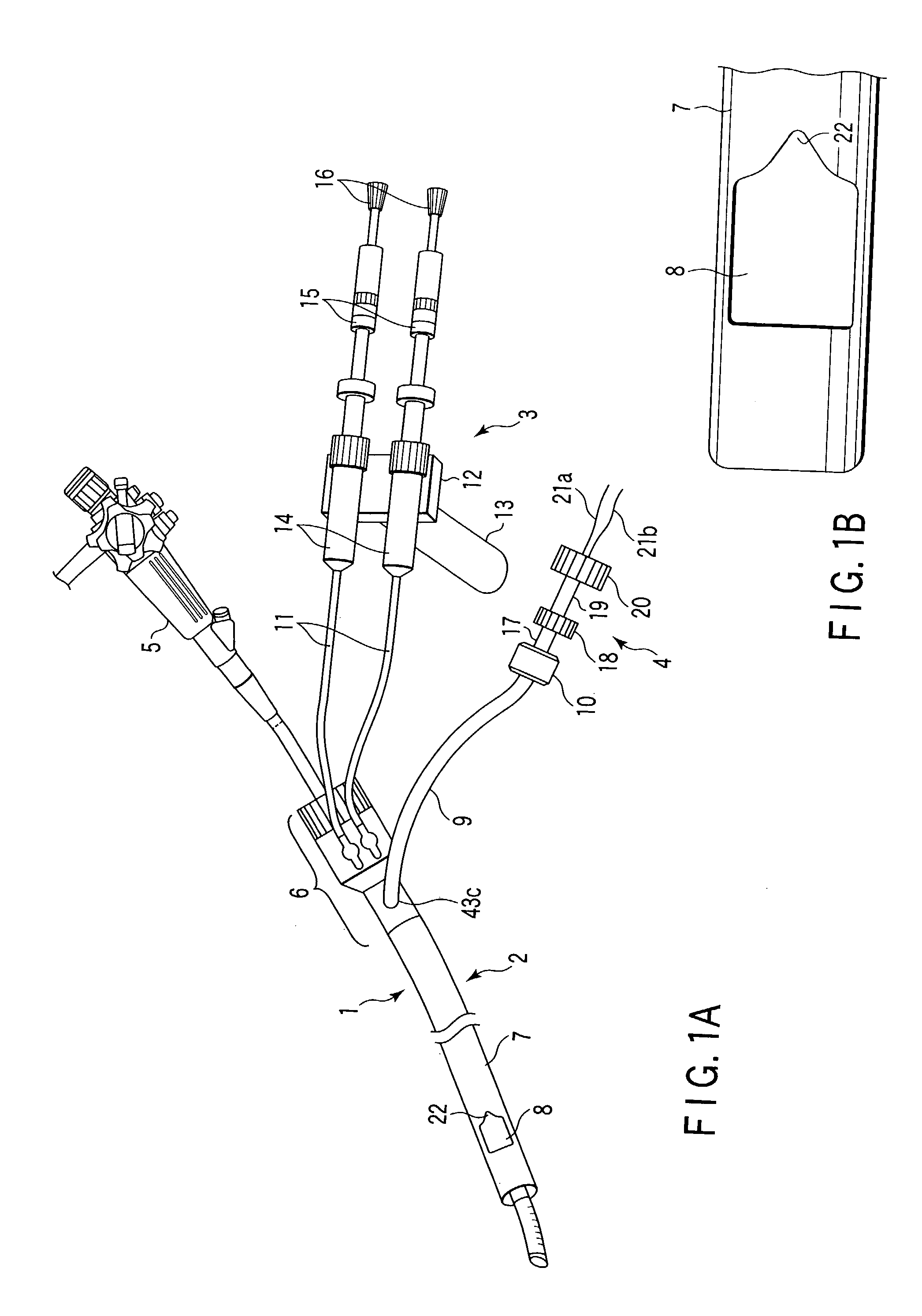
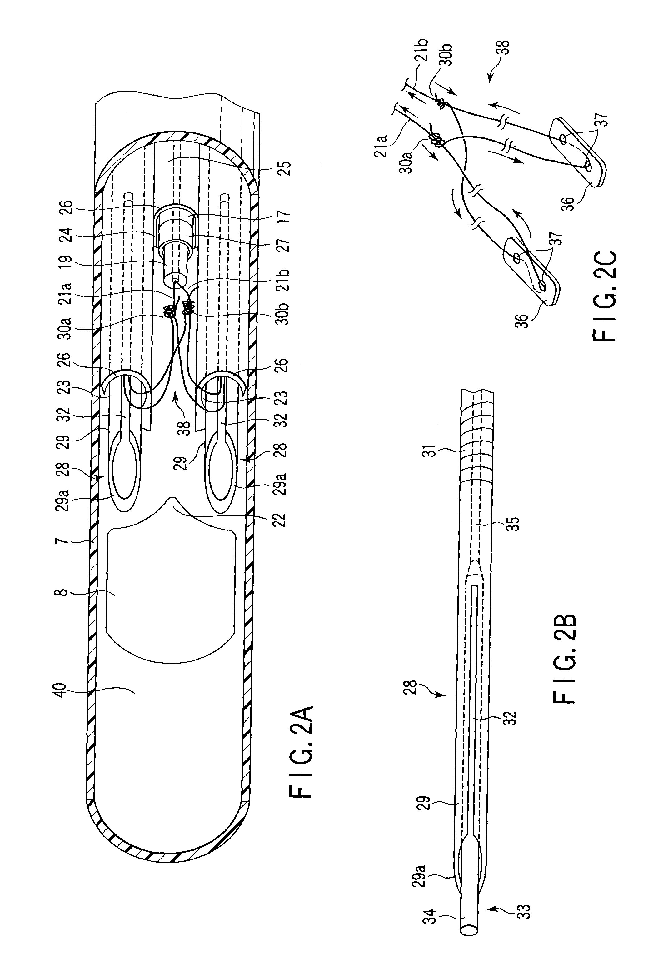
[0145]Hereinafter, the present invention will be described with reference to FIG. 1A to FIG. 12. FIG. 1 shows an appearance of an entire suturing apparatus 1 according to the present embodiment. This suturing apparatus 1 comprises an over-tube 2, a needle manipulating section 3, a ligating device 4, a needle 28 (refer to FIG. 2A), and a ligating unit 38 (refer to FIG. 2C).

[0146]An elongated sheath section 7 is provided at the over-tube 2. This sheath section 7 is set to about 0.3 m to 2 m, for example, and preferably set to about 1 m. As shown in FIG. 1B, a side opening 8 is formed at a distal end section of the sheath section 7. Further, a connecting section 6 is arranged at a proximal end section of the sheath section 7.

[0147]Further, as shown in FIG. 3, the sheath section 7 is formed of a multi-lumen tube, i.e., in this embodiment, a four-lumen tube comprising four lumens (one endoscope lumen 25, two needle lumens 23, and one ligating lumen 24). Then, the two needle lumens 23, th...

second embodiment

[0223]FIG. 14 to FIG. 20B show the present invention. According to the present embodiment, a part of the suturing apparatus 1 according to the first embodiment (refer to FIG. 1A to FIG. 12) has been modified as follows. In the present embodiment, like elements identical to the suturing apparatus 1 of the first embodiment are designated by like reference numerals. A duplicate description is omitted here. Here, only elements different from those of the first embodiment will be described.

[0224]As shown in FIG. 14, in the over-tube 2 of the present embodiment, a suction port 53 is provided at the connecting section 6. This suction port 53 communicates with an endoscope lumen 25. One end of a connecting tube 55a of a suction apparatus 54 is removably connected to the suction port 53.

[0225]A suction source 59 is provided at the suction apparatus 54. One end of a connecting tube 55d is removably connected to this suction source 59. The other end of this connecting tube 55d is connected to ...

sixth embodiment

[0345]FIG. 30A to FIG. 36B each show the present invention. In the present embodiment, a constitution of the over-tube 2 of the suturing apparatus 1 according to the first embodiment (refer to FIG. 1A to FIG. 12) is changed as follows.

[0346]That is, in the present embodiment, the sheath section 7 of the over-tube 2 is formed of a proximal end sheath 109, an intermediate sheath 110, and a distal end sheath 111, as shown in FIG. 30A. The inner and outer diameters of the proximal end sheath 109, the intermediate sheath 110, and the distal end sheath 111 are dimensionally defined to be substantially equal to each other.

[0347]The proximal end section of the proximal end sheath 109 is connected to the distal end of the connecting section 6. The proximal end section of the intermediate sheath 110 is connected to the distal end of this proximal end sheath 109 by means of bonding or the like. The distal end sheath 111 is removably connected to the distal end of the intermediate sheath 110.

[0...


PUM


Abstract

In the case where the living tissues in the patient's body is resected as well, it is possible to suture the resected portion immediately without replacement with another treatment device after resection. Thus, prevention of bleeding from the resected portion or earlier repair of an ulcer that occurs with the resected portion can be carried out for the patent lowly invasively and easily.

Description

CROSS-REFERENCE TO RELATED APPLICATIONS[0001]This application claims the benefit of U.S. Provisional Application No. 60 / 378,548, filed May 8, 2002.BACKGROUND OF THE INVENTION[0002]The present invention relates to an apparatus for ligating / suturing living tissues and a system for resecting / suturing living tissues, for carrying out endoscopic repair such as resecting / suturing or ligating or suturing living tissues in the patient's body in combination with an endoscope. More particularly, the present invention relates to a repair system for carrying out the following repairs in a digestive tract including:[0003]suturing for repair of a damaged site of tissues or reliable hemostasis of a bleeding part;[0004]forming an artificial valve depending on tissues for Gastroesphageal Reflux Disease (GERD); and[0005]resecting a lesion that exists in an organ wall such as mucous membrane having a surface existing lesion part or sub-mucous tumor.[0006]Currently, in the case of carrying out repair o...

Claims


Application Information

Patent Timeline
Patent Type & Authority Patents(United States)
IPC IPC(8): A61B17/10G02B23/24A61B1/00A61B17/04A61B17/06A61B17/22A61B17/221A61B17/28A61B17/32A61B18/14
CPCA61B17/0469A61B17/0482A61B17/0483A61B17/0467A61B17/0487A61B17/32056A61B2017/0404A61B2017/0409A61B2017/0414A61B2017/0417A61B2017/0419A61B2017/0458A61B2017/0464A61B2017/0472A61B2017/0474A61B2017/0475A61B2017/0477A61B2017/0488A61B2017/0496A61B2017/06052A61B2017/061A61B2017/2905
Inventor ONUKI, YOSHIOMIYAMOTO, SATOSHIKAWASHIMA, KOICHIPASRICHA, PANKAJ JAY
Owner APOLLO ENDOSURGERY INC
Why Eureka
Try Eureka