Optical tomography system using short-pulse laser for early cancer diagnostics

a short-pulse laser and tomography technology, applied in the field of optic tomography, can solve the problems of high energy ionization of the tissue, inability to distinguish between malignant and benign tumors, and ineffective for younger women with dense, fibrous breasts, and may be potentially harmful

Inactive Publication Date: 2012-05-29
FLORIDA INST OF TECH
View PDF2 Cites 6 Cited by
  • Summary
  • Abstract
  • Description
  • Claims
  • Application Information

AI Technical Summary

Benefits of technology

[0007]Time-resolved optical tomography systems using a short pulse laser according to the invention provides great advantage in detection and treatment of cancer and tumors, one of the leading causes of mortality in the U.S. and other parts of the world. By using a combination of a short-pulse light source and an ultrafast light detecting system, cancer and tumors can be detected in early stages prior to metastasis, when they are small, and hence can then be irradiated (killed) in situ.
[0011]Time-resolved optical tomography is a novel biomedical imaging technology by which one can detect small tumors in tissue in earlier stages normally not detected by other techniques. Optical technologies have the potential capability of performing in situ diagnosis on tissue without the need for sample excision and processing. This diagnostic information can be available in real time. In addition, since removal of tissue is not required for optical diagnostics, a more complete examination of the organ of interest can be achieved than with excisional biopsy or cytology. The experimental techniques will aid in predicting how the measured temporal radiation signal changes with property distributions. Optical tomography using a short-pulse laser will provide detailed morphological and physiological information about the state of the tissue without any adverse side effects associated with some of the techniques mentioned above.

Problems solved by technology

X-ray mammography, the modality commonly used for breast cancer screening, cannot distinguish between malignant and benign tumors, and is less effective for younger women with dense, fibrous breasts.
Moreover, their high energy can ionize the tissue and may be potentially harmful if used too often for routine screening or at high intensity.
However, both of these diagnostic procedures require physical removal of specimens followed by tissue processing in the laboratory.
As such, these procedures incur a relatively high cost because specimen handling is required and, more importantly, diagnostic information is not available in real time.
Moreover, in the context of detecting early neoplastic changes, both excisional biopsy and cytology can have unacceptable false negative rates, often arising from sampling errors.
Another technique, radioisotope imaging, exposes the body to radioactivity that is potentially harmful.
Ultrasound lacks the resolution to detect objects with linear dimensions smaller than a few millimeters, and like x-rays, do not provide any information about tissue chemistry.
MRI is a powerful technique with sub-millimeter spatial resolution but the cost of superconducting magnets needed for its operation makes it very expensive.
From the standpoint of detecting early cancer and tumors, a primary technological challenge arises from the fact that laser light is highly scattered in all directions by tissue.

Method used


Image

  • Optical tomography system using short-pulse laser for early cancer diagnostics
  • Optical tomography system using short-pulse laser for early cancer diagnostics
  • Optical tomography system using short-pulse laser for early cancer diagnostics

Examples


Embodiment Construction

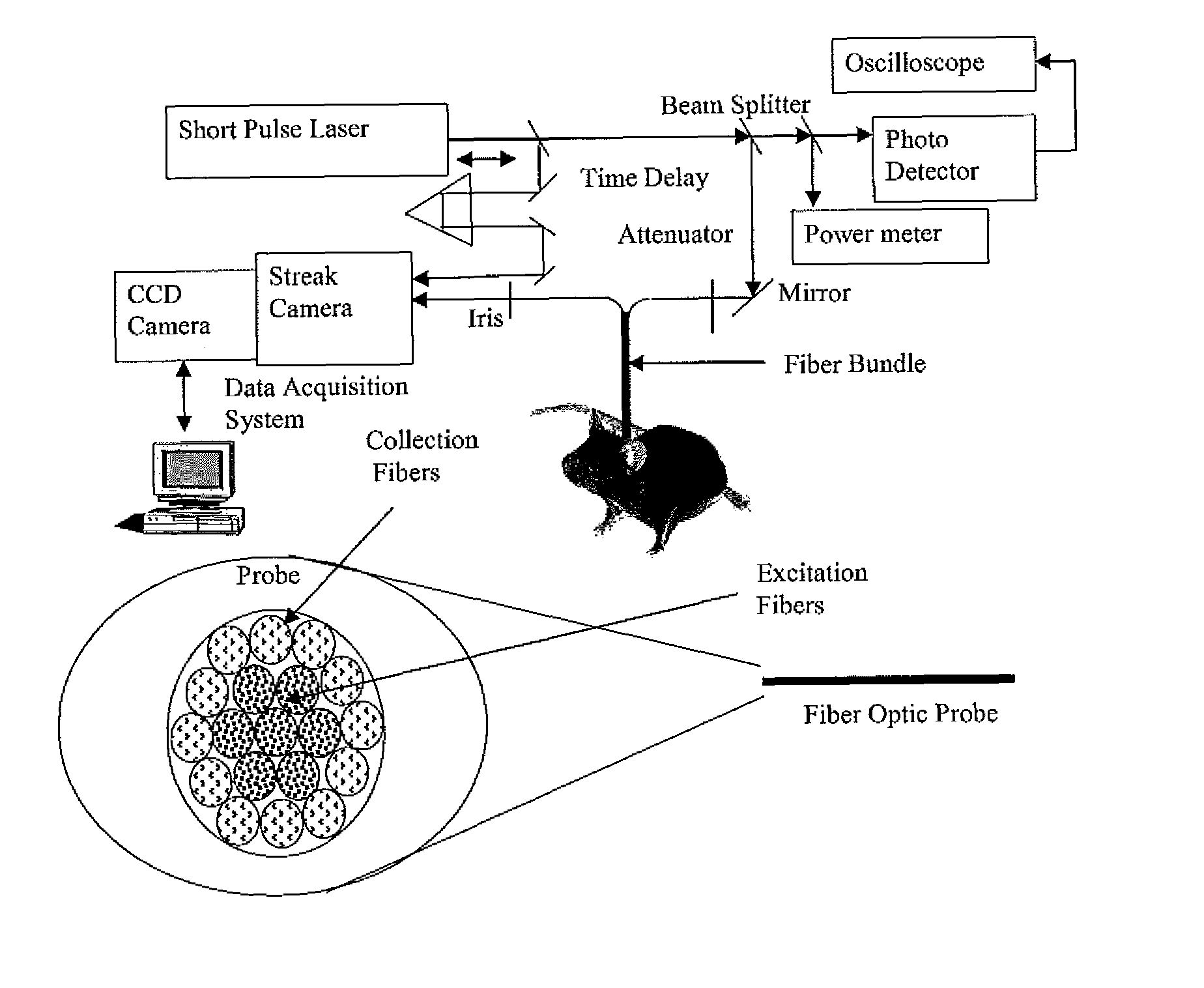
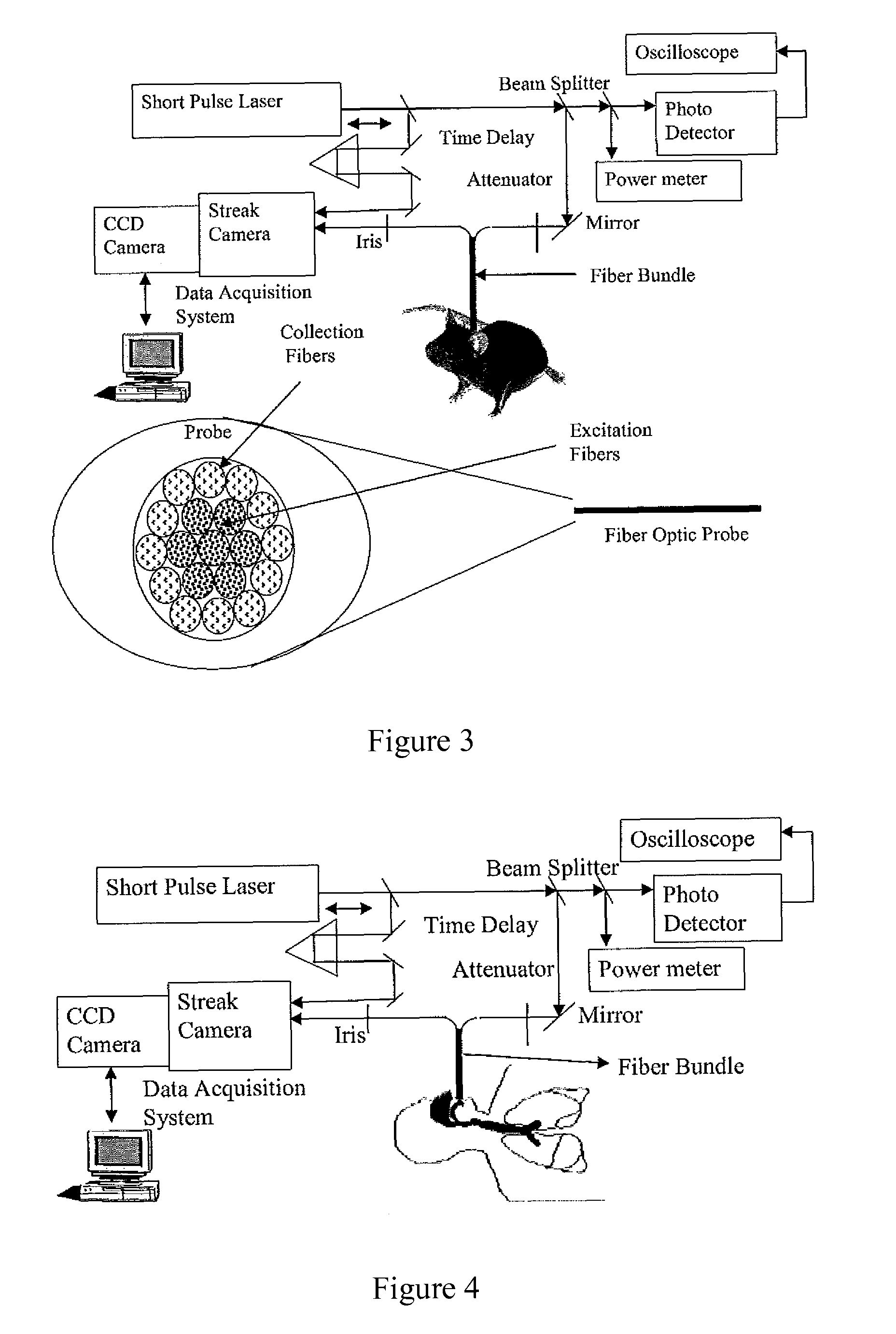
[0029]The invention is an optical tomography system for detection of early tumors and cancer using time-resolved intensity measurements. The location, size, and optical properties of tumors are determined from measured temporal reflected and transmitted signals. Coupling and delivery studies of short-pulse laser energy into tissue using hollow waveguides and tapers have been developed. Software based on transient radiative transport models is used for reconstructing the images from measured reflected and transmitted signals.

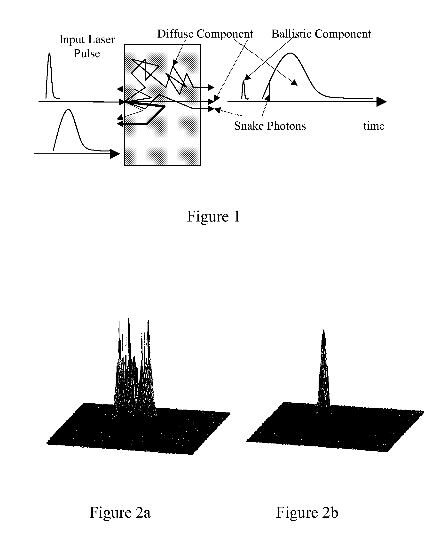
[0030]A short-pulse laser system is used for detection of tumors in tissue. Steps to further the development and understanding of time-resolved optical tomography for biomedical imaging include determination of optical properties of tissue medium, validation of experimental data using numerical models, better understanding of short-pulse laser transport processes, and gaining insight about laser-tissue interaction characteristics via careful experimentation. Tran...


PUM


Abstract

Time-resolved optical tomography systems using a short pulse laser, may be used in detection and treatment of cancer and tumors. Information is conveyed about tissue interior by the temporal variation of the observed scattered, and reflected measured when short-pulse lasers interact with scattering-absorbing media like tissue. Multiple scattering-induced temporal signature that persists for time periods greater than the duration of the source pulse and is a function of the source pulse width, the scattering and absorbing properties of the medium, and the location in the medium where the properties undergo changes. If the detection is carried out on the same short time scale (comparable to the order of the pulse width), the signal continues to be observed even for long durations after the pulse has been off due to the time taken for the photons to migrate to the detector after multiple scattering in the tissue media.

Description

CROSS-REFERENCE TO RELATED APPLICATION[0001]This is a divisional application of U.S. application made under 35 U.S.C. 121 and claims the benefit under 35 USC §120 of U.S. patent application Ser. No. 11 / 693,955, filed Mar. 30, 2007.FIELD OF THE INVENTION[0002]The invention relates to methods of performing cancer diagnostic procedures, in particular diagnostic procedures for early cancer detection.BACKGROUND OF THE INVENTION[0003]Currently, different techniques are used for detection of tumors and other evidence of cancer. For example, cancerous tumors in the lungs are usually located by using conventional x-rays. X-ray mammography, the modality commonly used for breast cancer screening, cannot distinguish between malignant and benign tumors, and is less effective for younger women with dense, fibrous breasts. Moreover, their high energy can ionize the tissue and may be potentially harmful if used too often for routine screening or at high intensity. If a tumor is suspected from an x-...

Claims


Application Information

Patent Timeline
Patent Type & Authority Patents(United States)
IPC IPC(8): A61B6/00
CPCA61B5/0073
Inventor MITRA, KUNAL
Owner FLORIDA INST OF TECH
Why Eureka
Try Eureka