Multi-modality mammography imaging device for improved imaging conditions

a mammography and imaging device technology, applied in the field of medical imaging systems, can solve the problems of low detection capability of specific tissues (tissue properties), difficult detection of tumor mass, early detection of breast cancer, etc., and achieve the effect of improving examination efficiency and detection accuracy of the lesion par

Active Publication Date: 2012-06-05
FUJIFILM CORP
View PDF13 Cites 13 Cited by
  • Summary
  • Abstract
  • Description
  • Claims
  • Application Information

AI Technical Summary

Benefits of technology

[0016]According to one aspect of the present invention, on the basis of the information obtained by performing an examination using the first imaging means (e.g., one of the X-ray mammography apparatus and the ultrasonic imaging apparatus), imaging condition in an examination using the second imaging means (e.g., the other of the X-ray mammography apparatus and the ultrasonic imaging apparatus) are automatically set, and thereby, medical images imaged under appropriate imaging condition according to the characteristics of the respective imaging means and the object status can be obtained. Therefore, for example, in breast cancer screening using both the X-ray mammography apparatus and the ultrasonic imaging apparatus, the detection accuracy of the lesion part such as calcification or tumor mass can be improved and the examination efficiency can be improved regardless of the skills of examiners.

Problems solved by technology

Therefore, finding the lesion parts as earlier in minimal size as possible leads to early detection of breast cancer.
However, the X-ray imaging has a disadvantage that detection capability of specific natures of tissues (tissue properties) is low.
Further, in an X-ray image, mammary glands are expressed in homogeneous soft tissue density, and thus, the detection of tumor mass is difficult in the case where mammary glands have developed (so-called, “dense breast”) as is the case of adolescent to premenopausal women.
On this account, even when a tumor mass is found, it is difficult to take a sample for determination of benign or malignant lesion.
However, ultrasonic imaging examination often depends on the skill of an examiner (an operator such as a doctor) in accuracy and low in reproducibility.
Further, it is difficult to observe minute calcification in an ultrasonic image.
However, in U.S. Pat. No. 5,479,927, by the combination of an X-ray mammography apparatus and an ultrasonic imaging apparatus, only two kinds of examination images correlated with respect to positions of the object or their synthesized images can be generated, but no effort is made to mutually utilize information obtained by the respective examination apparatuses.
However, appropriate imaging condition varies according to object statuses, and thus, it is often difficult to set appropriate imaging condition depending on skills of examiners.

Method used


Image

  • Multi-modality mammography imaging device for improved imaging conditions
  • Multi-modality mammography imaging device for improved imaging conditions
  • Multi-modality mammography imaging device for improved imaging conditions

Examples


first embodiment

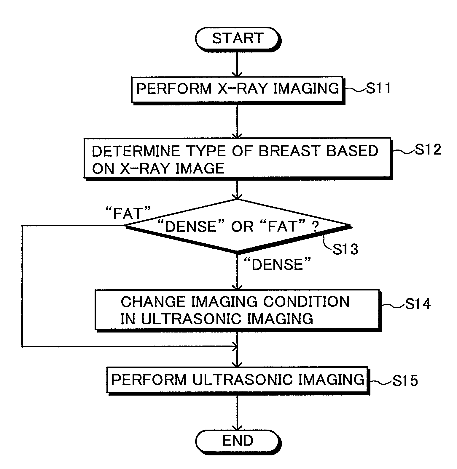
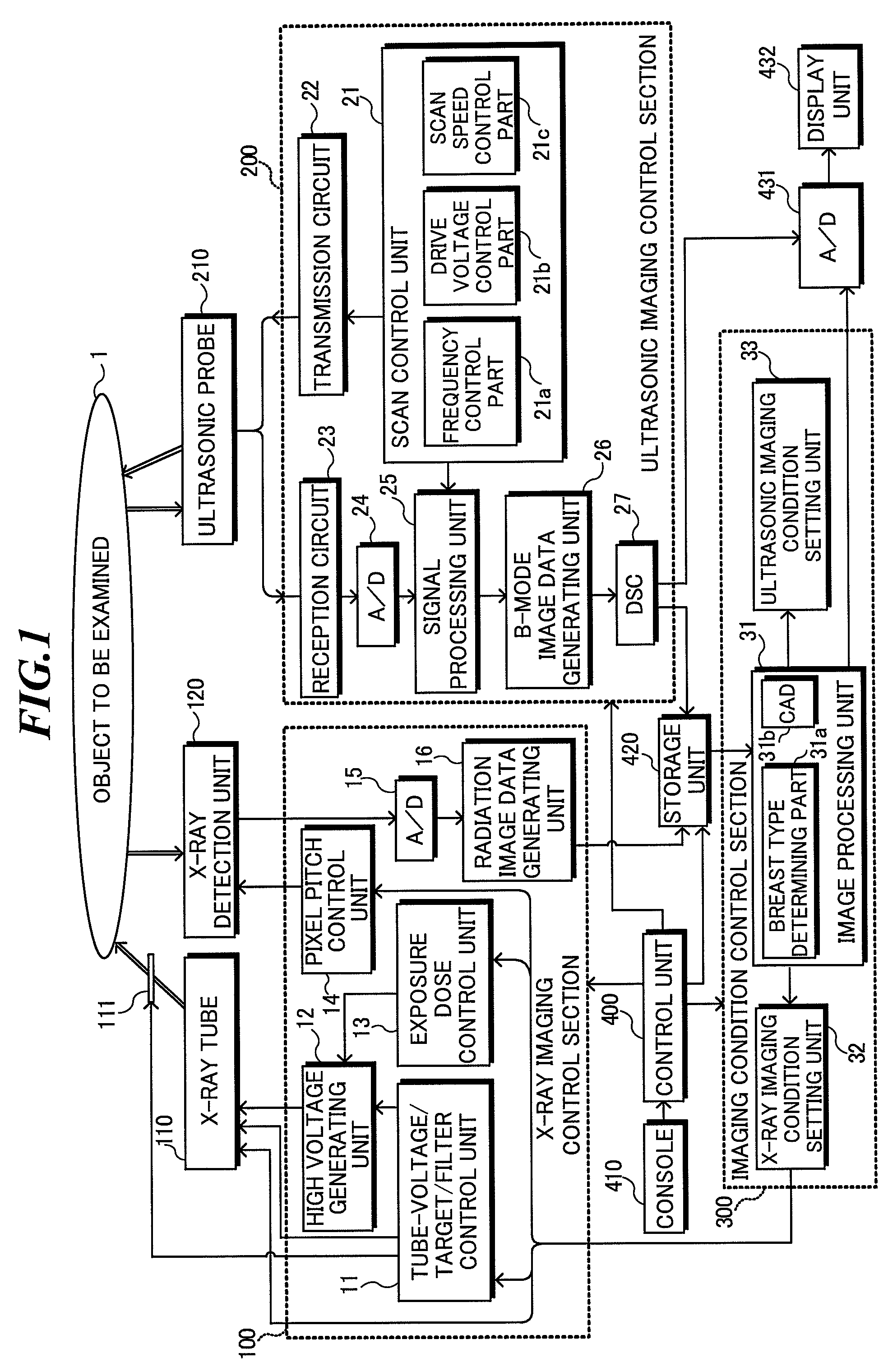
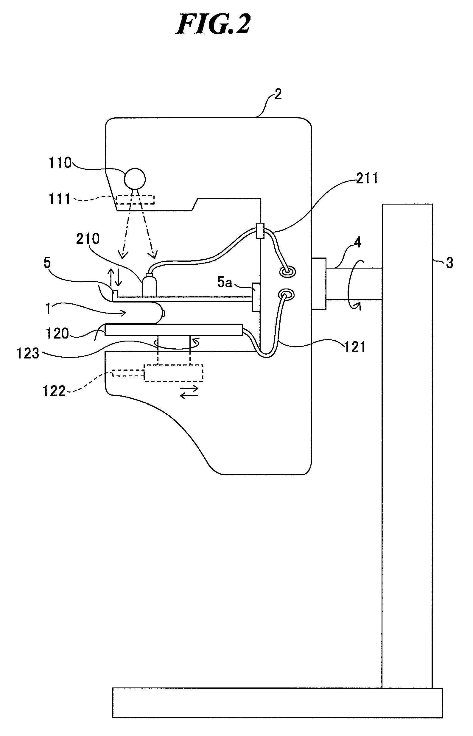
[0032]FIG. 1 is a block diagram showing a configuration of a medical imaging system according to the present invention. The medical imaging system includes a combination of a radiation mammography apparatus for applying radiation to a breast and generating radiation images by detecting radiation transmitted through the breast, and an ultrasonic diagnostic apparatus for transmitting ultrasonic waves to the breast and generating ultrasonic images by receiving ultrasonic waves reflected within the breast. In the embodiment, X-rays are used as radiation.

[0033]The medical imaging system shown in FIG. 1 includes an X-ray imaging control section 100, an X-ray tube 110, a filter unit 111, an X-ray detection unit 120, an ultrasonic imaging control section 200, an ultrasonic probe 210, an imaging condition control section 300, a control unit 400, a console 410, a storage unit 420, a digital / analog converter (D / A) 431, and a display unit 432. Here, the X-ray tube 110, the filter unit 111, the ...

fourth embodiment

[0107]Next, a first modified example of the medical imaging method according to the present invention will be explained.

[0108]In the fourth embodiment, if a tumor mass is detected at step S45 in FIG. 11, the imaging condition is changed and X-ray imaging is performed (step S46), while, in the first modified example, imaging for generating an energy subtraction image is performed in place of typical X-ray imaging.

[0109]Here, the energy subtraction image is an image generated by generating two kinds of X-ray images based on X-rays generated under different condition and subtracting pixel values of the second X-ray image from pixel values of the first X-ray image. In typical X-ray imaging, normal mammary gland tissues, other soft tissues, tumors, and so on overlap, and it may be difficult to separate and observe the lesion part. However, those respective tissues are different from one another in X-ray absorption characteristics, and thus, particular tissues (structures) can be extracte...

second embodiment

[0125]Next, a medical imaging system according to the present invention will be explained.

[0126]FIG. 14 is a block diagram showing a configuration of a medical imaging system according to the second embodiment of the present invention. As shown in FIG. 14, the medical imaging system includes plural kinds of imaging modalities (imaging apparatuses) 501-507 each for imaging an object to be inspected to generate a medical image, an imaging condition setting apparatus 510 for setting imaging condition in one imaging modality based on the medical image generated by another imaging modality, a medical image server 520 for storing medical image data, a plurality of medical image display apparatuses 531 and 532 for displaying medical images, an image interpretation report creating apparatus 540 to be used by an image interpretation doctor when creating an image interpretation report, an image interpretation report server 550 for storing image interpretation report data. Those apparatuses ar...


PUM


Abstract

In a medical imaging system including plural imaging section, detection accuracy and examination efficiency for breast cancer are improved by setting appropriate imaging condition. The system includes: a first imaging section for imaging an object to be inspected by using one of radiation, ultrasonic waves, nuclear magnetic resonance, positron, infrared light and fluorescence to generate a medical image; a second imaging section for imaging the object by using another one of radiation, ultrasonic waves, nuclear magnetic resonance, positron, infrared light and fluorescence to generate a medical image; and an imaging condition setting section for setting imaging condition in the second imaging section based on the medical image generated by the first imaging section.

Description

[0001]The present application claims priority from Japanese Patent Application No. 2006-351634 filed on Dec. 27, 2006, the contents of which are incorporated herein by reference in their entirety.BACKGROUND OF THE INVENTION[0002]1. Field of the Invention[0003]The present invention relates to medical imaging system and method for generating medical images to be used for medical diagnoses, and specifically, relates to medical imaging system and method employing plural imaging apparatus such as a radiation mammography apparatus and an ultrasonic imaging apparatus.[0004]2. Description of a Related Art[0005]Recent years, breast cancer patients have been increasing not only in Europe and the United States but also in Japan. However, even when a breast has a cancer, early treatment can cure the breast cancer relatively easily without the need for breast amputation in many cases. Accordingly, apparatuses and systems for early detection of breast cancer are desired.[0006]When a breast has a ...

Claims


Application Information

Patent Timeline
Patent Type & Authority Patents(United States)
IPC IPC(8): A61B8/00G06V10/10G06V10/25G06V10/98
CPCA61B5/0077A61B6/037A61B6/485A61B6/502A61B6/5247A61B8/0825A61B8/4416G06K9/03G06K9/20G06K9/3233A61B6/4417G06K2209/05A61B5/0035G06V10/98G06V10/10G06V10/25G06V2201/03
Inventor SENDAI, TOMONARI
Owner FUJIFILM CORP
Why Eureka
Try Eureka