Methods and systems for segmentation using boundary reparameterization

a segmentation algorithm and boundary reparameterization technology, applied in image analysis, image enhancement, instruments, etc., can solve the problems of inability to segment medical ultrasound images, time-consuming and laborious, and medical ultrasound images are intrinsically difficult to segmentation algorithms, so as to facilitate cleaning of binary images

Active Publication Date: 2012-06-12
ELEKTA AB
View PDF6 Cites 81 Cited by
  • Summary
  • Abstract
  • Description
  • Claims
  • Application Information

AI Technical Summary

Benefits of technology

[0007]In general, a contour within an image of a lesion or organ of interest is identified, and a region or “band” around the initial contour is then defined. The band is transformed into a boundary image in the form of a rectangular array. The boundary image comprises sampling lines, such as columns within the image. For example, each sampling line can originate on the inside of the organ and end on the outside of the organ, in which case each line contains at least one point (an edge point) of the organ boundary. Features of the boundary image are found, and these features are then transformed back into the original image, thus resulting in improved image segmentation.
[0009]The methods described herein may include using a constant local threshold (CLT) approach, which employs a threshold estimate based on the combination of local and global statistics to binarize the boundary image and further facilitates cleaning of these binary images to find the edge curve in boundary space.

Problems solved by technology

However, because of the complexity of these systems and various characteristics of the resulting images, interpretation typically requires a skilled and highly-trained physician.
The process, however, is time-consuming and prone to user variability.
In particular, medical ultrasound images are intrinsically difficult for segmentation algorithms.
This complexity leads to high failure rates for conventional automatic segmentation algorithms.

Method used


Image

  • Methods and systems for segmentation using boundary reparameterization
  • Methods and systems for segmentation using boundary reparameterization
  • Methods and systems for segmentation using boundary reparameterization

Examples


Embodiment Construction

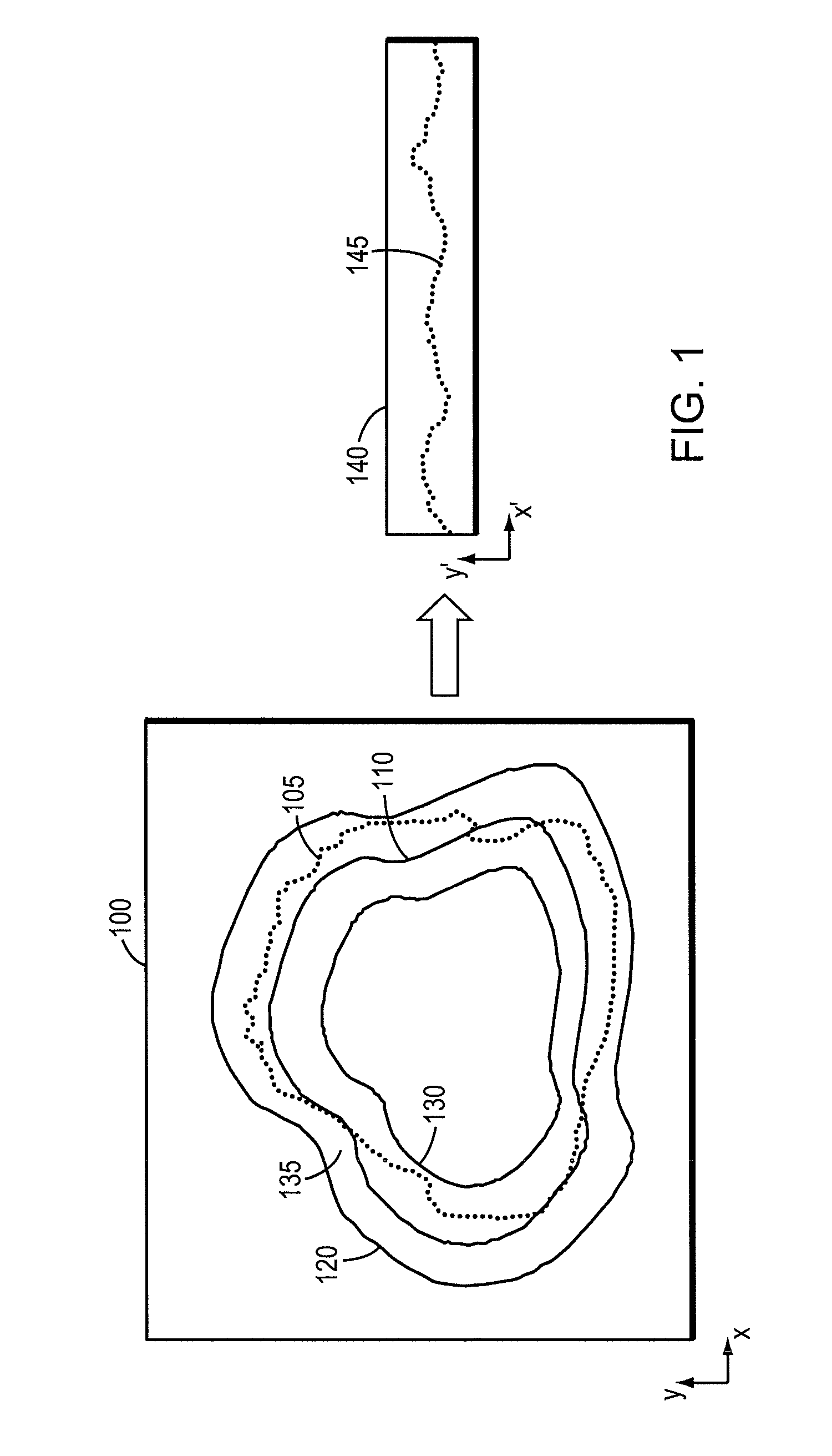
[0028]Referring to FIG. 1, an image 100 of an organ or lesion 105 is used to plan and / or assist with various medical procedures. The image 100 can be an individual two-dimensional image, a two-dimensional sectional image or slice through a three-dimensional image, a three-dimensional image, or any combination thereof. The image 100 can be obtained using one or more devices such as a CT scanner, an ultrasound device, an MRI device, a PET scanner, and / or an x-ray device, or any other suitable imaging modality as commonly used in the art. The image 100 may be used by an oncologist, physician, or radiation technician to determine a diagnosis, the location and shape of the lesion 105 to be treated and / or to determine the parameters of a radiation treatment plan such as beam angle, beam shape, the number of beams needed to administer a sufficient radiation dose to eradicate the target lesion 105, the dose level for each beam, as well as patient positioning parameters.

[0029]In accordance w...


PUM


Abstract

Representations of a segmented, contoured organ or lesion are obtained from two-dimensional or three-dimensional images. A contour within the image of the lesion or organ of interest is used to identify a region around the initial contour and transform it into a boundary image comprising sampling lines that contain points identifying the organ boundary.

Description

CROSS-REFERENCE TO RELATED APPLICATION[0001]This application claims priority to and the benefit of, and incorporates herein by reference, in its entirety, provisional U.S. patent application Ser. No. 60 / 801,317, filed May 18, 2006.TECHNICAL FIELD[0002]This invention relates to methods and systems for identifying anatomical features in medical images, and more specifically to using various segmentation and mapping techniques to accurately identify boundaries in the images.BACKGROUND INFORMATION[0003]Imaging modalities such as computed tomography (CT), magnetic resonance imaging (MRI), ultrasound, positron emission tomography (PET) and single-photon emission computed tomography (SPECT) provide various representations of anatomical features of a subject. Image sets generated with any of these modalities can be used for diagnosis or to guide various treatments, such as surgery or radiation therapy. The images can consist of two-dimensional representations, three-dimensional voxel images...

Claims


Application Information

Patent Timeline
Patent Type & Authority Patents(United States)
IPC IPC(8): G06K9/00G06V10/44
CPCG06K9/4604G06T3/0043G06T7/0083G06K2209/05G06T2207/10132G06T2207/20104G06T2207/30096G06T7/12G06V10/44G06V2201/03
Inventor KOPTENKO, SERGEILACHAINE, MARTINHUANG, XINGFALCO, TONY
Owner ELEKTA AB
Why Eureka
Try Eureka