Systems and methods for cardiac tissue electroporation ablation

a technology of electroporation ablation and cardiac tissue, which is applied in the field of cardiac tissue treatment, can solve the problems of disrupting the normal path of depolarization events, disrupting the normal propagation of electrical impulses, and disrupting the normal activation of atria or ventricles

Active Publication Date: 2012-07-17
MEDTRONIC INC
View PDF22 Cites 170 Cited by
  • Summary
  • Abstract
  • Description
  • Claims
  • Application Information

AI Technical Summary

Benefits of technology

[0008]Some aspects in accordance with principles of the present disclosure relate to a method of performing an electroporation ablation procedure on tissue of a patient. At least one electrode is positioned on the tissue, and pulsed, high voltage biphasic energy is delivered to the electrode to induce electroporation of cells of the tissue followed by cell rupturing. In some applications, the pulsed, biphasic energy has a cycle time of not more than 500 microseconds, and is delivered at an output voltage in the range of 200-700 volts and a pulse width in the range of 50-200 microseconds. In other embodiments, the electroporation ablation procedure is performed on heart tissue.
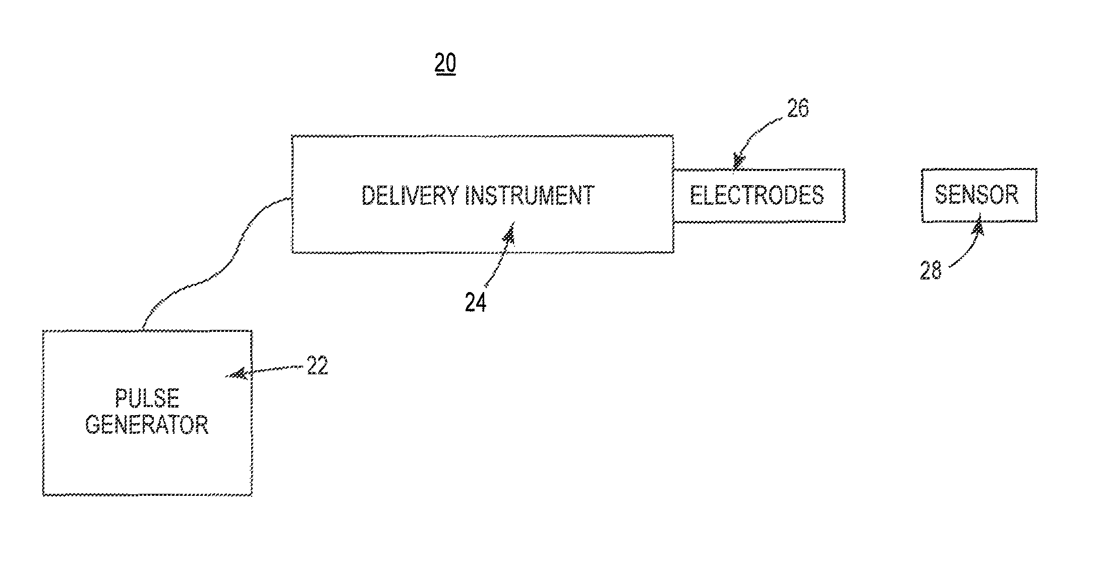
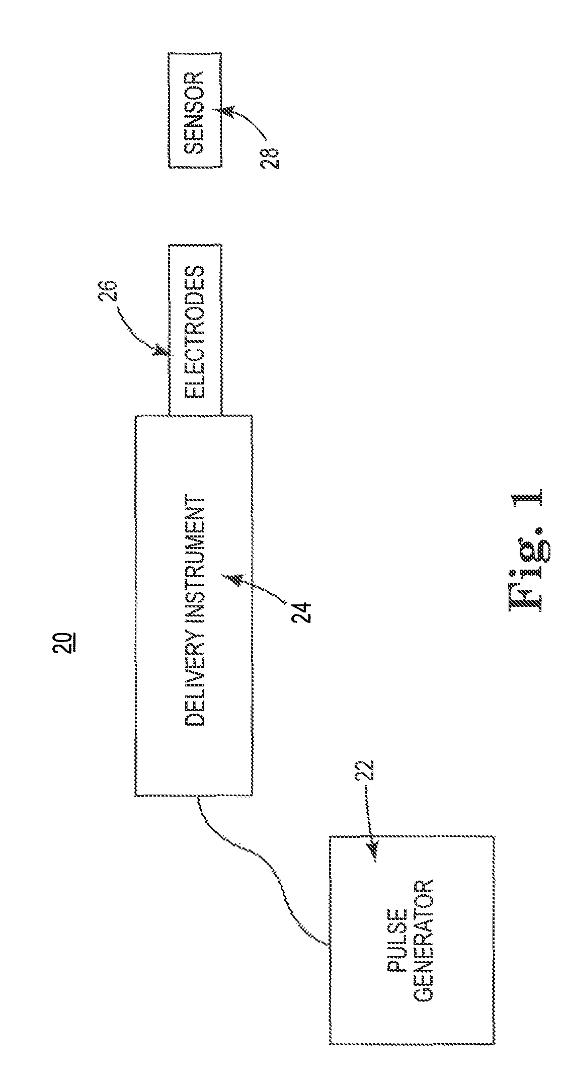
[0009]Other aspects in accordance with principles of the present disclosure relate to a cardiac tissue electroporation ablation system including a delivery instrument and an electrical pulse generator. The delivery

Problems solved by technology

Sometimes, aberrant conductive pathways develop in heart tissue, which disrupts the normal path of depolarization events.
For example, anatomical obstacles in the atria or ventricles can disrupt the normal propagation of electrical impulses.
These wavelets, called “reentry circuits,” disrupt the normal activation of the atria or ventricles.
In cer

Method used


Image

  • Systems and methods for cardiac tissue electroporation ablation
  • Systems and methods for cardiac tissue electroporation ablation
  • Systems and methods for cardiac tissue electroporation ablation

Examples


examples

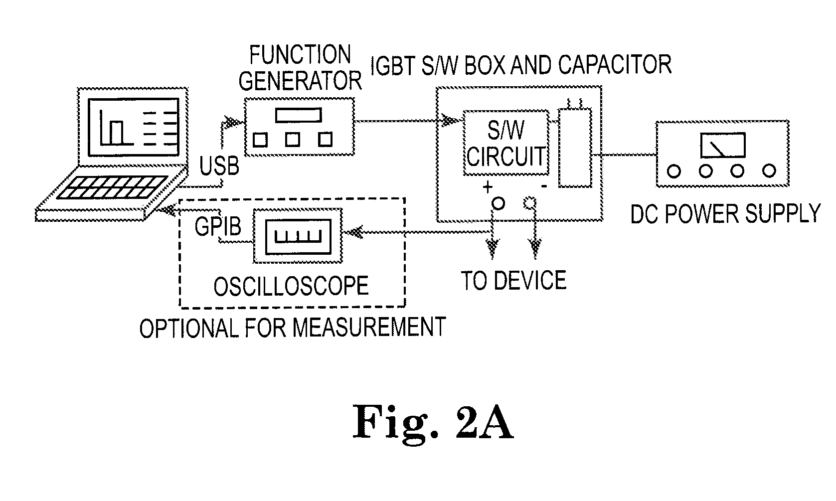
[0054]Extensive efforts have been made by the inventors to confirm an ability of systems and methods in accordance with the present disclosure to effectuate viable cardiac ablation via IEP.

Ex Vivo Study

[0055]An ex vivo swine skeletal muscle model was developed for evaluating cell death mechanism by measuring stimulated muscle force up to 24 hours. Skeletal muscle specimens from porcine rectus abdominis muscle biopsies were obtained and placed in oxygenated Krebs buffer. Immediately after removal, the muscle was placed in a dissection dish that was continuously oxygenated with carbogen (95% O2 and 5% CO2) at room temperature. The muscle specimens were affixed with small pins to the bottom of a dish lined with sylgard in order to fix the position of the muscle while it was further prepared. Connective tissue and fat was removed from the muscle with a fine scissors and forceps while the preparation was viewed through a dissecting microscope. Several smaller muscle bundles, with approxi...


PUM


Abstract

Cardiac electroporation ablation systems and methods in which pulsed, high voltage energy is delivered to induce electroporation of cells of cardiac tissue followed by cell rupturing. In some embodiments, the delivered energy is biphasic, having a cycle time of not more than 500 microseconds.

Description

CROSS-REFERENCE TO RELATED APPLICATION[0001]This application claims priority under 35 U.S.C. §119(e)(1) to U.S. Provisional Patent Application Ser. No. 61 / 084,149, filed Jul. 28, 2008, entitled “Systems and Methods for Cardiac Tissue Electroporation Ablation”, and bearing the entire teachings of which are incorporated herein by reference.BACKGROUND[0002]The present disclosure relates generally to the treatment of cardiac tissue of a patient with ablative energy. More particularly, it relates to ablation of cardiac tissue using high voltage ablation, for example via high voltage electroporation ablation or irreversible electroporation (IEP) ablation.[0003]There are many medical treatments that involve instances of cutting, ablating, coagulating, destroying, or otherwise changing the physiological properties of tissue. These techniques can be used beneficially to change the electrophysiological properties of tissue, for example by ablation of cardiac tissue to cure various cardiac con...

Claims


Application Information

Patent Timeline
IPC IPC(8): A61B18/18
CPCA61B18/1442A61B18/1492A61B2018/00291A61B2018/00351A61B2018/00613A61B2018/00642A61B2018/00702A61B2018/00738A61B2018/00839A61B2018/00875A61B2018/122A61B2218/002A61B2218/007
Inventor FRANCISCHELLI, DAVIDSTEWART, MARKHONG, JINBACKNIKOLSKI, VLADIMIRHAMLEN, CUSHINGCHEEK, DANIELBONNER, MATTHEW
Owner MEDTRONIC INC
Why Eureka
Try Eureka