Tissue anchor for annuloplasty device

a tissue anchor and annuloplasty technology, applied in the field of tissue anchors, can solve the problems of reducing cardiac output, increasing total stroke volume, and ultimate weakening of the left ventricle, and achieve the effect of facilitating the implantation of the implant and facilitating the coupling of the implan

Active Publication Date: 2012-10-02
VALTECH CARDIO LTD
View PDF194 Cites 192 Cited by
  • Summary
  • Abstract
  • Description
  • Claims
  • Application Information

AI Technical Summary

Benefits of technology

[0027]In some applications of the present invention, a tissue anchor is provided that is configured for receiving an implant and facilitating implantation of the implant. The anchor comprises a distal tissue coupling element, e.g., a helical anchor, which penetrates tissue of a patient. The anchor also comprises a proximal implant-penetrating element which receives and facilitates coupling of the implant to the tissue anchor. The implant-penetrating element comprises a post, which extends between the proximal tip and the proximal end of the distal tissue coupling element. For some applications, the proximal tip of the implant-penetrating element comprises a barb which punctures and receives the implant.
[0060]a flexible contracting member that is coupled to the spool and the sleeve, such that winding the contracting member around the spool tightens the device.
[0073]restraining the implant from separating from the implant-penetrating element by allowing the one or more arms to expand and rest against an outer surface of the implant following the penetrating of the barb through the implant.

Problems solved by technology

Mitral regurgitation of blood from the left ventricle into the left atrium results in increased total stroke volume and decreased cardiac output, and ultimate weakening of the left ventricle secondary to a volume overload and a pressure overload of the left atrium.

Method used


Image

  • Tissue anchor for annuloplasty device
  • Tissue anchor for annuloplasty device
  • Tissue anchor for annuloplasty device

Examples


Embodiment Construction

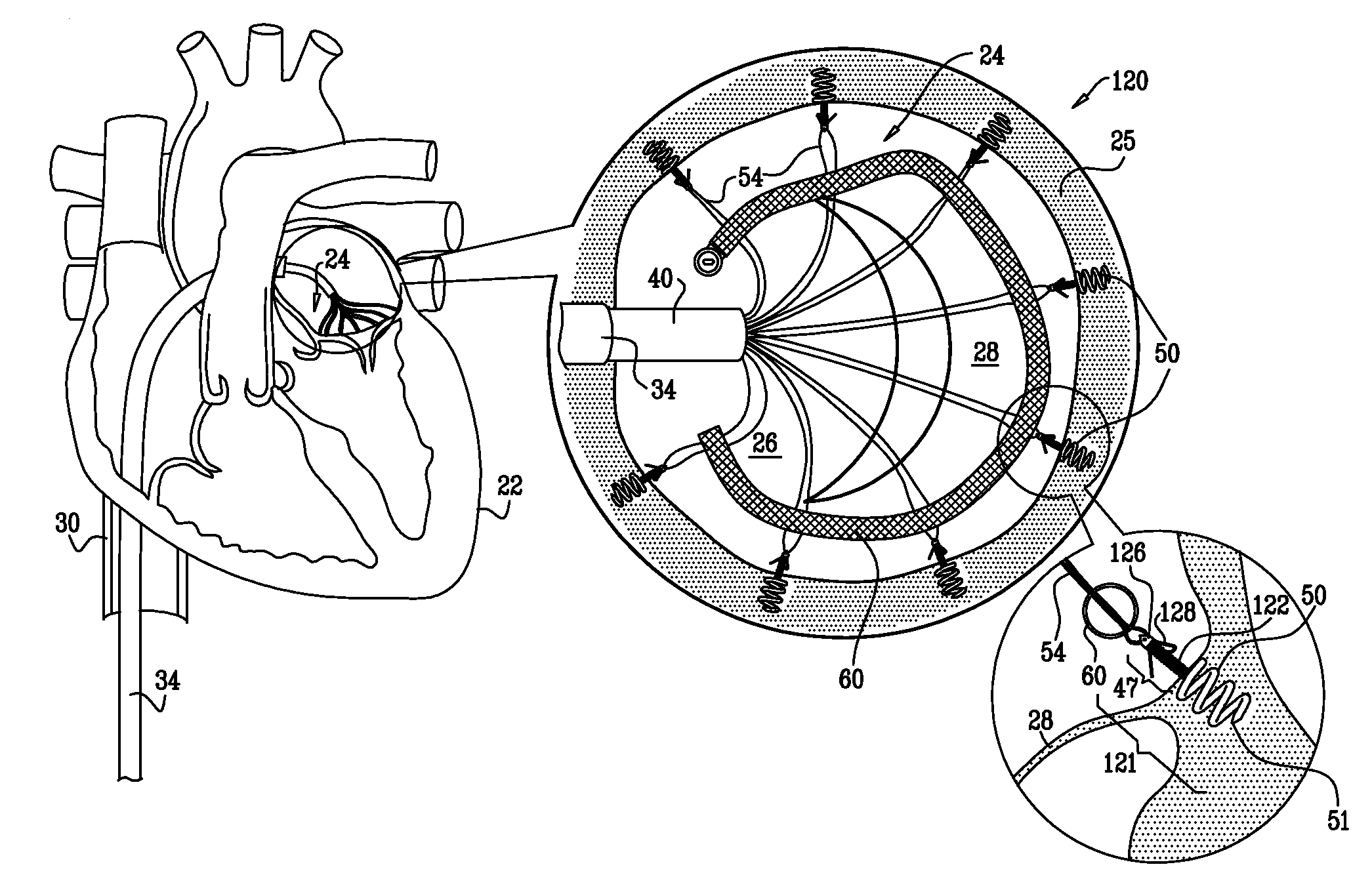
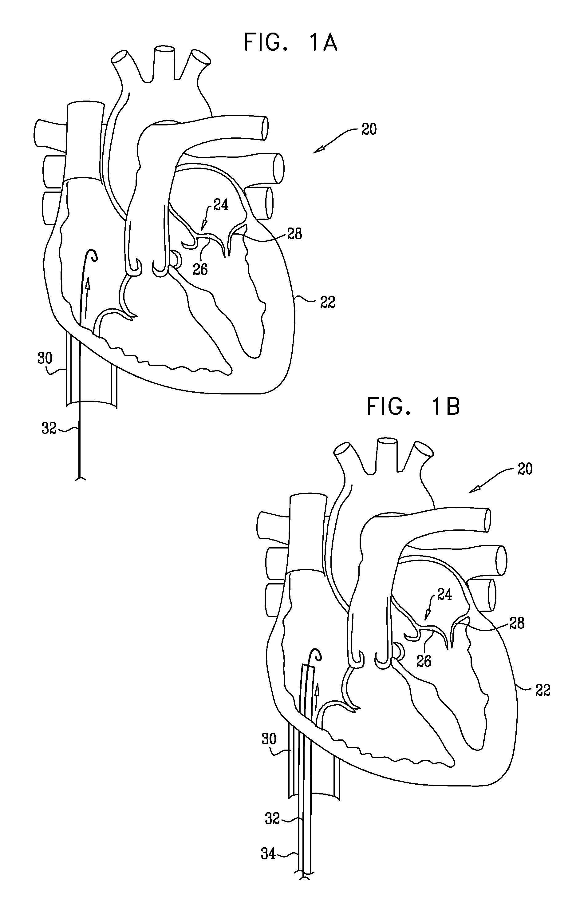
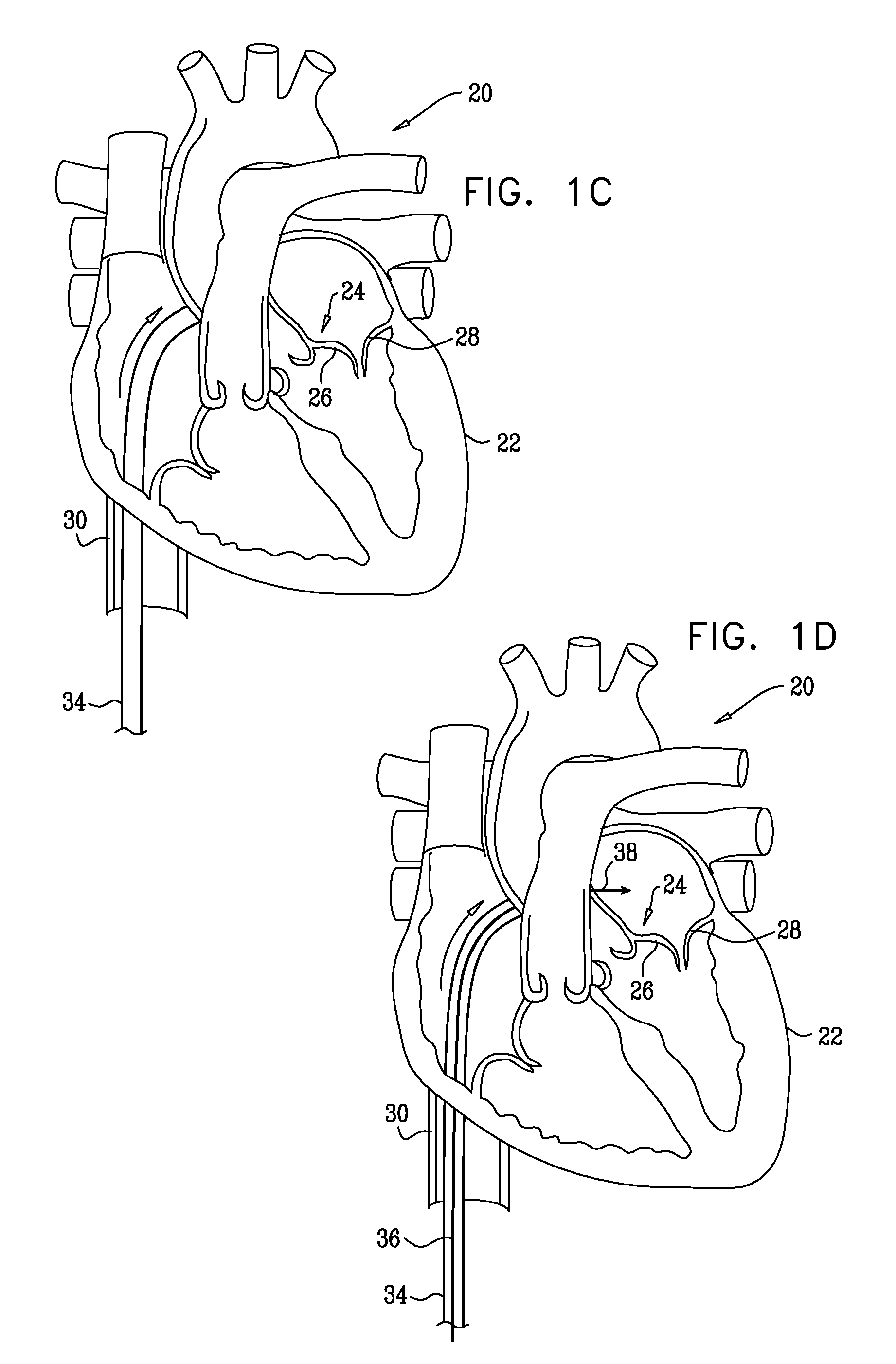
[0095]Reference is now made to FIGS. 1A-F, 2A-C, and 3, which are schematic illustrations of a system 20 for implanting a tissue anchor 49, in accordance with some applications of the present invention. FIGS. 1A-F show a transcatheter procedure for implanting tissue anchor 49. FIGS. 2A-C show a transcatheter delivery tool 42 for delivering toward and implanting anchor 49 at an implantation site, e.g., an annulus 25 of a heart 22 of a patient, as shown. Typically, the implantation site includes an annulus of an atrioventricular valve, e.g., a mitral valve or a tricuspid valve. It is to be noted that the implantation site is not limited to a heart valve of the patient, and anchor 49 may be implanted in other tissue of the patient, e.g., a portion of the inner wall of the heart of the patient, in a stomach of a patient, etc. Tissue anchor 49, as shown in FIG. 2B comprises a distal tissue coupling element 50, e.g., a helical tissue anchor, and a proximal implant-penetrating element 47. ...


PUM


Abstract

A method is provided, including coupling, to a first portion of tissue of a patient, a distal tissue coupling element of a tissue anchor. The tissue anchor further includes (a) an implant -coupling element, and (b) a cord, which is removably coupled to the implant-coupling element. The method comprises advancing an implant over the cord until the implant reaches and is coupled to the implant-coupling element. The method further includes restraining the implant from separating from the implant-coupling element, with an implant-restraining element of the implant-coupling element. Other applications are also described.

Description

FIELD OF THE INVENTION[0001]Some applications of the present invention relate in general to tissue anchors. More specifically, some applications of the present invention relate to tissue anchors for repair of an atrioventricular valve of a patient.BACKGROUND OF THE INVENTION[0002]Dilation of the annulus of the mitral valve prevents the valve leaflets from fully coapting when the valve is closed. Mitral regurgitation of blood from the left ventricle into the left atrium results in increased total stroke volume and decreased cardiac output, and ultimate weakening of the left ventricle secondary to a volume overload and a pressure overload of the left atrium.[0003]US Patent Application 2004 / 0236419 to Milo describes methods for reconfiguring an atrioventricular heart valve that may use systems comprising a partial or complete annuloplasty rings proportioned to reconfigure a heart valve that has become in some way incompetent, a pair of trigonal sutures or implantable anchors, and a plu...

Claims


Application Information

Patent Timeline
Patent Type & Authority Patents(United States)
IPC IPC(8): A61F2/06
CPCA61B17/0401A61B17/064A61B17/068A61F2/2445A61F2/2466A61B2017/0409A61B2017/0414A61B2017/0427A61B2017/00243A61B2017/0448A61B2017/0464A61B2017/0647A61B2017/0649A61B2017/0441
Inventor MILLER, ERANCABIRI, OZ
Owner VALTECH CARDIO LTD
Why Eureka
Try Eureka