Apparatus and method for imaging a medical instrument

a medical instrument and apparatus technology, applied in the field of medical instruments, can solve the problems of headache, failure rate of 6 to 20%, other procedures are difficult to perform without additional guidance, etc., and achieve the effect of enhancing stability

Inactive Publication Date: 2014-04-15
THE UNIV OF BRITISH COLUMBIA
View PDF15 Cites 4 Cited by
  • Summary
  • Abstract
  • Description
  • Claims
  • Application Information

AI Technical Summary

Benefits of technology

[0017]The mount may further have a second contact that is to be in contact with the body in addition to the probe. In this way, the second contact point can provide additional stability for the apparatus with respect to the patient.
[0018]The mount may also have markings representing the inferior-superior and left-right axes of the body thereby indicating the desired position of the apparatus on the body. In this way, the operator can easily figure out the orientation and position the apparatus onto the body.
[0025]The size of the volume acquired by the probe can be determined in a manner that it minimally encompasses the maximum extents of the thick slice used to create the image. In this way, the probe can acquire the smaller volume faster and the smaller volume can be processed by the circuit faster.
[0028]The target can be an epidural space. The probe can be placed at a paramedian location with respect to the spine. Moreover, the probe can be placed over spinae erector muscles of the patient. In this way, the muscle tissue may serve as a “window” that transmits ultrasound particularly well.

Problems solved by technology

Other procedures are difficult to perform without additional guidance because the target is deep, the target is small, sense of feel is inadequate for recognizing when the needle's tip has reached the target, or there is a lack of visual landmarks on the body surface.
This method can result in failure rates of 6 to 20% depending on the experience and training of the health care provider.
Complications include inadvertent dura puncture resulting in loss of cerebral spinal fluid and headache, as well as nerve injury, paralysis and even death.
While pre-puncture scanning is a useful advance, doctors still face challenges associated with performing needle insertion procedures without information provided by real-time imaging.
However, none of these approaches have proven to be entirely satisfactory.
Problems include overly limiting views of the images of the target and needle due to poor reflection of ultrasound waves, and / or inherent limitations in the ultrasound equipment.

Method used


Image

  • Apparatus and method for imaging a medical instrument
  • Apparatus and method for imaging a medical instrument
  • Apparatus and method for imaging a medical instrument

Examples


Embodiment Construction

[0051]Other aspects and features of the present invention will become apparent to those ordinarily skilled in the art upon review of the following description of illustrative embodiments in conjunction with the accompanying figures.

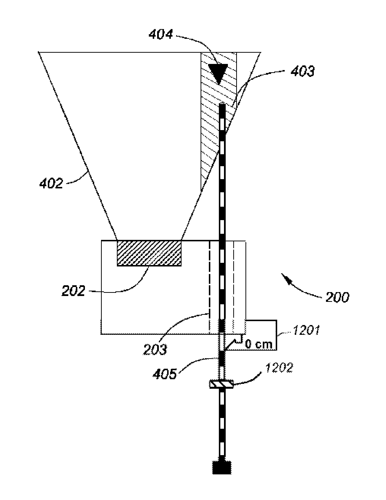
[0052]Ultrasound imaging is a technique for imaging the interior of the body with high frequency sound waves. A standard ultrasound probe comprises a set of transducer elements emitting sound waves into the body. The sound waves reflect on tissue or bone in the body and the reflected sound (echo) is detected by the same transducer elements. By calculating the time from emission to detection of the sound waves at each transducer and measuring the intensity of the reflected sound wave, an ultrasound image can be constructed that shows various anatomical features in the ultrasound probe's field of view.

[0053]Ultrasound scanning during a needle insertion procedure enables the observation of both the needle and the target on a real-time ultrasound display. One...


PUM


Abstract

The invention provides an ultrasound imaging and medical instrument guiding apparatus, a system for acquiring and displaying ultrasound medical images and methods of using the apparatus and system in epidural anesthetic procedure. The apparatus comprises a hand-held ultrasound probe, configured to acquire a volumetric dataset representing a 3-D depiction of a volume; a mount to which the probe is mounted; and a medical instrument guide positionable relative to the ultrasound probe and configured to receive and guide a medical instrument along a propagation axis to a target in a body such that the target and the propagation axis intersect in the volume. The volumetric dataset acquired by the hand-held ultrasound probe comprises information about the medical instrument's position relative to the target in three dimensions and the medical instrument guide has a visible mark from which the depth of the medical instrument insertion along the propagation axis can be referenced.

Description

CROSS-REFERENCE TO RELATED APPLICATIONS[0001]This application claims the benefit of U.S. Provisional Application No. 61 / 484,373 filed 10 May 2011, the entire contents and substance of which are hereby incorporated by reference.BACKGROUND OF THE INVENTION[0002]1. Field of the Invention[0003]This invention relates generally to medical imaging, and particular to an apparatus and method for imaging a medical instrument, particularly while being inserted inside a patient.[0004]2. Description of Related Art[0005]Some medical procedures require a needle or needle-like instrument to be inserted into a patient's body to reach a target. Examples of these procedures include tissue biopsies, drug delivery, drainage of fluids, ablation for cancer treatment, and catheterization. Some of these procedures can be done manually without any additional guidance other than the sense of feel and visualization of the surface of the body. Other procedures are difficult to perform without additional guidanc...

Claims


Application Information

Patent Timeline
Patent Type & Authority Patents(United States)
IPC IPC(8): A61B8/14
CPCA61B2017/3413A61B2019/5276A61B8/0841A61B19/5225A61B8/461A61B17/3401A61B8/4483A61B8/4245A61B8/463A61B8/483A61B90/37A61B2090/378
Inventor ROHLING, ROBERT
Owner THE UNIV OF BRITISH COLUMBIA
Why Eureka
Try Eureka